Arthroscopic Anterior Cruciate Ligament (ACL) Reconstruction Using Hamstring Tendon at the Order of Malta's Hospital Center in Dakar
Abstract
The Anterior Cruciate Ligament (ACL) rupture constitutes the main injury in modern sport. We have been interested in an arthroscopic reconstruction of the ACL using hamstring tendons. This is a prospective study covering a period of 58 months. Eighty-one patients aged 28.60 ± 8.77 years were included. The objective functional evaluation was performed using the IKDC and Lysholm scales; radiographic evaluation using the Aglietti method and laximetry using the GNRB® arthrometer. The mean follow-up for the evaluation was 12.80 ± 3.27 months.
The anatomo-clinical results obtained were very satisfactory and allow us to conclude that this is a reliable, low-iatrogenic technique with low morbidity and rapid functional recovery, compatible with the requirements of high-level sports.
Keywords
Reconstruction, Anterior cruciate ligament, Arthroscopy, Hamstring tendons
Introduction
The knee ligament lesion is a frequent pathology. Among all the possible knee ligament ruptures, the lesion of the Anterior Cruciate Ligament (ACL) is the most common. It results in a chronic anterior laxity of the knee that will inevitably evolve into knee osteoarthritis if it is not taken care of [1].
The diagnosis is based mainly on clinical examination (Lachman test, anterior drawer test, and pivot shift test) [2]. These tests have very different sensitivities and specificities depending on the experience of the examiner, the patient's body type, and the delay between the accident and examination [3]. Recently, a new arthrometer, the GNRB (Genourob, Laval, France) was developed to alleviate the difficulties of using the KT-1000 with partially trained examiners. The arthrometer is powered and incorporates pressure and movement sensors facilitating more accurate measurements.
There are many techniques for reconstructing ACL. Arthroscopic reconstruction of the ACL is a common procedure that requires great rigor at each step of the procedure, from installation to closure. Any failure at the end of a step of the intervention may compromise the final result.
The increasingly precise repair procedures can allow a return to sporting activities of the same level. To achieve this goal, two major stages have marked the research. First, the identification of ACL rupture and its functional repercussions were studied in the 1970's to 1980s.
Then, therapeutic solutions by surgery appeared efficient and reproducible thanks to the publications of Kenneth Jones [4], Dejour, et al. [5-7]. The choice of the transplant is usually made on several arguments, of which the personal preferences of the surgeon are often at the forefront. The most used transplants to date are the patellar ligament and the hamstring tendons. The use of the Hamstring Tendons (HT) is a preferred method for reconstructing the Anterior Cruciate Ligament.
The aim of this study was to evaluate prospectively the clinical, radiometric and radiological findings in a series of 81 patients who underwent ligament reconstruction of the Anterior Cruciate Ligament using the hamstring tendons.
Material and Methods
This is a prospective study of a continuous series of 81 patients operated after an Anterior Cruciate Ligament rupture by the same technique, by two senior operators from January 1, 2011 to October 31, 2014, i.e. a 58 months period.
The inclusion criteria were a postoperative follow-up of at least six (6) months. The criteria for non-inclusion were represented by a previously operated knee, bifocal ligament reconstruction, ligament reconstruction of the ACL using a technique other than the semitendinosus-gracilis one.
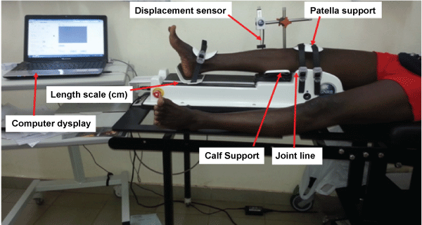
The GNRB system is shown in Figure 1. The patient is lying on a standard examination table in the supine position with the arms placed along the body, each knee being comparatively tested; the healthy knees are investigated first. The lower limb is placed in a single thermoformed support, which does not pose any problem for the installation of different sized adult patients or variation in the positioning of the support. An electric actuator increasing loads according to the examiner: 67, 89, 134, 150, or 250 N on the upper aspect of the calf. Testing is repeated on both knees, and the amount of tibial translation is compared between the 2 limbs. Motion data obtained from the displacement transducer produce a force-displacement curve. The slope (lm/N) of the curve obtained determines the ligamentous elasticity of the patient's ACL. All the data are collected on a remote computer. A laxity file is built up for each patient including measurement conditions (pressure applied to the thigh, load forces) and results (the displacement- load curve, the side-to-side difference in mm and the slope in l m/N) (Figure 2).
This series included 81 patients with an average age of 28.60 years [17-60], distributed as follows: 71 (87.65%) men and 10 (12.35%) women. Sixty-one (75.30%) patients had their ACL rupture following a football-type sports accident. Twenty-eight (34.57%) patients were professional athletes. The mean interval between trauma and surgery was 16.74 months. The reconstruction with the semitendinosus-gracilis technique under arthroscopy was exclusive in 53 (65.43%) patients and associated either with a meniscal gesture or a high tibial osteotomy in the other cases. We used exclusively the arthroscopic reconstruction of the ACL by hamstring tendons autograft. The tibial tunnel was performed with an Acufex® director guide set between 45 and 55 degrees. For the femoral tunnel, the drilling is performed from the tibial tunnel with an extension of the knee. The femoral tunnel is drilled 5 to 6 mm from the posterior border of the external condyle, at 11 hours for the right knees and at 13 hours for the left knees. A first blind tunnel with the caliber of the graft is realized and then, in a second time, an auger of 5 mm is used to drill the femoral cortex and to allow the passage of the Endobutton. The femoral fixation is made using an Endobutton® and the tibial one with a resorbable interference screw and a clamp clip. The length of the Endobutton® was chosen according to the length of the femoral tunnel. The remaining part of the anterior cruciate ligament was preserved. After the drilling of the tibial and femoral tunnels, the transplant moved from the tibial tunnel to the femoral tunnel. The absence of conflict of the graft with the roof of the intercondylar notch and with the medial edge of the external condyle is verified in extension of the knee. The isometry of the transplant is tested before the tibial fixation by the evaluation of the swallowing at the passage of the bending from 90° to the extension. We perform repetitive flexion-extension movements to recycle the transplant, avoiding the intrinsic secondary relaxation of the four beams. We then proceed to the placement of the tibial fixation screw, knee bent at 40°. We used a total of 14 (17.30%) fixation by femoral and tibial interference screw and 67 (82.70%) fixation by femoral Endobutton® and tibial interference screw. Rehabilitation was undertaken the next day. All patients were examined at 45 days, 3, 6 and 12 months postoperatively and at their evaluation date. At the results were evaluated by the IKDC score 1999 (International Knee Documentation Committee) and the clinical level, the Lysholm score. Radiological calculation of the positioning of the tunnels was done according to Aglietti's method [8] on a profile incidence of the knee.
The evaluation of the anterior laxity was done in pre and postoperative in all the patients by arthrometric measurement with GNRB®.
The data were entered in Excel and the analysis was made with the R version of the software version 3.1.0. The ANOVA (Analysis of Variance) test was used for the comparison of the Lysholm score with respect to the laximetry and the t Student test for the other comparisons. The decision to use the parametric test was based on the Shapiro-Wilk normality test with a coefficient greater than or equal to 0.95. The difference between the values of a parameter whose p value < 0.05 was estimated statistically significant.
Results
The mean follow-up of the evaluation was 12.80 ± 3.27 months. Two patients presented complications. The table below summarizes all the complications and their management [9-12] (Table 1).
The mean subjective IKDC was 81.73 ± 19.67 [3.44-100]. At the objective level, 64 (79.01%) patients were classified as A, 13 (16.05%) classified as B, 4 (4.94%) classified as C and none classified as D.
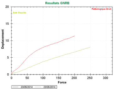
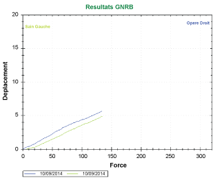
The average LYSHOLM score at the last follow-up was 87.52 ± 12.88% [46-100]. 67.90% of our patients had good or excellent results, 23.46% average results, and 8.64% bad results. The mean differential at GNRB test in preoperative was 5.18 ± 1.68 mm [2.6 and 7.8]. The mean postoperative differential was 2.73 ± 1.10 mm with extremes ranging from 0.7 to 4 mm. The Aglietti index was calculated with an average of 69.07% [55-83%] at the femoral level and 42.57% [33-60%] at the tibial level. The positioning of the tunnels was correct in 85.71% at the femoral level and 78.57% at the tibial level.
Parametric analysis
Influence of the delay of surgical management and the appearance of the amyotrophy
The average delay between trauma and surgery was 22.40 months for the group with amyotrophy versus 12.21 months for the group without amyotrophy and the difference between these two means is statistically significant with t = 2.1221 and p = 0.03919.
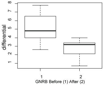
Comparison between GNRB before and GNRB after intervention
The comparative analysis between these two means is statistically significant with t = 4.7328 and p = 0.001. The following Box plot represents the variation in GNRB results before and after the intervention.
Influence of laximetry on the IKDC and Lysholm scores
There is a relationship between the postoperative GNRB differential value and the objective IKDC score. Indeed, the lower this differential, the better the IKDC score; p = 0.01894.
The mean postoperative GNRB for patients with a good and excellent Lysholm score was 2.33 mm, that for patients with an average score was 2.76 mm and that for patients with a poor score was 4.15 mm. The ANOVA test shows that this difference is significant.
Discussion
The study presented is a 58-month prospective study. It covers a relatively large number of subjects and a homogeneous population in terms of surgical gestures and surgeons. This study reported the anatomo-clinical and laximetry results of a consecutive series of 81 patients with anterior cruciate ligament rupture. All cases were treated by the same technique; there was no conversion either on the transplant level or on the approach route.
The primary endpoint of this study was the analysis of patient's functional scores, using the two questionnaires, IKDC and Lysholm.
Our results are satisfactory on the subjective IKDC score compared to the data of the literature and whatever the technique (Table 2).
Several studies have focused on the objective IKDC score. Our results of the objective IKDC are better than those obtained in a recent study [13], taking 11 randomized studies comparing the techniques with the PT and the hamstring (775 patients): 74% A or B versus 26% C and D. are similar to those of Messerli [14], Plaweski [15], Bedin [11] and El Andaloussi [16]. In a prospective, non-randomized comparative multicenter study, Plaweski, et al. [15] noted that there was no significant difference between IKDC objective A and B between the control group (patellar ligament) and the NAV group (semitendinosus-gracilis by assisted navigation). Charlton, et al. [17] in a retrospective study showed that the results of ligament reconstructions using semitendinosus-gracilis tendons fixed by bioabsorbable interfering screw were comparable to the results of other methods of ligament reconstruction of the anterior cruciate ligament in terms of satisfaction, knee stability and function. This could explain the similar results of different techniques at the IKDC.
Comparing the ways of fixing the semitendinosus-gracilis, Ma, et al. [18] analyzed in a prospective, nonrandomized clinical study at the two-year setback minimum, femoral fixation by bioresorbable interference screw and by Endobutton. In conclusion, the authors noted that the clinical results, between an anatomical fixation by interference screw and an Endobutton femoral fixation, were similar (results at 24 and 40 months of recoil). Table 3 summarizes all of these data [19,20].
At the Lysholm score, Alidrissi [12] scored 86.84% of good and excellent results and El Andaloussi [16] had 86% good and excellent results, 10% average results and 4% poor results. Our results are less conclusive than those of Alidrissi [12] and Andaloussi [16] and this could be explained by our short delay in retreat.
The correct position of the tunnels is an important anatomical element in the reconstruction of the anterior cruciate but also plays an element of failure, as shown by some studies. H. Robert [21] in the analysis of the anatomic failure factors of 50 surgeries of the anterior cruciate ligament found technical errors in 44 cases including 4 early releases and 40 femoral or tibial or mixed malpositions. These results show a very high proportion of failures related to the location of the tunnels and their fixing (88%); the other causes remaining marginal. Thus, all studies agree on the importance of the correct location of the tunnels. In our series, the positioning of the tunnels was correct in 85.71% at the femoral level and 78.57% at the tibial level. Regarding tunnels, it is their positioning precision that ensures the success of this surgery. According to Aglietti [22], the best results are obtained when the reconstruction is practically anatomical. A tibial tunnel placed too much earlier leads to a conflict between the graft and the intercondylar notch, causing a deficit of extension, anterior pain accompanied by residual effusion, instability, and ultimately ruptures of graft [23]. Similarly, an overly anterior femoral tunnel will be responsible for an increased elongation of the graft during flexion of the knee and will cause its rupture rather rapidly [22,23].
Our radiographic analysis did not include the analysis of the enlargement of the tibial and femoral tunnels. This absence is justified by the fact that no correlation has been found between the widening of the tunnels and the stability of the knee [11,23,24].
Differential values injured side/healthy side at 250 N postoperatively showed a difference of 2.73 ± 1.10 mm (0.7-4) which is located in the normal (< 3 mm), which is satisfactory. Alidrissi [12] found an average differential of 1.9 mm. Since GNRB® is the latest arthrometer, few studies have been published compared to KT 1000® and Telos®. The results of the study by M. Collette, et al. [25] demonstrate that the intra-observer reproducibility of GNRB® is good in both experienced and inexperienced operators. Moreover, this tool presents a very low variability of the measurements, whatever the experimental conditions.
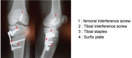
Three patients had an internal tibial valgus osteotomy (Figure 3) at the same operative time with an average IKDC average score of 84.72 and all of these patients were IKDC A or B. The OTV associated with ACL plasty is not a frequent intervention as indicated by Raguet but it protects the plasty in case of large varus and avoids the iterative rupture [26]; in fact, it represents less than 10% of the ACL plasties.
Roussignol [27] noted that the mean subjective IKDC score increased from 52.5 before the intervention to 71.4 after and the IKDC objective score of 0% knees graded A or B to 80%.
The laxity and instability induced by an ACL lesion are arthrogens. If in addition, there is an unrecoverable meniscal lesion and deformity in the frontal plane, osteoarthritis is inevitable. The OTV, combined with an ACL plasty, aims to avoid or stop this evolution. The results are still good and in the Raguet series as in ours; all the patients have resumed their work.
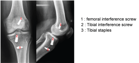
The comparative analysis of the differential values healthy side/injured side, at 250 N pre and postoperative, is statically significant. We conclude that this result attests of the good anatomical reconstruction of the ACL in our series (Figures 4 and Figure 5). Also, there is not only a significant relationship between the GNRB® differential value and the objective IKDC score but also between the GNRB® and the Lysholm score. In other words, the better the GNRB® differential value, the better the IKDC and Lysholm scores. In sum, our good differential value to GNRB® justifies our good score at IKDC and Lysholm.
The use of international instruments and standards, the relative effect of our sample (81 patients), and the rigor of the statistical tests used and their results give a certain quality to our analysis.
Many studies have emphasized the role of anatomic reconstruction in restoring knee stability [28].
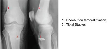
The Over the Top technique has many advantages vs. anatomical techniques: The creation of the femoral tunnel, respecting the ACL native footprint, represents the most challenging aspect of ACL reconstruction; in fact, incorrect positioning of the femoral tunnel accounts for the majority of technical failures following ACL reconstruction [29]. In addition, the Over the Top technique spares the bone stock on the femoral side, which assumes importance in cases of revision surgery involving the femoral tunnel (Figure 6).
This technique has been demonstrated to be safe, simple, reproducible, and inexpensive [30,31] requiring only one Endo button femoral and a screw and a tibial clip which is all its use in our countries (Figure 7).
Verdano, et al. [32] reported comparable clinical outcomes between the OTT (Over the Top) technique and double-bundle ACL reconstruction in a series of 40 patients evaluated at 4-year follow-up.
We used Non-anatomical ACL reconstruction using the Over the Top technique provided satisfactory results in terms of control of both static and dynamic instability at long-term follow-up. However, the 2SHG group showed better subjective and functional outcomes with fewer degenerative changes at long-term follow-up compared with the 1SHG group [33].
Conclusion
The choice of the transplant in reconstructive surgery of the ACL remains a topical problem. The most commonly used transplants are the hamstrings (double-bundle semitendinosus-gracilis technique) and the patellar tendon (KJ), each of which has its own advantages and disadvantages.
This study not only demonstrated the feasibility of reconstructing the ACL by the semitendinosus-gracilis technique under arthroscopy but also its effectiveness compared to other techniques. The authors advocate the promotion of arthroscopic surgery and indications expansion to other joints.
References
-
Lerat Jl, Moyen B, Garin C, et al. (1993) Laxité antérieure et arthrose du genou. Résultats de la reconstruction du LCA associée à une ostéotomie tibiale. Rev Chir Orthop 79: 365-374.
-
Lubowitz JH, Bernardini BJ, Reid JB (2008) Comprehensive physical examination for instability of the knee. Am J Sports Med 36: 577-594.
-
Anderson AF, Synder RB, Federspiel CF, et al. (1992) Instrumented evaluation of knee laxity: A comparison of five arthrometers. Am J Sports Med 20: 135-140.
-
Jones KG (1963) Reconstruction of the anterior cruciate ligament. A technique using the central one-third of the patellar ligament. J Bone Joint Surg 45: 925-932.
-
Dejour H, Dejour D, Ait Si Selmi T (1999) Laxités antérieures chroniques du genou traitées par greffe libre du tendon rotulien avec plastie latérale extra-articulaire: 148 cas revus à plus de 10 ans. Rev Chir Orthop 85: 777-789.
-
Chol C, Ait Si Selmi T, Chambat P, et al. (2002) Seventeen year outcome after anterior cruciate ligament reconstruction with a intact or repaired medial meniscus. Rev Chir Orthop Reparatrice Appar Mot 88: 157-162.
-
Lerat JL, Chotel F, Besse JL, et al. (1998) Les résultats après 10 à 16 ans du traitement de la laxité chronique antérieure du genou par une reconstruction du ligament croisé antérieur avec une greffe de tendon rotulien associée à une plastie extra-articulaire externe: À propos de 138 cas. Rev Chir Orthop 84: 712-727.
-
Aglietti P, Buzzi R, Zaccherotti G, et al. (1994) Patellar tendon versus doubled semitendinosus and gracilis tendons for anterior cruciate ligament construction. Am J Sports Med 22: 211-217.
-
Agletti P, Giron F, Losco M, et al. (2010) Comparison between single-and double-bundle anterior cruciate ligament reconstruction: A prospective, randomized, single-blinded clinical trial. Am J Sports Med 38: 25-34.
-
Aglietti P, Giron F, Cuomo P, et al. (2007) Single-and double-incision double-bundle ACL reconstruction. Clin Orthop Relat Res 454: 108-113.
-
Bedin Bertand (2010) Evaluation de la reconstruction du ligament croisé antérieur selon 3 techniques: Fascia Lata, ischio-jambier, tendon patellaire. Thèse Méd Limoges.
-
Alidrissi N, Elyaacoubi M, Berrada MS, et al. (2011) Ligamentoplastie du LCA aux ischiojambiers sous arthroscopie avec fixation de l'implant par le système TLS. Principes et résultats de 38 cas. J Traumatologie du Sport 28: 159-164.
-
Lewis PB, Parameswarn AD, Rue JP, et al. (2008) Systematic review of single bundle anterior cruciate ligament reconstruction outcomes: A baseline assessment for consideration of double-bundle techniques. Am J Sports Med 36: 2028-2036.
-
Messerli Guy (2008) Reconstruction du ligament croisé antérieur assisté par ordinateur: étude prospective. non randomisée avec résultats à 12 mois des 30 premiers cas. Thèse Méd Genève 10503.
-
Plaweski S, Davaid TS, Dumas J, et al. (2012) Evaluation d'un système de reconstruction du ligament croisé antérieur par navigation assistée par ordinateur: Etude d'une cohorte prospective non randomisée. Rev Chir Orthop et Traumatologique 98: S203-S209.
-
Ei Andaloussi Y, Arssi M, Potel JF, et al. (2011) La reconstruction du ligament croisé antérieur aux tendons ischiojambiers chez les sportifs de haut niveau. Étude prospective de 30 cas. Journal de Traumatologie du Sport 28: 3-7.
-
Charlton WPH, Randolph DA, Lemos S, et al. (2003) Outcome of anterior cruciate ligament reconstruction with quadrupled hamstring tendon graft and bioabsorbable interference screw fixation. Am J Sports Med 31: 518-521.
-
Ma CB, Francis K, Towers J, et al. (2004) Anterior cruciate ligament reconstruction: A comparison of bioabsorbable interference screw and endobutton-post fixation. Arthroscopy 20: 122-128.
-
Mouhamed Massoundi Ben Ali (2012) Ligamentoplastie au ligament patellaire selon kenneth Jones modifiée par mini arthrotomie. Corrélation entre les résultats anatomiques et les résultats fonctionnels. A propos de 27 cas. Mém DES Ortho-trauma Dakar, N° 638.
-
Plaweski S, Rossi J, Merloz P (2009) Reconstruction du ligament croisé antérieur du genou : évaluation d'un procédé de fixation fémorale (Endobutton CL®) des tendons de la patte d'oie (Gracilis-semi tendinosus) : A propos d'une série prospective continue de 105 cas revus à plus de quatre ans. Rev Chir Orthop et Traumatologique 95: 734-742.
-
H Robert, P Calas, D Bertin, et al. (2004) Analyse des facteurs d'échec anatomique de 50 plasties du ligament croisé antérieur. 90: 78.
-
Aglietti P, Buzzi R, Giron F, et al. (1997) Arthroscopic-assisted anterior cruciate ligament reconstruction with the central third patellar tendon: A 5-8-year follow-up. Knee Surg Sports Traumatol Arthrosc 5: 138-144.
-
Aglietti P, Giron F, Buzzi R, et al. (2004) Anterior cruciate ligament reconstruction: Bone-patellar tendon-bone compared with double semitendinosus and gracilis tendon grafts. A prospective, randomized clinical trial. J Bone Joint Surg AM 86: 2143-2155.
-
Baumfeld JA, Diduch DR, Rubino LJ, et al. (2008) Tunnel widening following anterior cruciate ligament reconstruction using hamstring autograft: A comparison between double cross-pin and suspensory graft fixation. Knee Surg Sports Traumatol Arthrosc 16: 1108-1113.
-
Collette M, Courville J, Forton M, et al. (2011) Objective evaluation of anterieur knee laxity; comparison of the KT 1000 and GNRB® arthrometers. Knee Surg Sports Traumatol Arthrose 20: 2233-2238.
-
Raguet M Plastie du ligament croisé antéro-externe et ostéotomie tibiale Clinique PriolletCourlancy, 2, avenue du Général-de-Gaulle, 51000 Châlons-en-Champagne, France.
-
Roussignol X, Sfez J, Courage O, et al. (2012) Ostéotomie de valgisation et ligamentoplastie de type DIDT dans le cadre des laxités antérieures chroniques. Rev Chir Orthop et Traumatologique 98: S138-S143.
-
Markatos K, Kaseta MK, Lallos SN, et al. (2013) The anatomy of the ACL and its importance in ACL reconstruction. Eur J Orthop Surg Traumatol 23: 747-752.
-
Taketomi S, Inui H, Sanada T, et al. (2014) Eccentric femoral tunnel widening in anatomic anterior cruciate ligament reconstruction. Arthroscopy 30: 701-709.
-
Asai S, Maeyama A, Hoshino Y, et al. (2014) A comparison of dynamic rotational knee instability between anatomic single-bundle and over- the-top anterior cruciate ligament reconstruction using triaxial accelerometry. Knee Surg Sports Traumatol Arthrosc 22: 972-978.
-
Buda R, Ruflli A, Parma A, et al. (2013) Partial ACL tears: Anatomic reconstruction versus nonanatomic augmentation surgery. Orthopedics 36: e1108-e1113.
-
Verdano MA, Pedrabissi B, Lunini E, et al. (2012) Over the top or Endo- button for ACL reconstruction? Acta Biomed 83: 127-137.
-
Ruffilli A, Buda R, Pagliazzi G, et al. (2015) Over-the-Top anterior cruciate ligament reconstruction using single or double-strand hamstrings autograft. Feature Article 38: e635-e643.
Corresponding Author
Kinkpe CVA, Orthopaedic Department, Order of Malta's Hospital Center in Dakar, Senegal, Tel: +0022-1774-502646.
Copyright
© 2017 Kinkpe CVA, et al. This is an open-access article distributed under the terms of the Creative Commons Attribution License, which permits unrestricted use, distribution, and reproduction in any medium, provided the original author and source are credited.