Effects of Surface Tissue Compression on Muscle Tone of Stroke Patients
Abstract
Aim
To analyze the effects of surface tissue compression on the muscle activity of elderly patients with stroke and hypertonia of the upper limbs.
Methods
Fourteen male individuals, 10 with right spastic hemiparesis and 4 with left spastic hemiparesis, aged 60 to 80 years (± 72.33-years-old), with clinical diagnosis of stroke were selected to participate in the study. The muscle activity of the participants was detected by surface electromyography before and immediate after the application of the surface pressure device. For the treatment procedure, the surface compression equipment used was made of plastic and was similar to a buoy, with a lateral opening to couple the equipment to the upper limbs of the individual. The EMG signal normalized values obtained during MVC were tabulated and analyzed using the t-test of paired samples.
Results
In the analysis, it was observed that, following the application of the surface pressure technique, the evaluated muscles, showed a decrease of muscle activity. However, the results obtained were statistically significant (p < 0.05).
Conclusion
It can be concluded that the surface compression technique did show statistically significant differences when the muscle activity values were compared before and after treatment intervention. The reduction in myoelectric activity of the brachii biceps and triceps brachii muscles was observed.
Keywords
Electromyography, Muscle spasticity, Surface tension, Stroke, Aged
Abbreviations
GTOs: Golgi Tendon Organs; EMG: Electromyography; BB: Biceps Brachii; TT: Triceps brachii; SCT: Surface Compression Technique; MVC: Maximal Voluntary Contraction
Introduction
There are two types of cerebrovascular accident, or stroke. An ischemic stroke is caused by a blockage of blood flow, and a hemorrhagic stroke is caused by a breakage in a blood vessel [1]. Elderly individuals have more chances to be affected due to formation of atherosclerotic plaques that block blood flow, leading to acute loss of brain function [2]. Those individuals with neurological conditions, such as stroke, are subject to neuromuscular disorders that include spastic paralysis of the upper and lower limb muscles in the hemisphere contralateral to a brain lesion [3].
Neuromuscular changes are associated with sensory stimuli generated by mechanoreceptors that determine the position and direction of the limbs during the movements, differentiating the neuromuscular function. These receptors are called Golgi tendon organs (GTOs) and muscle spindles. GTOs detect and respond to changes in muscle tension that are caused by muscular contraction. This reflex is called the inverse myotatic reflex, which is a muscle contraction in response to stretching within the muscle [4].
These receptors relay information from skin, joints and muscles. Each sense organ contains different receptors: general receptors are found throughout the body with higher concentration in the skin tissue; and special receptors include chemoreceptors found in the mouth and nose, photoreceptors found in the eyes, and mechanoreceptors found in the ear. The sensory receptors differ from the rest because they capture specific types of stimulion certain parts of the body, such as pressure [5]. The effect of pressure on the skin tissue mayreflect changes in the activities of muscles, which are detected by surface electromyography (EMG) to obtain information about the anatomical and functional state of neuromuscular components and to observe possible axonal damages not seen by a physical examination [6]. Therefore the aim of this study was to analyze the effects of surface tissue compression on the muscle activity of elderly patients with stroke and hypertonia of the upper limbs.
Materials and Methods
Sample
Fourteen male individuals, 10 with right spastic hemiparesis and 4 with left spastic hemiparesis, aged 60 to 80 years (± 72.33-years-old), with clinical diagnosis of stroke were selected to participate in the study. The inclusion criteria were individuals in attendance at the Clínica Escola de Fisioterapia - UNIFAFIBE, clinical diagnosis of spastic hemiparesis with grades 2 and 3 on the Ashworth scale, treatment period of two to five years, and no cognitive deficits. The exclusion criteria were individuals undergoing treatment for over five years, with grades lower than 2 or greater than 3 on the Ashworth scale, and with cognitive deficits.
The research was approved by the Research Ethics Committee of the University Center UNIFAFIBE (CAAE 23316314.2.0000.5387). All subjects were informed about the purpose and stages of the research and agreed to participate by providing their free and informed consent according to resolution 466/12 of the Health National Council.
Equipment
For the surface electromyography, eight channels of the portable Myosystem Br1 apparatus were used with a sampling rate of 4000 Hz, with its own battery and connected to a laptop. The electromyography device was used in partnership with the Ribeirao Preto School of Dentistry (FORP-USP).
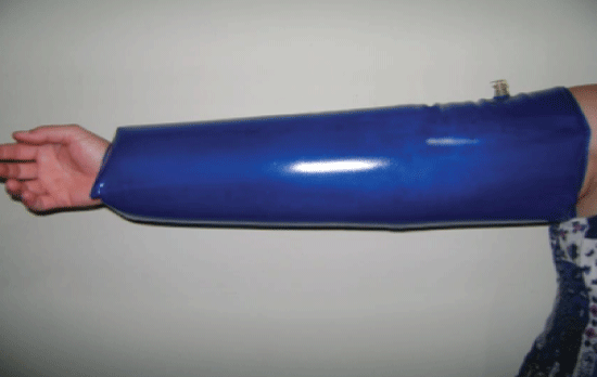
For the treatment procedure, the surface compression equipment used was made of plastic and was similar to a buoy, with a lateral opening to couple the equipment to the upper limbs of the individual, as shown in (Figure 1 and Figure 2) digital pressure gauge (Penalty) was used to measure the nominal surface tensile strength of 1.0 psi.
Procedures
For the functional analysis of the upper limbs, the electrodes were positioned over the bellies of the following muscles: the biceps brachii (BB) and the triceps brachii (TB) of the hemiparetic side. The measurement and demarcation of the best point to collect the surface electromyographic signals were performed following the standardization of the international protocol, SENIAM - Surface Electromyography for the Non-invasive Assessment of Muscles. The muscle activity evaluation was performed using the electromyographic recordings obtained pre and post intervention, with the surface compression technique (SCT), in clinical conditions of rest and during maximal voluntary contraction (MVC - normalization factor). The data collection was made in each muscle condition for 10 seconds.
During the application of the SCT, the subjects remained with the "buoy" to compress the tissue surface of the affected limb. The SCT was applied for 15 minutes, with the subjects sitting on a chair, with the upper limbs positioned according to the limitations imposed. A pressure gauge was used to measure the nominal surface tensile strength of 1.0 psi.
The gross EMG signal was used to derive EMG amplitude values by calculating the root mean square. The EMG signal normalized values obtained during MVC were tabulated and analyzed using the t-test of paired samples. The statistical analysis was performed using the SPSS software (SPSS Inc.; Chicago, IL, USA), version 21.0 for Windows.
Results and Discussion
In the analysis, it was observed that all evaluated muscles, showed a decrease in muscle activity when the pre and post intervention values were compared. However, the results showed statistical significance (p < 0.05) when subjected to the "t"-test for paired samples, as shown in Table 1.
The GTOs are also capable of generating muscle inhibition when the generated tension is higher than the capacity of the muscle, leading to a sudden relaxation of all muscle fibers. This is called stretch reaction. This explains the fact that the SCT reduces the potential action of BB and TB muscles, indicating that spasticity changes the function of the GTOs and of the muscle spindles. Pressure stimulation enables the normalization of this function, according to the exerted compression, by sending stimuli to the cerebral cortex, which activate the inhibitory effects on the hypertonic muscles.
The mechanoreceptors respond to mechanical changes generated within the cutaneous and subcutaneous tissue, providing a slow and sustained adjustment as a result of the compression strength on the tissue, which will be transmitted to the nerve endings. These receptors contribute to proprioception and indicate the sense of body position [7]. These findings corroborate those from our study since the SCT causes mechanical changes within the tissue by stimulating the mechanoreceptors through the nerve endings, and by inducing the reduction of hypertonia.
If left untreated, hypertonia can lead to musculoskeletal disorders and negatively affect posture and motor function of the individual. The loss of muscular elasticity and connective tissue generate deformations due to imbalance between the tensile forces surrounding the joint. All these alterations cause the individuals to lose the ability to perform tasks in their daily living and working environments [8]. The action potential on the muscles of the upper limbs suggests that individuals can benefit from the SCT treatment by a time window, where a reduction of hypertonia occurs, therefore, the therapeutic use of this device can facilitate their daily living activities.
Conclusion
It can be concluded that the use of surface tissue compression reduced hypertonia for up to thirty minutes in individuals with stroke. This study can help guide future research on surface tissue compression devices, especially with regard to the technique, the application time and the number of sessions.
Conflict of Interest
Competing Interests: None declared. This research received no specific grant from any funding agency in the public, commercial, or not-for-profit sectors.
References
- Taylor RA, Sansing LH (2013) Microglial Responses after Ischemic Stroke and Intracerebral Hemorrhage. Clin Dev Immunol 2013: 1-10.
- Brito ES, Rabinovich EP (2008) Messed up everything! The impact of stroke in the family. Health and Society 17: 153-169.
- Zamberlan AL, Kerppers II (2007) Neural Mobilization as a Physiotherapeutic Resource in the Rehabilitation of Patients with Stroke - Review. Revista Salus 1: 185-192.
- Bauer N, Preis C, Neto LB (2013) The Importance of Proprioception in the Sportive Kinetic-Functional Prevention and Recovery. RBRAF 2: 28-37.
- Mochizuki L, Amadio AC (2006) As Informações Sensoriais para o Controle Postural. Revista Fisioterapia e Movimento 19: 11-18.
- Fialho RA, Herrera EM (2004) Neurophysiology in the study of neuromuscular diseases, development and current limitations. Rev Cub Med Mil 33: 11-14.
- Kandel E, Schwartz J, Jessell TM, et al. (2014) Princípios de neurociências. (5th edn), Porto Alegre: AMGH, 1544.
- Teixeira Lf, Olney Sj, Brauwer B (1998) Mecanismos E Medidas De Espasticidade. Rev Fisioter Univ Sao Paulo 5: 4-19.
Corresponding Author
Saulo Fabrin, Department of Morphology, Physiology and Basic Pathology, Electromyography Laboratory, University of São Paulo, Ribeirão Preto; Department of Physiotherapy, Claretiano University Center, Biomechanics and Movement Analysis Laboratory - Labim, Brazil, Avenue Bandeirantes 3900, Brazil, Tel: +55-16-3602-0281.
Copyright
© 2017 de Lima C, et al. This is an open-access article distributed under the terms of the Creative Commons Attribution License, which permits unrestricted use, distribution, and reproduction in any medium, provided the original author and source are credited.


