Chronic Papillomatous Dermatitis in a Patient with a Urinary Ileal Diversion: A Case Report and Review of the Literature
Introduction
Gastrointestinal surgical procedures result in approximately 100,000 ostomies per year in the United States, with over 10,00,000 people living with intestinal stomas [1]. Proper use and management of a stoma appliance relies heavily on peristomal skin integrity; however abdominal stoma patients can develop various dermatologic complications including allergic contact dermatitis, mechanical dermatitis from appliance stripping, infections, pyoderma gangrenosum and irritant contact dermatitis. Peristomal irritant contact dermatitis is partially attributed to leakage around the stoma due to ill-fitting ostomy equipment. Chronic exposure of peristomal skin to urine and fecal matter can manifest as an acanthomatous inflammatory reaction, known as Chronic Papillomatous Dermatitis (CPD). CPD has been classically associated with urostomies, while few ileostomy-associated CPD reports exist [1]. Specifically, it has been attributed to the alkaline pH of urine mechanical irritation and epidermodysplasia verruciformis Human Papilloma Virus (HPV) types [2]. 9.5% of stoma patients reportedly develop CPD after the stoma has been present for an average of 6.5 years [3].
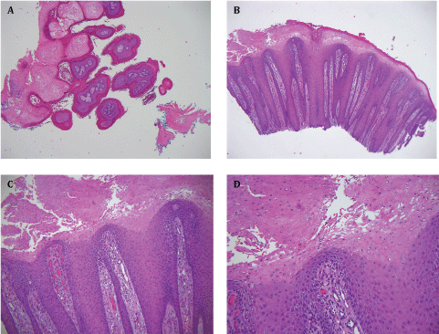
Histopathological descriptions of CPD have ranged broadly in various urologic and wound care literatures including hyperkeratotic stenosis, stomal keratinization, pseudoverrucous, pseudoepitheliomatous, acanthomatous epidermal hyperplasia and reactive acanthosis. The majority of CPD lesions have been previously described to fall under the realm of reactive changes resulting from a chronic irritant contact dermatitis. Furthermore, the lack of CPD reports in dermatologic literature, and the rarity of specifically ileostomal associated CPD cases indicate a need for further clinical and histologic description (Table 1). To this end, we report a case of ileostomy-associated CPD and a review of the literature for clinical and therapeutic interest (Figure 1).
Keywords
Chronic papillomatous dermatitis, Stoma dermatitis, Peristomal skin
Abbreviations
Chronic Papillomatous Dermatitis (CPD), Human Papilloma Virus (HPV)
Case Presentation
A 74-year-old Caucasian male was referred to dermatology for an exophytic, peristomal skin lesion suspected to be peristomal dermatitis. Patient history was significant for prostate cancer treated with Cyber Knife radio surgery and subsequent cystoprostatectomy with ileal conduit urinary diversion and nephrectomy. One year later, the patient presented with a 3 cm tender, flesh-colored, exophytic papillomatous nodule around the ostomy site. The mass progressively enlarged until the primary stoma site was completely occluded, necessitating catheter insertion proximal to the ostomy site for urine release. Shave biopsy of the nodule was performed to exclude neoplastic growth. Microscopic examination revealed papillomatosis and massive parakeratosis with significant regular epidermal hyperplasia and vertical papillary dermal fibrosis with pallor of upper layer keratinocytes. There was a mild superficial lymphocytic, neutrophilic and eosinophilic perivascular infiltrate with foci of occasional neutrophil exocytosis. These histopathologic findings were consistent with CPD. The nodule was excised and the patient was advised to perform vinegar soaks of the affected area. No complications were noted following the advised vinegar soaks.
Discussion
We report a case of CPD, a distinct cutaneous eruption comprised of exuberant, wart-like papules due to stomal site chronic irritant contact dermatitis. Similar lesions are described as hypertrophic scarring, hyperkeratotic stenosis, stomal keratinization, pseudoepitheliomatous hyperplasia, and reactive acanthosis in urologic and wound care literature [1,4]. Stomal irritant reactions have similar histopathologic features, though clinical appearance is contingent on stoma type and source of irritation. The higher incidence of CPD with urostomies reported in the literature may be due to larger cutaneous surface area exposed to urine irritation. Ileostomies contain high concentrations of degradative enzymes and bile acids, which can lead to severe dermatitis and erosions [1].
HPV's role in the etiology of CPD has been contended. HPV can lead to a number of benign and malignant epithelial tumors, due to viral integration and expression of oncoproteins E6 and E7 [5,6]. The resulting genomic instability has been theorized to be involved in the development of rare-stoma related cutaneous pathologies [3]. Ileostomal-associated papillomatous lesions have been reported positive for epidermodysplasia verruciformis HPV strains, as well as HPV-16 [5]. Still, the role of HPV in CPD remains unclear as not all lesions are positive for HPV and cutaneous symptoms resolve following removal of irritant source [5]. Furthermore, amongst reported cases of HPV-positive CPD, clinical presentation, histopathology, and treatment do not differ from HPV negative CPD cases [2,3,5]. As such, HPV testing was not performed, as it would not have altered patient care. Nevertheless, the potential spread of cutaneous HPV to mucosal surfaces of the intestine highlight the need for timely diagnosis of peristomal lesions.
Primary management involves refitting the stomal apparatus and reducing bag change intervals. If urine leakage occurs due to a receding stoma, convex-backed appliances can lengthen urostomies and stop leakage. Construction of a protruding stoma, i.e. classic Brooke ileostomy, can be used to prevent effluence of stomal contents [7]. Non-invasive treatment includes topical silicone, hydrocortisone and 5-fluorouracil creams [8]. Vinegar soaks at each bag change may be effective in resolution of lesions [2]. If CPD lesions result in stomal stenosis, extirpation and revision may be indicated. Maintaining stomal hygiene is necessary to minimize irritant dermatitis from fecal and urinary soiling.
Recognition and awareness of peristomal lesions among dermatologists, urologists and wound care specialists is necessary to improve patient quality of life.
Conflict of Interest Disclosure
None to declare.
Funding Source
PHR and RZC are supported by the NIH 5 T32 AR 7569-22 National Institute of Health T32 grant.
References
- Agarwal S, Ehrlich A (2010) Stoma dermatitis: Prevalent but often overlooked. Dermatitis 21: 138-147.
- Lyon CC, Smith AJ, Griffiths CE, et al. (2000) The spectrum of skin disorders in abdominal stoma patients. Br J Dermatol 143: 1248-1260.
- Raghu P, Tran TA, Rady P, et al. (2012) Ileostomy-associated chronic papillomatous dermatitis showing nevus sebaceous-like hyperplasia, HPV 16 infection, and lymphedema : a case report and literature review of ostomy-associated reactive epidermal hyperplasias. Am J Dermatopathol 34: e97-e102.
- Borglund E, Nordstrom G, Nyman CR, et al. (1988) Classification of peristomal skin changes in patients with urostomy. J Am Acad Dermatol 19: 623-628.
- Wieland U, Gross GE, Hofmann A, et al. (1998) Novel Human Papillomavirus (HPV) DNA sequences from recurrent cutaneous and mucosal lesions of a stoma-carrier. J Invest Dermatol 111: 164-168.
- Stanley MA (2012) Epithelial cell responses to infection with human papillomavirus. Clin Microbiol Rev 25: 215-222.
- Williams CM, Wieland U, Rodning CB, et al. (2003) Human papillomavirus-negative ileostomal chronic papillomatous dermatitis. J Cutan Pathol 30: 271-274.
- Nybaek H, Jemec GB (2010) Skin problems in stoma patients. J Eur Acad Dermatol Venereol 24: 249-257.
- Kitamura S, Hata H, Inamura Y, et al. (2016) Chronic papillomatous dermatitis around an ileostoma. J Dermatol 43: 220-221.
- Fernandez IS, Moreno C, Vano-Gavan S, et al. (2010) Pseudoverrucous irritant peristomal dermatitis with an histological pattern of nutritional deficiency dermatitis. Dermatol Online J 16: 16.
- Mattoch IW, Pham N, Robbins JB, et al. (2008) Reactive eccrine syringofibroadenoma arising in peristomal skin: an unusual presentation of a rare lesion. J Am Acad Dermatol 58: 691-696.
- Kazakov DV, Mikyskova I, Mukensnabl P, et al. (2005) Reactive syringofibroadenomatous hyperplasia in peristomal skin with formation of hybrid epidermal-colonic mucosa glandular structures, intraepidermal areas of sebaceous differentiation, induction of hair follicles, and features of human papillomavirus infection: a diagnostic pitfall. Am J Dermatopathol 27: 135-141.
- Clarke LE, Ioffreda M, Abt AB (2003) Eccrine syringofibroadenoma arising in peristomal skin: a report of two cases. Int J Surg Pathol 11: 61-63.
- Goldberg NS, Esterly NB, Rothman KF, et al. (1992) Perianal pseudoverrucous papules and nodules in children. Arch Dermatol 128: 240-242.
- Nordstrom GM, Borglund E, Nyman CR (1990) Local status of the urinary stoma-the relation too peristomal skin complications. Scand J Urol Nephrol 24: 117-122.
- Bergman B, Knutson F, Lincoln K, et al. (1979) Chronic papillomatous dermatitis as a peristomal complication in conduit urinary diversion. Scand J Urol Nephrol 13: 201-204.
- Cordonnier JJ, Nicolai CH (1960) An evaluation of the use of an isolated segment of ileum as a means of urinary diversion. J Urol 83: 834-838.
- Burnham JP, Farrer J (1960) A group experience with ureteroileal-cutaneous anastomosis for urinary diversion: results and complications of the isolated ileal conduit (Bricker procedure) in 96 patients. J Urol 83: 622-629.
- Rattner WH, Moran JJ, Murphy JJ (1959) The histologic appearance of small bowel segments used as urinary conduits. The Journal of Urology 82: 236-239.
- Cordonnier JJ (1957) Ileal bladder substitution: an analysis of 78 cases. J Urol 77: 714-717.
Corresponding Author
Pooja H Rambhia, Department of Dermatology, University Hospitals Cleveland Medical Center, 2085 Cornell Road, Cleveland, OH 44106, USA, Tel: 516-870-8632.
Copyright
© 2017 Rambhia PH, et al. This is an open-access article distributed under the terms of the Creative Commons Attribution License, which permits unrestricted use, distribution, and reproduction in any medium, provided the original author and source are credited.


