Clinical Image: Olfactory Bulb and Tract Aplasia/Hypoplasia on MRI
Abstract
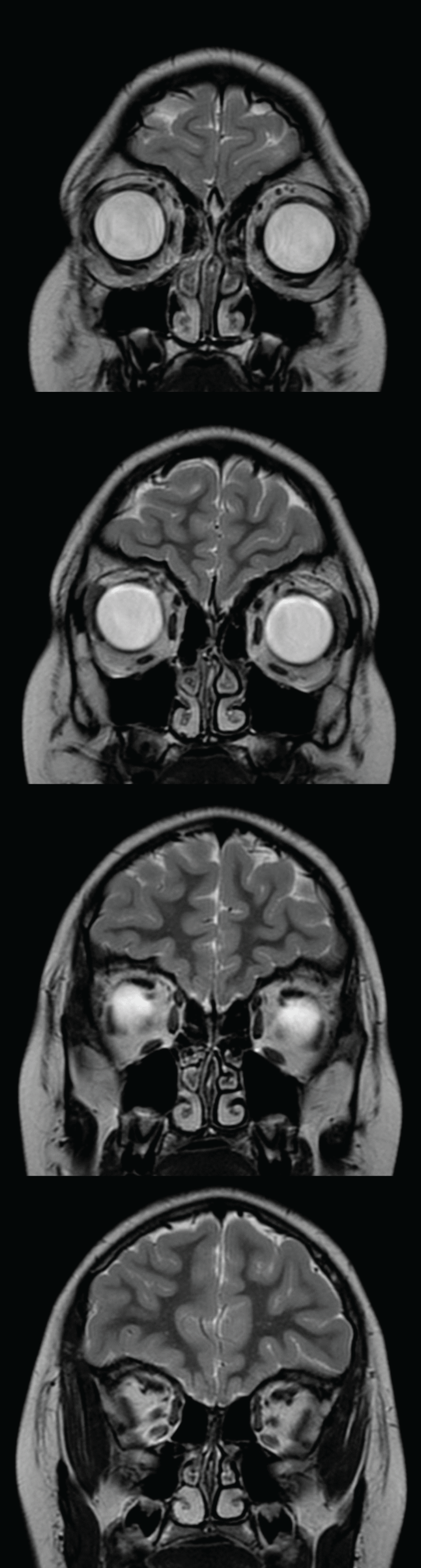
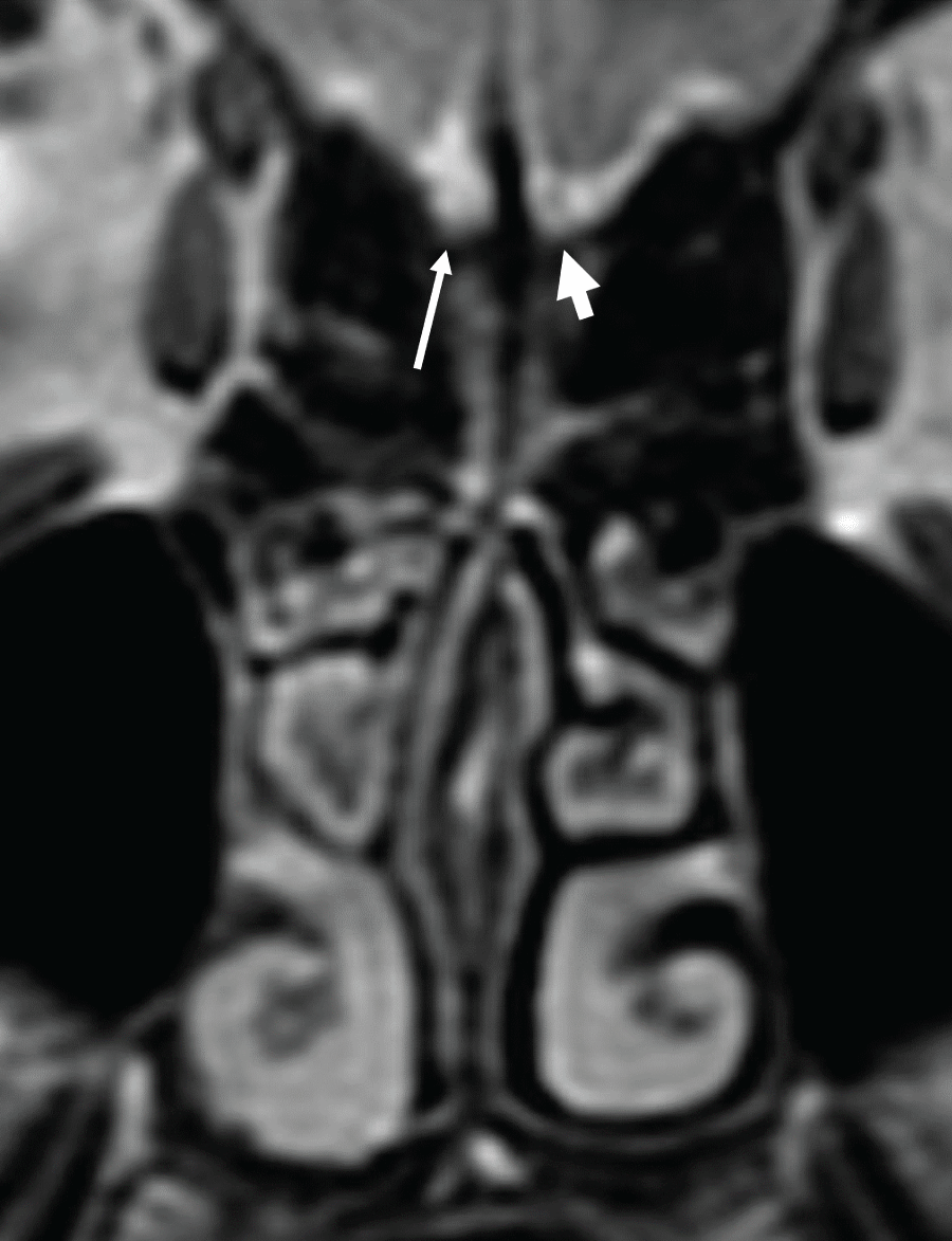
Magnetic resonance imaging (MRI) is commonly ordered in the workup of the anosmic patient. Anosmia is associated with relatively reduced olfactory bulb and tract (OBT) volumes on MRI in a variety of clinical settings, but congenitally anosmic patients will characteristically have olfactory nerve aplasia or hypoplasia. We present the case of an otherwise healthy 9-year-old male who presented to the Ear, Nose, and Throat clinic for evaluation of longstanding anosmia. On exam, the patient was a well-developed, healthy-appearing male, who was unable to smell an alcohol pad. Physical exam and endoscopic examination were unremarkable. An MRI Brain with and without contrast was ordered, demonstrating left OBT hypoplasia and right OBT aplasia, and these findings are discussed.
Keywords
Olfaction, Magnetic resonance imaging, Anosmia, Hyposmia
Case
A 9-year-old male presents to the Ear, Nose, and Throat clinic of a tertiary referral hospital on the recommendation of his pediatrician for anosmia beginning at least 5 years prior. He could taste sweets, but denied sensation of fragrant or pungent smells. He denied head trauma, headaches, or vision changes. He noted moderate right-greater-than-left nasal obstruction and snoring for which he was given over-the-counter antihistamines. He had no other past medical or surgical history. His birth history was also uncomplicated, being full-term, passing newborn screens, without prolonged stay in neonatal intensive care unit (NICU).
On exam, the patient was a well-developed, healthy-appearing male, who was unable to smell an alcohol pad. He had edematous, moderately enlarged turbinates right-greater-than-left, but no masses, lesions, or foreign bodies were noted. Cranial nerves II through XII were intact and symmetric.
Endoscopic evaluation of the nasal cavity confirmed edematous mucosa and turbinate enlargement and revealed moderate adenoid hypertrophy. No purulent discharge or polyps noted. An MRI Brain with and without contrast was ordered, as seen in Figure 1a and Figure 1b, demonstrating left olfactory bulb and tract (OBT) hypoplasia and right OBT aplasia. The patient was referred to Endocrinology for evaluation of Kallman's syndrome.
Magnetic resonance imaging (MRI) is the mainstay of evaluation of the anosmic or hyposmic patient [1]. MRI is specifically helpful in the measuring OBT volumes. This measurement is achieved through tracing of the tract either manually or in a computer-assisted fashion with three-dimensional volumetric calculation [2]. OBT volume has been shown to correlate with olfactory function in cases of post-traumatic anosmia, anosmia after upper respiratory infection [3]. Patient with chronic rhinosinusitis (CRS) with polyps before and after surgery show associated increase in OBT volume with improvement in olfactory threshold in objective testing [4].
In patients who complain of anosmia since birth or having no memory of normal olfaction, congenital anosmia should be suspected. Patients with congenital anosmia often have no other complaints or abnormalities, but nearly all will have absent or hypoplastic OBTs, the latter defined as an OBT that is less than half the volume of the average OBT for that age [5]. In one study of 16 patients with isolated congenital anosmia, 5 (31%) had bilateral OBT hypoplasia, 3 (19%) had unilateral aplasia and unilateral hypoplasia, and 8 (50%) had bilateral aplasia [6].
In the evaluation of the congenitally anosmic patient, an MRI is useful to evaluate the olfactory system, and OBT hypoplasia or aplasia should be high on the differential.
References
- Leboucq N, Menjot de Champfleur N, Menjot de Champfleur S, et al. (2013) The olfactory system. Diagn Interv Imaging 94: 985-991.
- Yousem DM, Geckle RJ, Bilker WB, et al. (1998) Olfactory bulb and tract and temporal lobe volumes: normative data across decades. Ann N Y Acad Sci 855: 546-555.
- Mueller A, Rodewald A, Reden J, et al. (2005) Reduced olfactory bulb volume in post-traumatic and post-infectious olfactory dysfunction. Neuroreport 16: 475-478.
- Sadeghi M, Amali A, Ezabadi SR, et al. (2015) Evaluation of the olfactory bulb volume and olfactory threshold in patients with nasal polyps and impact of functional endoscopic sinus surgery: A longitudinal study. Int Forum Allergy Rhinol 5: 356-360.
- Yousem DM, Geckle RJ, Bilker W, et al. (1996) MR evaluation of patients with congenital hyposmia or anosmia. AJR Am J Roentgenol 166: 439-443.
- Abolmaali ND, Hietschold V, Vogl TJ, et al. (2002) MR evaluation in patients with isolated anosmia since birth or early childhood. AJNR Am J Neuroradiol 23: 157-164.
Corresponding Author
Prasanth Pattisapu, MD MPH, Bobby R Alford Department of Otolaryngology-Head and Neck Surgery, Baylor College of Medicine, 1 Baylor Plaza, Mail Stop NA-102, Houston, TX 77030, USA, Tel: +713-798-7217.
Copyright
© 2017 Pattisapu P, et al. This is an open-access article distributed under the terms of the Creative Commons Attribution License, which permits unrestricted use, distribution, and reproduction in any medium, provided the original author and source are credited.