

Hydroxyapatite Tube Augmentation at the Lag Screw Entry Site Reduces Postoperative Anterior Wall Fracture After Intertrochanteric Fracture Fixation: A Propensity Score-Matched Study
Abstract
Background: Osteoporotic intertrochanteric fractures are increasing with population ageing, and mechanical complications after intramedullary fixation remain a clinical concern [1-3].
Purpose: To evaluate whether hydroxyapatite (HA) tube augmentation at the lag screw entry site reduces postoperative anterior wall fracture (POAWF) after InterTAN ® fixation for intertrochanteric fractures.
Methods: This single-center retrospective observational study compared cases treated with HA tube augmentation (augmentation group, AG) and without augmentation (control group, CT). Propensity score matching (PSM) was performed to balance baseline characteristics. The primary outcome was POAWF incidence in the matched cohort. Secondary outcomes included POAWF incidence in the subset with lateral wall fracture at the lag screw entry site (LWF-LS), postoperative telescoping, and regression analyses for factors associated with telescoping and POAWF.
Results: After PSM, 39 patients were included in each group (n = 78). POAWF occurred in 20.51% (8/39) of the CT group and 5.13% (2/39) of the AG group (p = 0.042). In the LWF-LS subset, POAWF occurred in 61.54% (8/13) of CT patients versus 12.50% (2/16) of AG patients (p < 0.001). Postoperative telescoping was greater in CT than AG (2.20 vs. 1.37 mm, p = 0.009). In multivariable logistic regression, HA augmentation was independently associated with lower odds of POAWF (OR 0.073, 95% CI 0.008-0.655; p = 0.019).
Conclusions: HA tube augmentation at the lag screw entry site was associated with a lower incidence of POAWF after PSM, including among high-risk cases with LWF-LS. These findings suggest that local augmentation may be a useful adjunct to reduce POAWF after intertrochanteric fracture fixation.
Introduction
With the rapid ageing of populations worldwide, osteoporotic hip fractures are increasing and represent a major driver of future healthcare burden [1,2]. In Japan, national surveys indicate that the incidence of proximal femoral fractures remains high, underscoring the importance of strategies to prevent and optimally treat fragility fractures [3]. Intramedullary nailing is widely used for intertrochanteric fractures; however, mechanical complications such as excessive sliding/telescoping and fixation failure continue to occur, particularly in unstable fracture patterns [4].
Mechanical failure is influenced not only by fracture morphology but also by the integrity of the lateral wall and reduced support around the implant entry pathway [4-6]. Technical factors, including appropriate lag screw placement (e.g., tip-apex distance), also contribute to fixation stability [7]. More recently, inadequate sagittal reduction (anterior malreduction) has been linked to an increased risk of cutout, highlighting the importance of anterior cortical support [8]. Nevertheless, the clinical significance of anterior wall fracture in intertrochanteric fractures has been debated, as some studies have reported inconsistent associations with telescoping or reoperation [9].
Cement augmentation has been explored to enhance head-neck fixation, and meta-analyses suggest potential reductions in complications and reoperations [10]. However, cement augmentation increases procedural complexity and cost and carries risks such as leakage and embolic events. Therefore, a biocompatible and simpler augmentation strategy is desirable. Hydroxyapatite (HA) augmentation at the lag screw entry site has been reported to improve the bone-implant interface, increase the insertion torque ratio, and reduce postoperative telescoping [11,12]. However, whether HA augmentation can reduce postoperative anterior wall fracture (POAWF), particularly in high-risk cases with lateral wall fracture at the lag screw entry site (LWF-LS), has not been sufficiently evaluated clinically.
The aim of this study was to assess the association between HA tube augmentation at the lag screw entry site and POAWF after intramedullary fixation for intertrochanteric fractures, using propensity score matching to adjust for baseline differences. A subset analysis was additionally performed, restricted to LWF-LS cases restricted to LWF-LS cases and regression analyses for factors associated with postoperative telescoping and POAWF.
Methods
Study design and participants
This was a single-center retrospective observational study of consecutive patients who underwent intramedullary fixation for intertrochanteric femoral fractures at a regional acute-care hospital in Japan between April 2020 and March 2023. Patients treated with HA tube augmentation at the lag screw entry site were assigned to the augmentation group (AG), and those without augmentation to the control group (CT). Propensity score matching (PSM) was used to adjust for baseline confounding, and the primary analyses were conducted in the matched cohort. PSM is a widely used approach for confounding control in observational studies [13-15]. Exclusion criteria included pathological fractures, polytrauma, ipsilateral refracture, metastatic bone tumors, inability to complete follow-up, and insufficient imaging for evaluation. This study was approved by the Institutional Review Board of a regional acute-care hospital in Japan (approval no. 22-2). An opt-out consent procedure was used.
Surgical procedure and HA augmentation
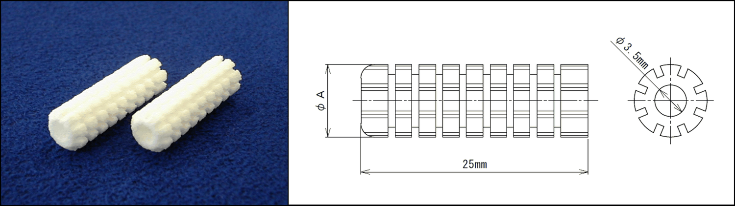
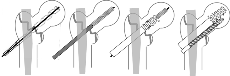
All procedures were performed on a traction table with fluoroscopic guidance. Intramedullary fixation was performed using the InterTAN ® system (Smith & Nephew, Memphis, TN, USA). The lag screw was inserted aiming for the center of the femoral head, and placement was optimized according to the tip-apex distance concept [7]. In the AG, two HA tubes (Neobrace S ® (Aimedic MMT Co., Ltd., Tokyo, Japan)) were inserted at the lag screw entry site (Figure 1 and Figure 2). This augmentation technique is intended to enhance fixation at the bone-implant interface and has been reported to improve the insertion torque ratio [11].
Imaging evaluation and definitions
Preoperative radiographs and computed tomography (CT), as well as postoperative follow-up radiographs, were reviewed.
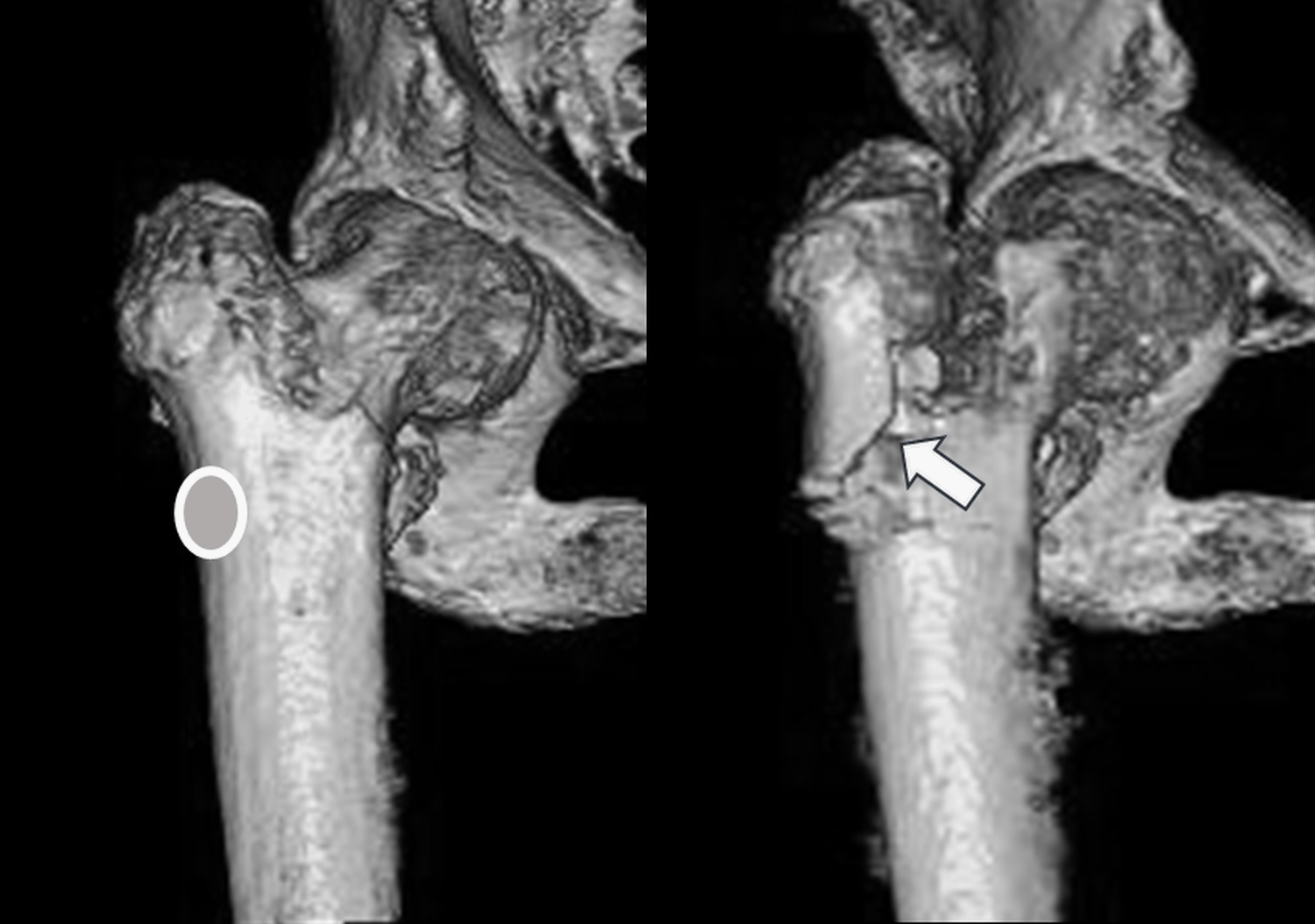
POAWF was defined as a newly identified fracture line or clear cortical discontinuity of the anterior wall (anterior cortex) on postoperative imaging [16](Figure 3).
LWF-LS was defined as a fracture line or cortical disruption of the lateral wall in the vicinity of the lag screw entry site (Figure 3).
Postoperative telescoping was calculated as the change in the lag screw (or head element) position between immediate postoperative and final follow-up radiographs, using a standardized measurement method.
Bone mineral density was assessed using the young adult mean (YAM, %) based on dual-energy X-ray absorptiometry of the lumbar spine and/or proximal femur. Final follow-up was defined as the last available postoperative radiograph during routine clinical care.
Outcomes
The primary outcome was POAWF incidence in the matched cohort. Secondary outcomes included (i) POAWF incidence in the LWF-LS subset, (ii) postoperative telescoping, and (iii) regression analyses for factors associated with telescoping and POAWF.
Statistical analysis
All statistical analyses were performed using JASP (JASP Team, 2025) [17]. Continuous variables are reported as mean (SD) or median [interquartile range], and categorical variables as n (%).
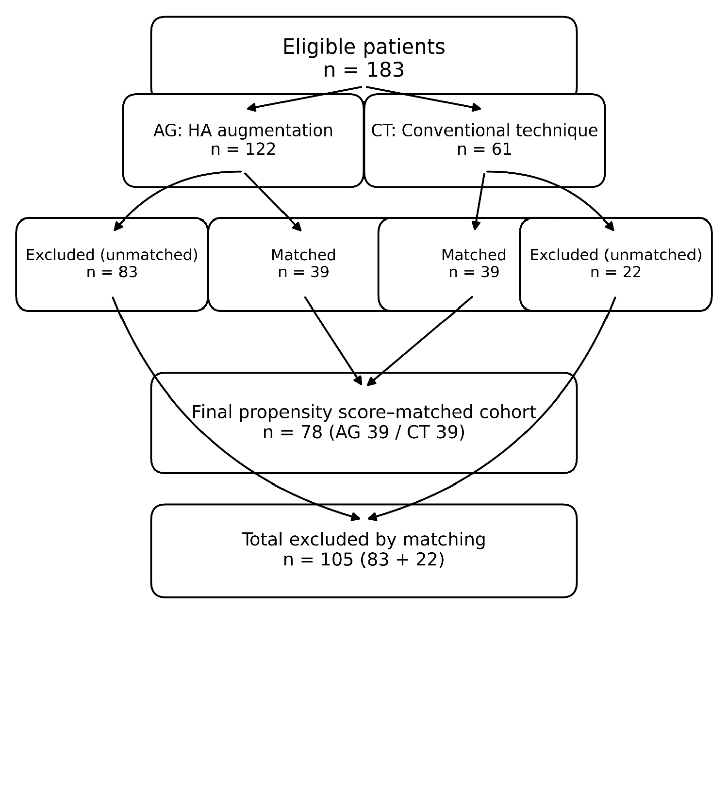
For PSM (Figure 4; Table 1), propensity scores were estimated using logistic regression with treatment group (AG vs. CT) as the dependent variable and age, sex, body mass index (BMI), age-adjusted Charlson comorbidity index (ACCI), and bone mineral density (YAM, %) as covariates [13-15]. One-to-one nearest-neighbor matching without replacement was performed using a caliper width of 0.2. Covariate balance after matching was assessed using standardized mean differences (SMDs); an SMD <0.1 was considered indicative of good balance 18.
Group comparisons of categorical outcomes were performed using chi-square tests or Fisher’s exact tests, as appropriate 19. A two-sided p value < 0.05 was considered statistically significant.
Factors associated with postoperative telescoping were evaluated using multiple linear regression, and standardized regression coefficients (β) and p values were reported. Factors associated with POAWF were evaluated using multivariable logistic regression, and odds ratios (ORs) with 95% confidence intervals (CIs) and p values were reported 20. Missing data, when present, were handled using complete-case analysis; no imputation was performed. No additional sensitivity analyses were performed beyond the prespecified subset analysis restricted to patients with LWF-LS.
Ethics statement
The study adhered to the principles of the Declaration of Helsinki. Approval was obtained from the institutional ethics committee. All patient data were anonymized, and confidentiality was maintained. Informed consent for surgery and its alternatives was obtained from all patients or their legal representatives.
Results
Study cohort after propensity score matching (Table 2)
After PSM, 39 patients in the AG and 39 patients in the CT group (total n = 78) were included in the analyses. PSM is a standard approach for confounding control in observational studies, and post-matching balance was assessed using SMDs [13-15,18]. No missing data were present for covariates used in propensity score estimation or for the primary outcome in the matched cohort. Final follow-up radiographs were available for all matched participants.
Intraoperative parameters and postoperative course (Table 3)
Table 3 summarizes intraoperative parameters and postoperative outcomes. Operative time was shorter in the CT group than in the AG (45.3 vs. 53.1 minutes, p < 0.001). Postoperative telescoping was significantly greater in the CT group than in the AG (2.20 vs.1.37 mm, p = 0.009). No significant between-group differences were observed in intraoperative blood loss (p = 0.468) or tip-apex distance (p = 0.858).
Primary outcome: POAWF incidence in the matched cohort (Table 4)
In the matched cohort, POAWF occurred in 20.51% (8/39) of CT patients versus 5.13% (2/39) of AG patients (p = 0.042). Because of small cell counts, Fisher’s exact test was used where appropriate [19].
Subset analysis: POAWF incidence in patients with LWF-LS (Table 4)
In the subset with LWF-LS, 13 CT patients and 16 AG patients were included. POAWF occurred in 61.54% (8/13) of CT patients compared with 12.50% (2/16) of AG patients (p < 0.001) [19].
Regression analyses for postoperative telescoping and POAWF (Table 5)
In multiple linear regression for postoperative telescoping, LWF-LS (β = 0.591, p < 0.001) and YAM (%) (β = 0.272, p = 0.040) were independently associated with greater telescoping. In multivariable logistic regression for POAWF, HA augmentation was independently associated with reduced odds of POAWF (OR 0.073, 95% CI 0.008-0.655; p = 0.019) [20].
Discussion
In this propensity score-matched comparison, hydroxyapatite tube augmentation at the lag screw entry site was associated with a lower incidence of postoperative anterior wall fracture (POAWF) after InterTAN ® fixation for intertrochanteric fractures. This association persisted in a clinically high-risk subset characterized by lateral wall fracture at the lag screw entry site (LWF-LS), and multivariable logistic regression identified HA augmentation as an independent protective factor for POAWF. Although this was an observational study, PSM was used to balance key baseline covariates, a widely accepted framework for confounding control [13-15].
The lateral femoral wall is a key stabilizing structure in intertrochanteric fracture fixation, and its compromise is strongly associated with reduced stability and higher complication rates [4-6]. The high POAWF incidence observed in the CT group among LWF-LS cases suggests that failure of lateral support around the entry site may facilitate micromotion and excessive telescoping, which can propagate to the anterior cortex. Notably, LWF-LS has been reported to precipitate POAWF and may be associated with postoperative deterioration in activities of daily living, indicating that LWF-LS is not merely a radiographic finding but a clinically meaningful marker of instability [16].
HA tube augmentation is designed to enhance the bone-implant interface, thereby improving fixation at the lag screw entry site. Iwata, et al. reported that HA tube use increases the insertion torque-to-BMD ratio, supporting improved interface fixation [11]. Usami, et al. further suggested, in a biomechanical study, that HA augmentation may increase resistance to rotational instability and reduce lag screw telescoping [13]. These findings are consistent with these reports and extend them by suggesting that entry-site augmentation may reduce a subsequent structural complication-POAWF-particularly when local support is compromised by LWF-LS.
Nevertheless, fixation failure is multifactorial. Technical fundamentals such as lag screw position (e.g., tip-apex distance) remain crucial determinants of stability [7], and sagittal reduction quality (anterior malreduction) has been linked to cutout risk [8]. Moreover, anterior wall fracture may not uniformly predict excessive telescoping or reoperation across studies [9]. Accordingly, HA augmentation should be considered an adjunct rather than a substitute for meticulous reduction and implant positioning, and may be most appropriately targeted to patients at higher risk of local instability, such as those with LWF-LS.
Cement augmentation is an alternative strategy with evidence from systematic reviews and meta-analyses [10]; however, cement and HA differ substantially in material properties and procedure-related risks. Therefore, the clinical role of HA augmentation should be evaluated independently. In this study, the effect was most pronounced in the LWF-LS subset, supporting a pragmatic strategy of using preoperative CT and/or early postoperative imaging to identify LWF-LS and concentrate augmentation in high-risk cases.
This study has several limitations. First, as a retrospective observational study, residual confounding from unmeasured variables cannot be fully excluded despite PSM [13-15]. Second, the number of POAWF events was limited, and effect estimates may therefore be imprecise. To strengthen causal inference in future work and in response to peer review, additional analyses could include (i) doubly robust adjustment by adding any residual imbalanced covariates in post-matching regression models, (ii) sensitivity analyses using alternative propensity score approaches such as inverse probability weighting or overlap weighting, and (iii) quantitative sensitivity analysis for unmeasured confounding (e.g., E-values) [21-23]. Prospective studies with standardized imaging follow-up and functional outcome assessment would further clarify the clinical implications of preventing POAWF.
In summary, LWF-LS identifies a high-risk phenotype for POAWF, and HA tube augmentation at the lag screw entry site may mitigate this risk. Further validation with robust sensitivity analyses and cost-effectiveness evaluation is warranted.
Conclusions
Among patients undergoing InterTAN ® fixation for intertrochanteric femoral fractures, hydroxyapatite tube augmentation at the lag screw entry site was associated with a significantly lower incidence of postoperative anterior wall fracture after propensity score matching. The association remained evident in the subset with LWF-LS, and multivariable logistic regression identified HA augmentation as an independent protective factor. Entry-site HA augmentation may represent a useful adjunct for high-risk intertrochanteric fractures.
Role of the Funders/Sponsors
Not applicable.
Funding
This research received no specific grant from any funding agency in the public, commercial, or not-for-profit sectors.
Disclosures / Conflicts of interest
None.
Funding / Support:
None.
IRB / Ethics
Institutional review board approval was obtained (approval no. 22-2). Consent: opt-out.
Prior presentation
None.
Data availability
De-identified data are available from the corresponding author on reasonable request, subject to institutional approval.
References
- Gullberg B, Johnell O, Kanis JA (1997) World-wide projections for hip fracture. Osteoporos Int 7: 407-413.
- Dhanwal DK, Dennison EM, Harvey NC, et al. (2011) Epidemiology of hip fracture: Worldwide geographic variation. Indian J Orthop 45: 15-22.
- Orimo H, Yaegashi Y, Hosoi T, et al. (2016) Hip fracture incidence in Japan: Estimates of new patients in 2012 and 25-year trends. Osteoporos Int 27: 1777-1784.
- Gotfried Y (2004) The lateral trochanteric wall: A key element in the reconstruction of unstable pertrochanteric hip fractures. Clin Orthop Relat Res 425: 82-86.
- Palm H, Jacobsen S, Sonne Holm S, et al. (2007) Integrity of the lateral femoral wall in intertrochanteric hip fractures: An important predictor of a reoperation. J Bone Joint Surg Am 89: 470-475.
- Hsu CE, Shih CM, Wang CC, et al. (2013) Lateral femoral wall thickness: A reliable predictor of postoperative lateral wall fracture in intertrochanteric fractures. Bone Joint J 95: 1134-1138.
- Baumgaertner MR, Curtin SL, Lindskog DM, et al. (1995) The value of the tip-apex distance in predicting failure of fixation of peritrochanteric fractures of the hip. J Bone Joint Surg Am 77: 1058-1064.
- Inui T, Watanabe Y, Suzuki T, et al. (2024) Anterior malreduction is associated with lag screw cutout after internal fixation of intertrochanteric fractures. Clin Orthop Relat Res 482: 536-545.
- Yamamoto N, Tamura R, Inoue T, et al. (2021) Radiological findings and outcomes of anterior wall fractures in pertrochanteric fractures. J Orthop Sci 26: 247-253.
- Rompen E, Wijffels M, Van Walsum ADP, et al. (2021) Cement augmentation for internal fixation of trochanteric fractures: A systematic review and meta-analysis. PLoS One 16: e0251894.
- Iwata H, Takada N, Kuroyanagi G, et al. (2021) Effect of hydroxyapatite tubes on the lag screw intraoperative insertion torque in intertrochanteric femoral fractures. Injury 52: 3377-3381.
- Usami T, Takada N, Iwata H, et al. (2023) A biomechanical analysis of the effect of hydroxyapatite augmentation for trochanteric femoral fractures. Exp Ther Med 25: 256.
- Rosenbaum PR, Rubin DB (1983) The central role of the propensity score in observational studies for causal effects. Biometrika 70: 41-55.
- Stuart EA (2010) Matching methods for causal inference: A review and a look forward. Stat Sci 25: 1-21.
- Austin PC (2011) An introduction to propensity score methods for reducing the effects of confounding in observational studies. Multivariate Behav Res 46: 399-424.
- Matsumoto T, Nakatani S, Inoue K, et al. (2025) Intertrochanteric femoral fracture with lateral wall fracture at the lag screw insertion site may cause postoperative anterior wall fracture and lead to deterioration in activities of daily living. J Orthop Sci 30: 358-363.
- JASP Team (2025) JASP (Computer software).
- Austin PC (2009) Balance diagnostics for comparing the distribution of baseline covariates between treatment groups in propensity-score matched samples. Stat Med 28: 3083-3107.
- Agresti A (2013) Categorical data analysis. (3 rd edn), Hoboken (NJ), Wiley.
- Hosmer DW, Lemeshow S, Sturdivant RX (2013) Applied logistic regression. (3 rd edn), Hoboken (NJ), Wiley.
- Funk MJ, Westreich D, Wiesen C, et al. (2011) Doubly robust estimation of causal effects. Am J Epidemiol 173: 761-767.
- Li F, Thomas LE (2019) Addressing extreme propensity scores via the overlap weights. Am J Epidemiol 188: 250-257.
- VanderWeele TJ, Ding P (2017) Sensitivity analysis in observational research: introducing the E-value. Ann Intern Med 167: 268-274.
Corresponding Author
Takuji Matsumoto, MD, PhD, Department of Orthopaedic Surgery, Nokami Kosei General Hospital, 198, Shobata, Kimino-Cho, Kaiso-Gun, Wakayama 640-1141, Japan.
Copyright
© 2025 Lima JM, et al. This is an open-access article distributed under the terms of the Creative Commons Attribution License, which permits unrestricted use, distribution, and reproduction in any medium, provided the original author and source are credited.