Closure Femoral Isthmoplasty in Patients with Femoral Defects III B and IV According to Paprosky's Classification
Summary
Introduction: Severe femoral defects remain a challenge for orthopedic surgeons, who face extensive proximal metaphyseal bone defects, extensive diaphyseal bone defects, and the need to achieve long-term survival of the implant. Femoral isthmoplasty is a surgical technique that reduces the diameter of the femoral isthmus by 2 to 4 mm.
Methods: A report of two cases. Case 1: A 63-year-old male with a Paprosky type 3B femoral defect. Case 2: A 72-year-old female patient with a Dorr C configuration of the femoral canal.
Conclusion: Closure femoral isthmoplasty in patients with femoral defects may be an appropriate surgical strategy for reconstructing a proximal femur.
Keywords
Closure femoral isthmoplasty, Femoral defects, Paprosky
Introduction
Revision total hip arthroplasty (rTHA) and exchange of the femoral component is a complex and technically demanding procedure. The term femoral isthmoplasty has not been clearly defined in the medical literature. It involves making specific cuts in the femoral stem to reduce the diameter of the femoral isthmus. This technique is used to treat severe femoral defects, such as Paprosky IIIB or IV, which are characterized by narrowing of the femoral canal [1]. The present research aims to describe a novel femoral isthmoplasty technique that can be employed to treat severe femoral defects as part of the surgical strategy.
Objective
To describe the closure femoral isthmoplasty technique and evaluate its application in treating severe femoral defects (Paprosky IIIB and IV) with cement less revision stems.
Methods
A report of two cases is presented, analyzing the surgical technique of femoral isthmoplasty as a means of reducing the diameter of the femoral isthmus by approximately 2 to 4 mm, achieving greater contact between the non-cemented prosthetic component and the femur [2]. This is particularly useful in cases where the diameter of the larger non-cemented distal fixation prosthetic component is only a few millimeters smaller than the diameter of the patient's femoral isthmus, a situation that frequently occurs in femurs with Paprosky IIIB and IV defects. This surgical practice would transform type III B femoral defects into III A and type IV defects into III A, avoiding the use of massive bone grafts, long cemented stems, or tumor prostheses.
Case 1
A 63-year-old male (BMI 37) with a 22-year-old Charnley prosthesis presented with a Paprosky IIIB defect. Revision with a modular uncemented metaphyseal-distally fixed stem was performed, using a distal stem with a diameter of 22 mm in a femur that had an approximate internal diameter of 24 mm. Case 2: A 72-year-old female (BMI 35) with a history of prosthetic joint infection had a Dorr C femoral canal with medial and lateral cortical loss and a stovepipe morphology. At the time of evaluation, she had an antibiotic spacer. The internal diameter of her femur was approximately 25 mm, and the largest stem available was of the same diameter.
Surgical technique of femoral isthmoplasty closure
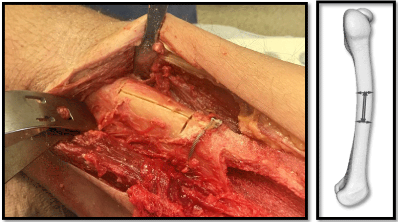
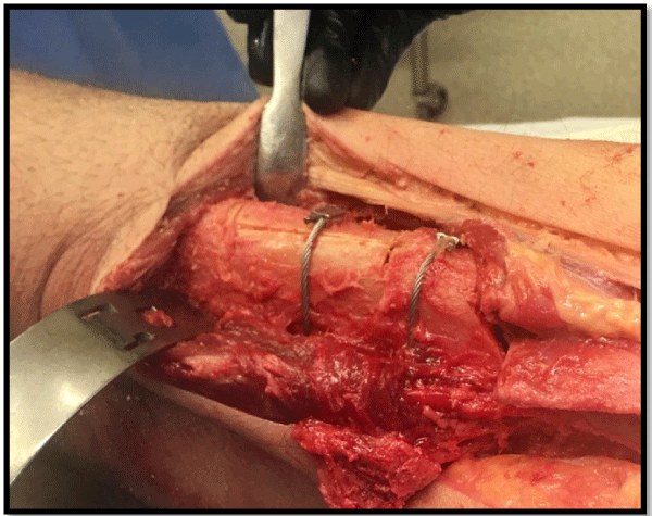
The surgical access route was performed extending distally in the case of using Gibson-Moore, opening the fascia lata and accessing the femoral isthmus retro vastus lateralis of the quadriceps. A longitudinal osteotomy was performed on the lateral side of the femur of 4 to 6 cm, with a variable width between 2 to 4 mm, depending on how much the diameter of the medullary canal needed to be reduced. To achieve this, two transverse osteotomies, proximal and distal to the longitudinal osteotomy, are performed, ranging from one-third to half of the femoral diameter. This allows for the closure of the longitudinal osteotomy as if they were two opposing doors, thus avoiding intraoperative femoral fractures. (Figure 1). To perform the closure, one or two multifilament wires or surgical wires of 1.25 mm should be used, and the osteotomy should preferably be closed with a Charnley-type wire tensioner (Figure 2).
The closure of the femoral osteotomy (isthmoplasty) is performed, and the corresponding prosthetic component is implanted, allowing for stable diaphyseal femoral fixation. Three femoral isthmoplasties of closure have been performed as part of the study to date, but we lost follow-up of one patient who was excluded from this research.
Results
Case 1
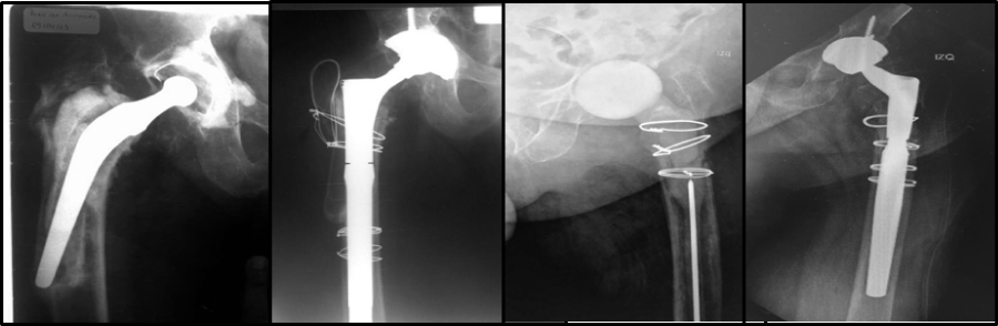
Radiographic control at 6 years postoperative showed no subsidence of the implant and adequate integration.
Case 2
A 4-year postoperative follow-up of the conversion revealed implant integration without radiographic subsidence. In both cases presented, a reduction in the diameter of the femoral isthmus was achieved, allowing good axial and rotational stability of the uncemented modular stem, with the respective follow-up of 6 and 4 years showing complete osseo integration without evidence of sinking radio graphically (Figure 3).
Discussion
Severe femoral defects remain a challenge for orthopedic surgeons, who face extensive proximal metaphyseal and diaphyseal bone defects, along with the desire to achieve long-term implant survival, biomechanical restoration of the joint, and stable fixation of the implant through osteointegration.
To recover the functionality of the hip joint in cases of severe proximal femoral defects, a variety of surgical techniques have been described, including cemented stems, uncemented stems with proximal porous coating, uncemented stems with total porous coating, uncemented modular distal fixation conical striated stems, allografts, and unconventional proximal femoral replacement. The best functional results reported in the literature were for uncemented modular distal fixation stems, as their conical design provides axial stability, their longitudinal grooves confer rotational stability, and their titanium material allows for better load distribution.
However, concerns persist regarding the risk of mechanical failure in the application of conical stems, particularly junction failure in the modular design, sinking rates greater than 10 mm reaching 20%, dislocations representing up to 14%, aseptic loosening, and the risk of periprosthetic fracture. These issues are mostly attributed to the surgical technique, along with the design of the implants, related to insufficient prosthetic sizes and the tendency to fix the stem based on leg length rather than prioritizing axial and rotational stability of the implant.
Femoral isthmoplasty, as a novel surgical technique, emerged from the need to achieve a reduction in diameter of the femoral isthmus by 2 to 4 mm, sufficient to ensure contact between the prosthetic system and the bone in severe femoral defects (Paprosky III B and IV). In the two cases presented, a reduction in the diameter of the femoral isthmus was achieved, allowing good axial and rotational stability of the uncemented modular stem, with follow-ups of 6 and 4 years showing complete osseointegration without radiographic sinking.
A wide variety of surgical techniques are described in the literature for this pathology due to the complexities of the surgeries and the high metabolic demand on the patient, who often presents a fragile skeletal system, poor bone reserve, and multiple comorbidities.
Femoral isthmoplasty could help avoid the need for cementing revision femoral stems (which are often the same uncemented stems that were being attempted to implant but not designed for that purpose), along with the risks associated with inadequate cementation techniques, which stem from difficulties in properly pressurizing the cement due to the inability to place a distal plug adequately in very poor-quality bone.
Another option to solve the problem of severe femoral defects is the technique published by Ling, et al. [3] which involves the impaction of cadaveric bone graft on the inner walls of the native femur, creating a neo-canal where a revision stem is then cemented. This technique is associated with a high risk of intraoperative fractures during bone impaction, secondary sinking of the stem, and is both technically and metabolically demanding for the surgeon and the patient, considerably increasing surgical time and associated risks of morbidity and mortality [4].
We acknowledge our limitations, as this is a case report with a small sample size and limited follow-up time. We suggest conducting a study with greater statistical power to thoroughly investigate the benefits of this intervention.
Conclusion
The use of the femoral closure isthmoplasty technique in femoral defects according to the Paprosky classification (IIIB and IV) could become a surgical strategy to maintain stable femoral fixation and adequate osteointegration between the cement less implant and the native bone.
References
- Sheth NP, Nelson CL, Paprosky WG (2013) Femoral bone loss in revision total hip arthroplasty: Evaluation and management. Am Acad Orthop Surg 21: 601-612.
- Jacob Wilkerson, Navin Fernando D (2020) Classifications in brief: The Dorr classification of femoral bone. Clin Orthop Relat Res 478: 1939-1944.
- Ding ZC, Ting Xian Ling, Ming Cheng Yuan, et al. (2020) Minimum 8-year follow-up of revision THA with severe femoral bone defects using extensively porous-coated stems and cortical strut allografts. BMC Musculoskelet Disord 21: 218.
- Matar HE, Veenesh Selvaratnam, Mikhil Jain, et al. (2021) Cortical strut allografts in salvage revision arthroplasty: Surgical technique and clinical outcomes. J Clin Orthop Trauma 17: 37-43.
Corresponding Author
Gonzalo Mur, Formación Académica, Universidad Católica del Ecuador, Hospital de Clínicas José de San Martín - Universidad de Buenos Aires, Argentina.
Copyright
© 2025 Mur G, et al. This is an open-access article distributed under the terms of the Creative Commons Attribution License, which permits unrestricted use, distribution, and reproduction in any medium, provided the original author and source are credited.