The Success of the Ponseti Method in the Treatment of Congenital Varus Bot Foot (Cvbp) in a Low-Income Country: Preliminary Results of a 125pbve Series in 82 Patients
Summary
Varus equines club foot (VEBF) is a complex, three-dimensional deformity characterized by equines and Varus of the hind foot, adduction of the forefoot and supination of the foot.
The purpose of this work was to evaluate the preliminary results of the treatment of equines varus clubfoot by the Ponseti method.
Methods: This was a prospective descriptive study over a 3-year period (January 2015 to December 31, 2017) including 125 EVBP in 82 patients aged 0 to 6 years. Syndromic and neurogenic club foot were not included.
Results: PBVE represented O, 69% of consultations, the average age of patients was 3.8 months; there were 46 boys and 36 girls. The damage was bilateral in 43 patients, tenotomy of the Achilles tendon was performed in 91.46% of the cases; the average number of casting sessions was 6.5 ± 1.54. The results were very good in 89.02% of the cases, good in 7.32% and bad in 3.36% of the cases.
Conclusion: The Ponseti method (PM) is an effective and inexpensive treatment adapted to low-income countries like ours. It allows the correction of PBVE from birth to late child hood for non-stiff feet. PD has allowed a correct functional future for potentially disabled children.
Keywords
Varus equines club foot, Congenital, Pirani score, Ponseti
Introduction
Varus equines club foot (VEBF) is a complex, three-dimensional deformity that combines equinus, varus of the hind foot, adduction of the forefoot and supination of the foot [1-3]. These deformities result in a stiffening in bad position of the foot and ankle. These deformities will be responsible for a physical handicap in the absence of an effective treatment [4]. It is the most common deformity in pediatric orthopedics [5]. Congenital varus equinus foot affects approximately 174,000 children each year worldwide, 91% of whom live in low and middle income countries [6]. In Africa its prevalence is estimated at 1.11 per 1000 births [7]. The treatment of PBVE used to be surgical from the first year of life. A radical change in the management of PBVE was made in view of the deterioration of the results of surgery from the outset with time, the good results of functional methods (French method) and especially the excellent results of the Ponseti method [1].
During the last decade, the Ponseti method has been accepted world wide as the most effective and cheapest treatment for PBVE [8].
This method consists of progressive and sequential correction of deformities by a series of casts, almost systematic tenotomy of the calcaneal tendon and relaying with derotation splints [9,10]. The preparation of these casts follows a very precise protocol that must be respected to avoid recurrence [11].
The total absence of management in certain developing countries leads to functional tragedies, with physical, psychological and social consequences [2].
To the best of our knowledge, no previous study has been conducted on equine clubfoot in our context. However, the EVLP is increasingly frequent in children's surgery consultations and poses a real problem of management. The lack of a precise treatment method and the poor results observed over the years led us to adopt the Ponseti method in our department in order to improve management.
The aim of this study was to evaluate the preliminary results of the treatment of equine varus clubfoot using the Ponseti method.
Material and Methods
This was a prospective descriptive study of 125 PBVE in 82 children, managed by the Ponseti method between January 2015 and December 2017 in the pediatric surgery department of the Donka National Hospital (CHU of Conakry). All children aged 0 to 6 years with unilateral or bilateral congenital varus equines clubfoot treated by the Ponseti method were included. Patients older than 6 years, syndromic and neurogenic clubfoot were not included.
Epidemiological, clinical, therapeutic and evolutionary parameters were studied.
A clinical examination was performed at the first consultation to look for other associated anomalies. An evaluation form (Ponseti form) was filled out. This form included general information and clinical parameters concerning the patient. It allowed a precise evaluation of the severity of the deformity and its evolution during the treatment.
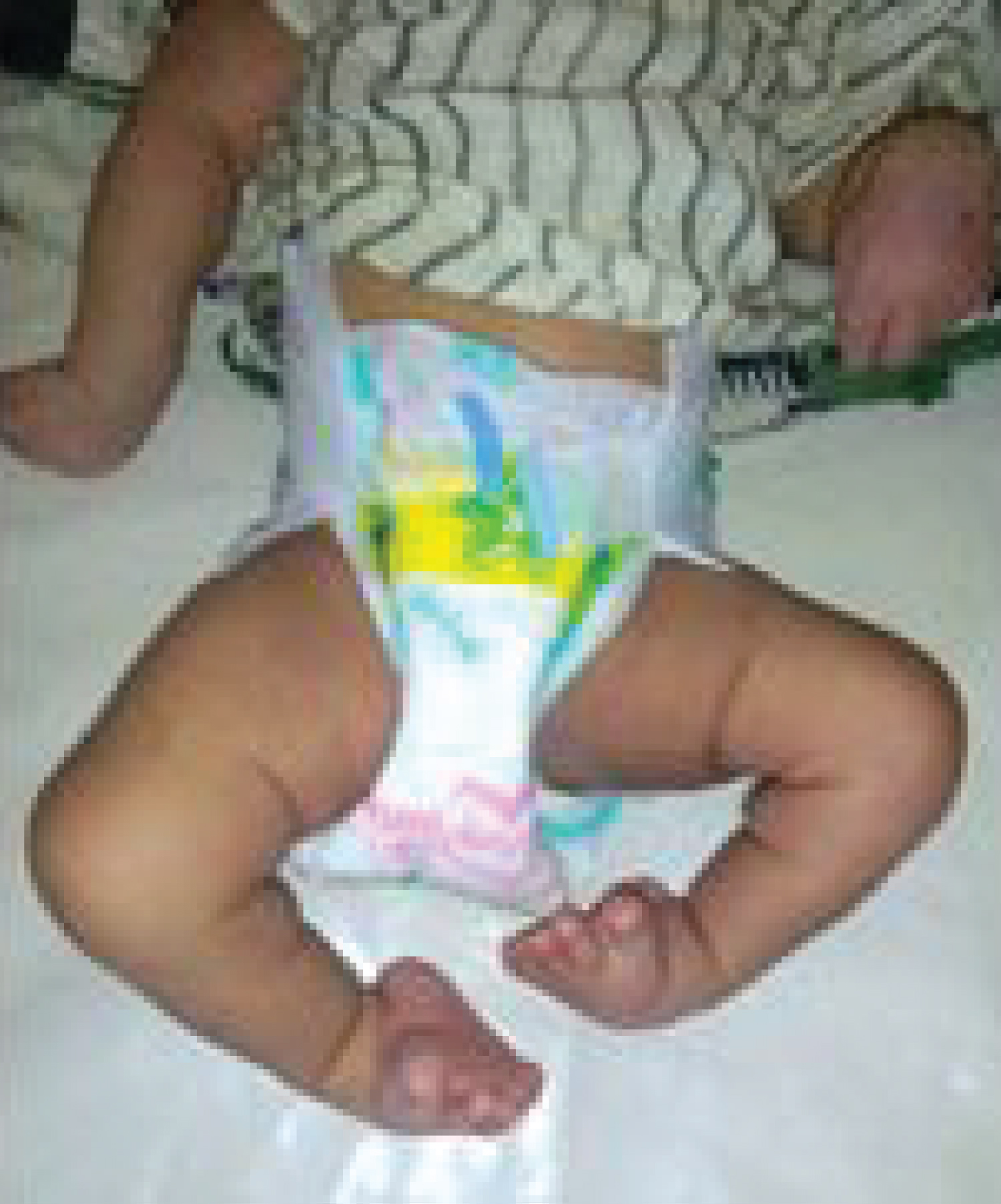
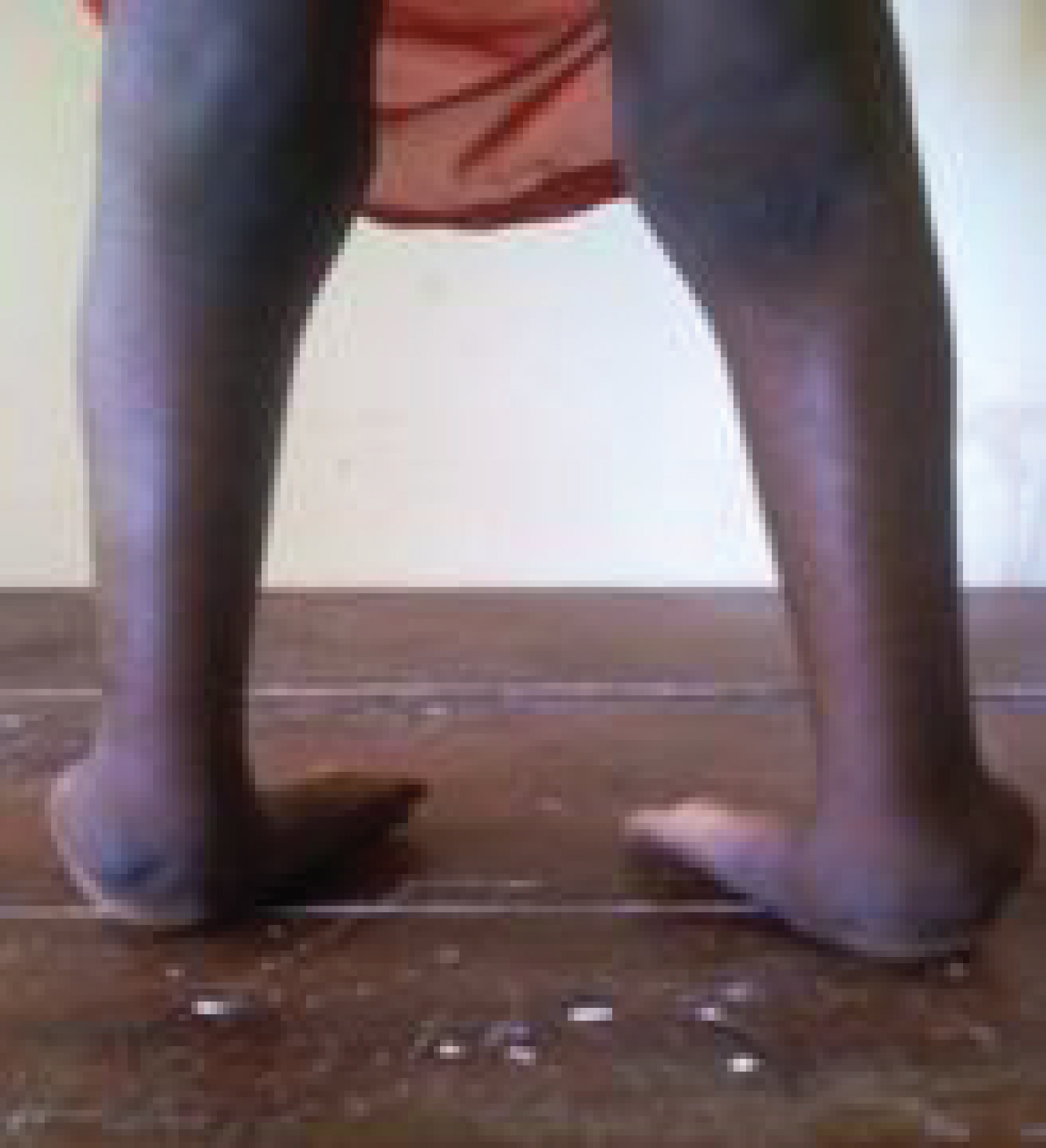
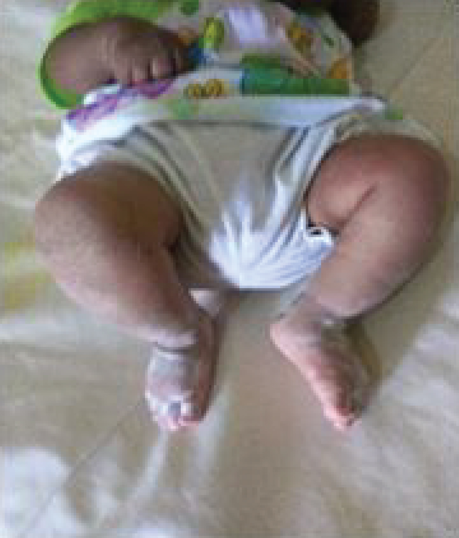
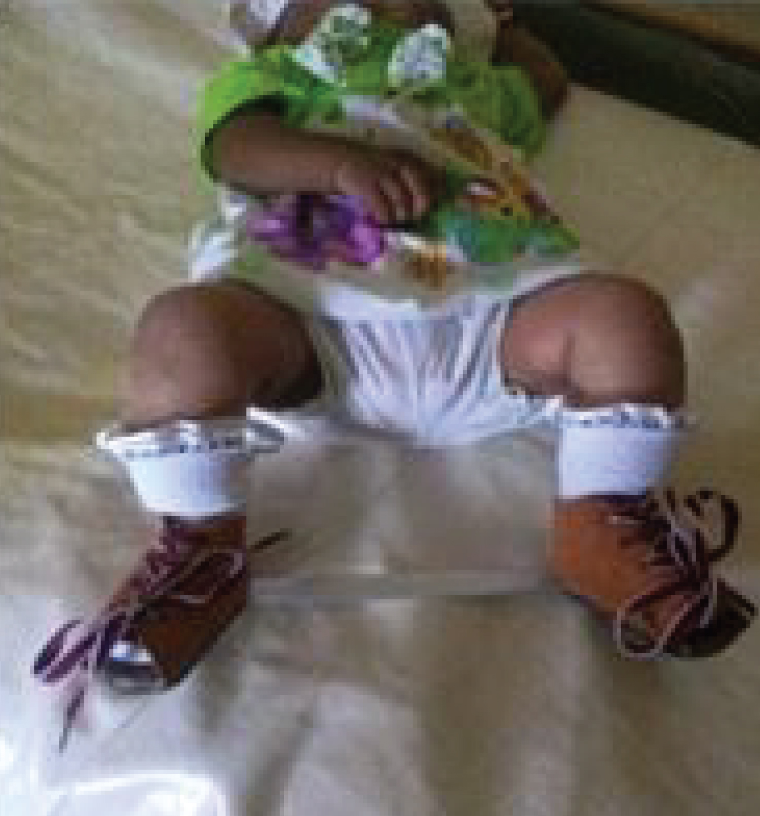
The Pirani score was used to quantify the severity of the deformity of the medial foot (lateral border curvature, medial groove, coverage of the head of the talus) and that of the hind foot (posterior groove, fixed equinus, vacuity of the heel). Each item was scored as 0 (normal foot), 0.5 (moderately abnormal), 1 (very abnormal). A PBVE with a total score = 0/6 was considered normal. A PBVE with score ≤ 3/6 was considered moderate. A PBVE with score between 4-5 was considered severe and a PBVE with score = 6 was considered very severe. This Score was performed at baseline, at removal of each cast, and at the end of deformity correction (Figure 1 and Figure 2).
Ponseti's method had two essential steps: The first step was the reduction of the deformity and the second was the maintenance of the correction by casts allowing a progressive and sequential correction of the formation.
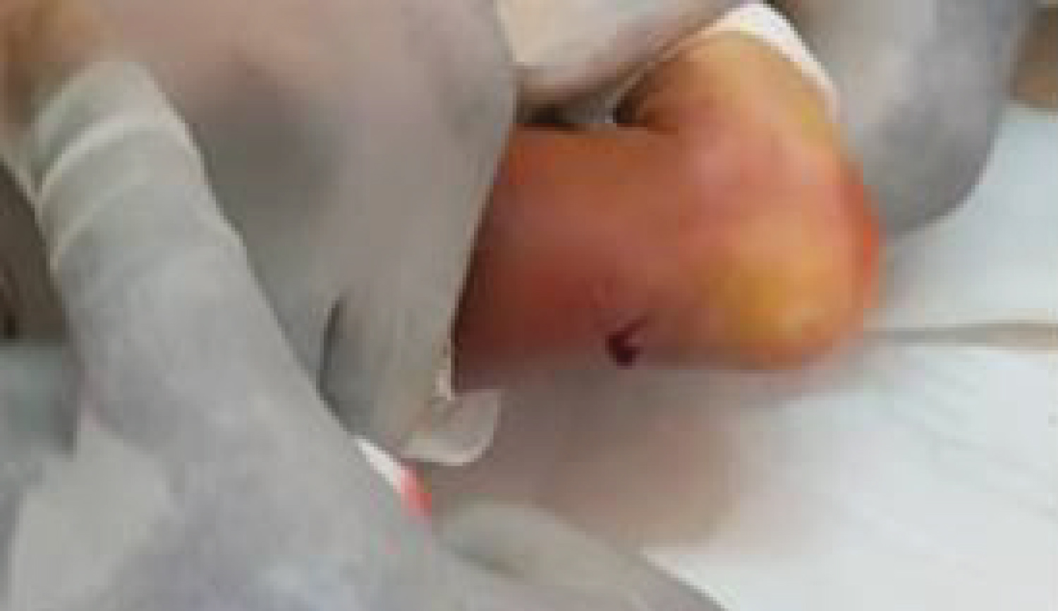
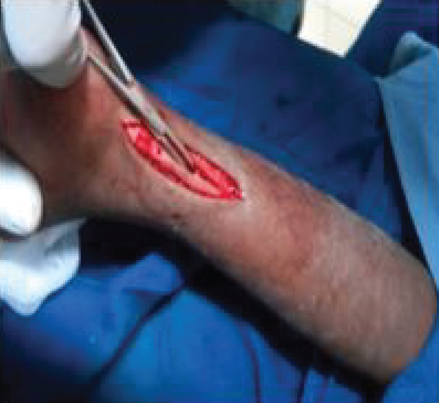
Percutaneous tenotomy of the calcaneal tendon was routinely performed under local anesthesia in all patients younger than 2 years of age when the dorsiflexion of the foot was less than 15° (Figure 3). In patients whose age was ≥ 2 years, a Z-lengthening of the calcaneal tendon was performed in the operating room under general anesthesia (Figure 4). A final cast with 15 to 20 degrees of ankle dorsiflexion with abduction and external rotation was placed for 21 days.
The maintenance of the correction began with the removal of the last cast by putting on shoes allowing the feet to be abducted and externally rotated by 70°.
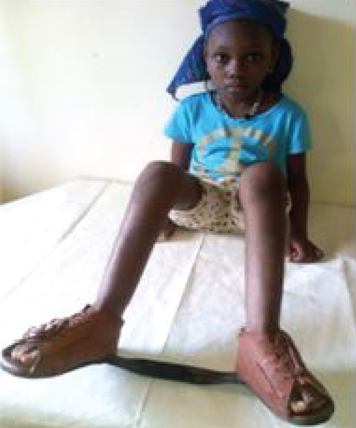
The shoes used in our study were SFAB (Steenbeek Foot Abduction Brace). These shoes were used full time for 3 months, at night and during naps until walking age in small infants (Figure 5a and Figure 5b). In patients over 2 years of age, the orthosis was used during the day and the shoes were used at night and during naps (Figure 6).
The evaluation of the result of the correction of the deformities was made according to the following morphological criteria:
-Very good: Any foot for which the correction of all deformities was obtained.
-Good: Any foot with a single persistent deformity.
-Poor: Any foot with at least two persistent deformities.
Patients were seen at follow-up once a month for the first three months when they started wearing shoes, then once every three months until they reached walking age. Our average follow-up was 3 years.
Data entry was done using Microsoft Word 2016 software, and analysis was done using Epi infos 3.5.1 software.
Results
Out of a total of 11873 patients seen in consultation, 82 patients were carriers of PBVE, i.e. a frequency of 0.69%. The average age at the beginning of treatment was 3.8 months with extremes of 0 and 72 months; the age range of 0 to 3 months was the most frequent, i.e. 76.83%. There was a male predominance with a sex ratio of 1.27. Thirty-six patients were from a consanguineous marriage; the involvement was bilateral in 43 patients and unilateral in 39 patients (23 right and 16 left). The initial Pirani score was very severe in 48 patients, severe in 28 patients and moderate in 6 patients. Percutaneous tenotomy of the Achilles tendon was performed in 75 patients (91.46%) and Z-stretching of the Achilles tendon was performed in 7 patients (8.54%). The average number of cast sessions was 6.5 ± 1.54 sessions. Cast complications were represented by 20 cases of skin damage and 4 cases of leg curvature (Table 1 and Table 2).
Discussion
Congenital varusequinus foot is a developmental anomaly of the foot occurring during the fetal period. It is one of the most common pediatric malformations [12,13]. The prevalence of 0.69% found in our study is purely hospital-based and cannot be generalized to the general population. However, it reflects the importance of this malformation in our consultation activity, although it is not a priority in the country's health policy. Epidemiological studies published in the last 55 years suggest a prevalence at birth of 0.5 to 2 cases/1000 live births. The higher prevalence seems to be associated with socio demographic, genetic and environmental risk factors; hence, its high prevalence in low and middle income countries [12]. The meanage found in our study corroborates that of Nyondika, et al. [5] who found a manage of 3 months 18 days. On the other hand, in the series of Trigui, et al. [4] the meanage was 61 days.
The average age found to be high in our study could be explained by several factors such as: The low socioeconomic level of the population, the use of traditional medicine, the negligence of parents, the lack of qualified personnel, the absence of information and awareness programs.
The clear predominance of males has been reported by several authors [12,14,15]. Krusse, et al. [16] in 2008 proposed a reason for the gender difference; however, the phenotypic variability in affected individuals is still poorly understood.
Many classification systems have been proposed to measure the severity of clubfoot deformity and the impact of treatment. The Pirani score and the Dimeglio score are the two most commonly used classification systems. The Pirani score is 0 to 6, where zero is a normal foot and six is the most severe deformity. The Dimeglio score has a maximum of 20 points and the deformity is classified as mild, moderate, severe or very severe. We opted for the Pirani score, which has the advantage of being simple, reproducible and very easy to handle in routine clinical practice [7]. The very severe form of PBVE was the most frequent in our series. This result is comparable to that found by Desai, et al. [17] with 81.25% of very severe form. This score allows an initial assessment of the deformities and the anatomical results obtained [18,19]. During treatment, Ponseti in his original method proposed a tenotomy of the calcaneal tendon (CT) when the ankle dorsiflexion was less than 15-20° or when the calcaneus appeared high before the last cast [20]. In our study, tenotomy of the calcaneal tendon was performed in 91.46% of cases and open Z-lengthening of the CT in 8.54% of cases. In 2002, Chotel, et al. [21] reported the preliminary results of 50 PBVEs. Tenotomy of the TC was performed in 95% of cases. In the series Niyondiko, et al. [5] in 2017, TC tenotomy was done for 147 for 162 feet. Malex, et al. [22] in their work on the clinical and ultrasound evaluation of a series of 221 PBVEs, proposed TC tenotomy as a routine procedure. This is a simple and minimally invasive procedure with exceptional complications [23], with rapid healing of the CT [24].
The number of casting sessions required for correction of the EVLP varies. It is most often correlated to the severity of the deformity and to the experience of the practitioner [25,26]. Classically, 5 to 6 casting sessions are necessary for correction of the deformity [27,28]. In our series, the average number of casting sessions required for correction was 6.5 ± 14 sessions. This result is comparable to that published by some authors.
In the series by Abass, et al. [29], the average number of casting sessions was 6.6. Nyondiko, et al. [5] used an average of 6.8 casting sessions to correct the deformity.
Clubfoot can be successfully treated in 95% of cases using a non-surgical method; the Ponseti treatment method [6].
The success rate in our study is comparable to the preliminary results of studies published by some authors. In the series of Nyondiko, et al. [5] the correction of the deformity was complete in 84.4%, moderate in 8.3% of cases; in complete in 7.3%. Faldini, et al. [15] treated 88 PBVE with MP before and after walking age with a mean follow-up of 5 years. In neonates, the results were excellent in 80.7%, moderate in 11.5%, and poor in 7.6%. In ambulant children, the results were excellent in 75% and good in 25% of cases. In ambulant children, the results were excellent in 20.8% of cases, good in 66.6% and poor in 12.5% of cases. In the study by Bronfen C, et al. [30], the results were good or very good in 61.3% of cases, poor in 17.33% of cases, and fair in 21.3% of cases. In our series, after correction of the deformity, the poor results (3.36%) that constitute the relapses were due at the beginning of our experience to the compliance of the abduction splints, and then to poor compliance in wearing the splints. Morcurendo, et al. [31] showed that noncompliance with abduction splinting was associated with a 17-fold increased risk of relapse.
Conclusion
Congenital varus equines clubfoot is a deformity commonly encountered in our pediatric orthopedic clinic. It is a public health problem in our country. PD is an effective treatment, inexpensive, adapted to our low-income countries and gives better results. It can correct club feet from birth to late childhood for non-stiff feet, thus reducing the rate of surgical correction. The cooperation of the parents is essential throughout the treatment for a better result. Ponseti's method has allowed a correct functional future for potentially disabled children.
References
- Bergerault F, Fournier J, Bodman C, et al. (2014) Initial management of varusequines club foot in 2012 in France. Journal of Orthopedic and Trauma Surgery 100: 87- 90.
- Chotel F, Parot R, Bérard J (2005) Congenital foot deformities. Arch Pediatrics 12: 797-801.
- Limpaphayom N, Sailohit P (2019) Factors related to early recurrence of idiopathic clubfoot post the ponseti method. Malays Orthop J 13: 28-33.
- Trigui M, Ayadi K, Ben Jmaa S, et al. (2010) Treatment of severe equine Varus clubfoot by the Ponseti method. Preliminary results of a 3-year prospective study. Orthopedic Tunisia 3: 27-34.
- Niyondiko JC, Havyarimana C, Ndayizeye G, et al. (2017) Morphological evaluation of the club foot after initial treatment using the Ponseti technique. J Afr Chir Orthop Traumatol 2: 70-75.
- Owen RM, Capper B, Lavy C (2018) Club foot treatment in 2015: A global perspective. BMJ Glob Health 3: e000852.
- Smythe T, Kuper H, Foster A, et al. (2017) Birth prevalence of congenital talipes equinovarus in low-and middle-income countries: A systematic review and meta-analysis. Trop Med Int Health 22: 269-285.
- Lynn S (2009) Clubfoot: The ponseti method. Global help. (3rd edn).
- Ponseti IV, Smoley EN (1963) Congenital club foot: The results of treatment. J Bone Joint Surg 45: 261-344.
- Scher DM (2005) The Ponseti method for clubfoot correction. Oper Tech Orthop 15: 345-349.
- Zhao D, Li H, Zhao L, et al. (2014) Results of clubfoot management using the Ponseti method: Do the details matter? A systematic review. Clin Orthop Relat Res 472: 1329-1336.
- Pavone V, Chisari E, Vescio A, et al. (2018) The etiology of idiopathic congenital talipes equinovarus: A systematic review. J Orthop Surg Res 13: 206.
- Ganesan B, Luximon A, Al Jumaily A, et al. (2017) Ponseti method in the management of clubfoot under 2 years of age: A systematic review. PLoS One 12: e0178299.
- Saltzman HM (2009) Foot focus: International initiative to eradicate clubfeet using the Ponseti method. Foot Ankle Int 30: 468-471.
- Faldini C, Traina F, Nanni M, et al. (2016) Congenital idiopathic talipes equinovarus before and after walking age: observations and strategy of treatment from a series of 88 cases. J Orthopaed Traumatol 17: 81-87.
- Kruse LM, Dobbs MB, Gurnett CA (2008) Polygenic threshold model with sex dimorphism in clubfoot inheritance: The carter effect. J Bone Joint Surg Am 90: 2688-2694.
- Desai S, Aroojis A, Metha R (2008) Ultrasound evaluation of club foot correction during Ponseti treatment. J Pediatr Orthop 28: 53-59.
- Jain S, Ajmera A, Solanki M, et al. (2017) Interobserver variability in pirani clubfoot severity scoring system between the orthopedic surgeons. Indian J Orthop 51: 81-85.
- Mejabi JO, Esan O, Adegbehingbe OO, et al. (2016) The Pirani scoring system is effective in assessing severity and mon2itoring treatment of clubfeet in children. Br J Med Med Res 17: 1-9.
- Ponseti IV (1992) Treatment of congenital club foot. J Bone Joint Surg 74: 448-445.
- Chotel F, Parot R, Durand JM, et al. (2002) Prise en charge initiale du pied bot varus équin congénital selon la méthode de Ponseti. Rev Chir Orthop 88: 710-717.
- Marleix S, Chapuis M, Fraisse B, et al. (2012) Intérêt de la ténotomie du tendon calcanéen dans le traitement du pied bot varus équin (PBVE) idiopathique selon la méthode de Ponseti. Evaluation clinique et Echographique à propos d’une série de 221 PBVE. Revue de chirurgie orthopédique et traumatologique 98: 174-178.
- Burghardt RD, Herzenberg JE, Ranade A (2008) Pseudoaneurysm after Ponseti percutaneous Achilles tenotomy: A case report. J Pediatr Ortho 28: 366-369.
- Barker SL, Lavy CB (2006) Correlation of clinical and ultrasonographic findings after Achilles tenotomy in idiopathic club foot. J Bone Joint Surg Br 88: 377-379.
- Aydin BK, Senaran H, Yilmaz G, et al. (2015) The need for Achilles tenotomy in the Ponseti method: Is it predictable at the initiation or during the treatment? J Pediatr Orthop 24: 341-344.
- Gao R, Tomlinson M, Walker C (2014) Correlation of Pirani and Dimeglio scores with number of Ponseti casts required for Clubfoot correction. J Pediatr Orthop 34: 639-642.
- Gunalan R, Mazelan A, Lee YPB, et al. (2016) Pattern of presentation and outcome of short-term treatment for idiopathic clubfoot/CTEV with Ponseti method. Malays Orthop J 3: 21-25.
- Dyer PJ, Davis N (2006) The role of the Pirani scoring system in the management of clubfoot by the Ponseti method. J Bone Joint Surg 88: 1082-1084.
- Abbas M, Qureshi OA, Jeelani LZ, et al. (2008) Management of congenital talipes equinovarus by Ponseti technique: A clinical study. J Foot Ankle Surg 47: 541-545.
- Bronfen C, Lebel B, Geffard B, et al. (2009) Traitements des pieds bots varus équins (PBVE) par la méthode de Ponseti. Etude rétrospective de 113 pieds chez 74 enfants. Rev de chirurgie orthopédique et traumatologique 95: 121-127.
- Morcurendo JA, Dolan LA, Dietz FR, et al. (2004) Radical reduction in the rate of extensive corrective surgery for clubfoot using the Ponseti method. Pediatrics 113: 376-380.
Corresponding Author
Dr. Balla Keita, Assistant Head of Clinic, Department of Pediatric Surgery, Donka National Hospital (CHU Conakry), Guinea, Tel: +224624911130
Copyright
© 2022 Keita B, et al. This is an open-access article distributed under the terms of the Creative Commons Attribution License, which permits unrestricted use, distribution, and reproduction in any medium, provided the original author and source are credited.