Paradoxical Presentation of a Calcaneonavicular Tarsal Coalition
Abstract
Case: Tarsal Coalition is a musculoskeletal deformity of the lower limb with a progression that is often asymptomatic until ossification of the calcaneoavicular coalition in the second decade. We report a novel case of a patient symptomatic before ossification of the coalition with resolution of symptoms coinciding with visual appreciation of the condition.
Conclusion: This case demonstrates two unique components of the development of tarsal coalition that are not well described in the literature currently: The rapid development of bilateral calcaneonavicular coalitions and transition from symptomatic to asymptomatic as the coalitions matured.
Introduction
Tarsal coalitions occur in less than 1% of the population, with calcaneonavicular (CN) and talocalcaneal (TC) patterns comprising some 90% of clinical presentations [1]. The condition often arises from an autosomal dominant inherited mutation that disrupts primitive mesenchyme with either a fibrous, cartilaginous, or bony manifestation, yet many patients have a negative family history of tarsal coalition [2]. Symptoms include mid-plantar ambulatory pain, pes planus, and limited subtalar motion, although the condition is commonly asymptomatic through early childhood or in its entirety [3]. If symptoms present, they generally coincide with ossification in the second decade of life [4]. Tarsal coalition that is both symptomatic prior to visual development and asymptomatic during ossification does not exist in literature.
We report a novel case of a symptomatic patient with no visible coalition on imaging who then asymptomatically progressed to pathology visible on imaging in 13 months. Tarsal coalitions often present as incidental findings on imaging due to the asymptomatic nature of the early stages of the pathology. In this case, symptoms developed before the progression was discernable on imaging. Literature suggests symptoms should have remained or worsened as the coalitions ossified. In this case, however, the coalitions fused in an ideal position-leading to symptom resolution without intervention.
Case Description
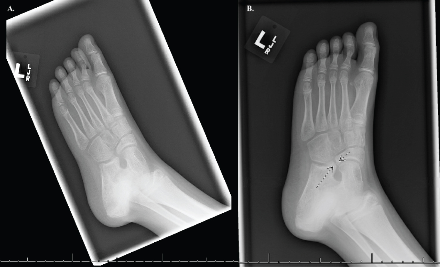
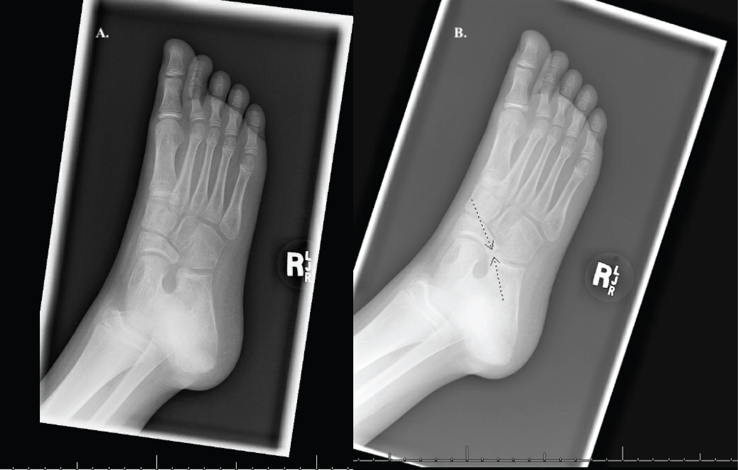
An 8-year-old female first presented with chronic foot and ankle pain to her pediatrician. The pain was described as achy sharp pain in the arch of both feet that increased with activity. Prior to this, she did not have any past medical history. Her biological father had a history of tarsal coalition. The plan during this time was to consult orthopedics for additional examination. The initial orthopedic examination showed increased tightness in her hamstring muscle and gastrocnemius bilaterally. Examination of the feet revealed flat straight feet with full range of motion of her hindfoot and midfoot. Gait, coordination, and balance were normal. X-ray radiographs were taken at this visit and were unremarkable (Figure 1A and Figure 2A). The patient's family was given instructions to start a hamstring and gastrocnemius stretching regime for the patient and orders to use orthotics in her shoes.
13 months later at age 10, the patient presents to the emergency department with an injury to her left great toe. X-ray radiographs taken during her ED visit did not show any structural damage to her great toe but did show anomalies in the tarsal bones that prompted imaging of the uninjured foot. Imaging was reviewed at her next orthopedic visit several months later and indicated bilateral calcaneonavicular coalition that were not present on radiographs during initial imagining 13 months prior (Figure 1B and Figure 2B). During this visit her mother stated the patient has been quite active and her foot and ankle pain have completely resolved through the stretching routine and use of orthotics in her shoes. Examination of her feet showed normal appearing arches with both heels in mild valgus position. Subtalar motion was limited in both feet. The diagnosis of tarsal coalition was made and the explanation of the natural history of this condition was given to family and patient with plans to continue wearing the orthotics.
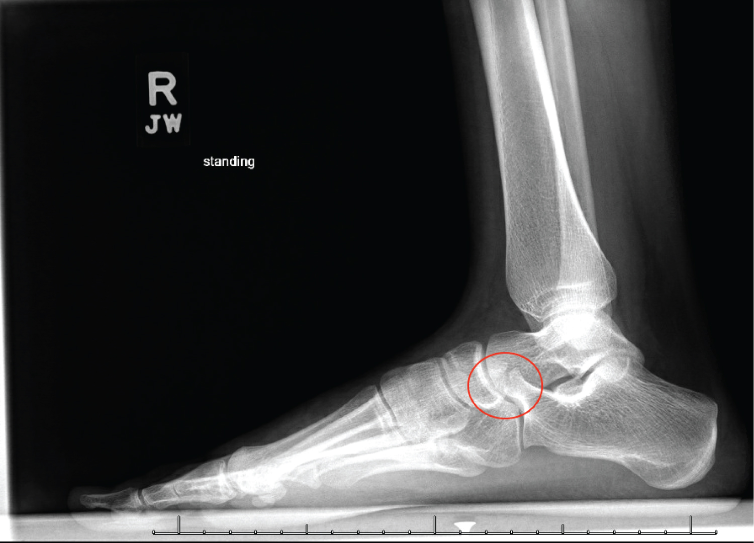
The last orthopedic visit to date for this patient occurred at age 16. She has not experienced any foot or ankle pain and has remained active in her theater, dance, and physical education classes without any restrictions to her activity. Examination of her feet was like prior visits, which showed straight flat feet with heels in slight valgus position with limited subtalar motion. X-ray radiographs taken at this visit revealed mature calcaneal navicular coalition of both feet (Figure 3 and Figure 4). The patient was instructed to continue wearing the orthotics and to schedule any additional appointments if she has any complications in the future.
Discussion
This case demonstrates two components of the development of tarsal coalition that are not well described in the literature currently, the rapid progression of bilateral calcaneonavicular coalition and transition from symptomatic to asymptomatic as the coalitions matured. The rapid development of bilateral calcaneonavicular coalition visible in a 10-year-old female child was found incidentally due to an ED visit for an injury to the great toe and was not present on imagining during the initial evaluation 13 months prior (Figure 1 and Figure 2). Although most cases of tarsal coalitions are asymptomatic, this case describes a patient who was symptomatic prior to visual development of the condition [5]. A possible explanation for this finding could be due to the composition of the coalitions prior to ossification not being visible on plain x-ray radiograph since their composition is usually fibrous or caliginous at birth [6].
Paradoxically, this patient's symptoms of foot and ankle pain diminished as the tarsal coalition developed and matured. This presentation seems to be contrary to reports of symptoms of foot pain presenting as the coalition mature and ossify and worsen as time goes on [6-10]. At age 16, approximately 7-years post diagnosis, that patient did not have any foot and ankle pain or restrictions on physical activity although x-ray radiographs of her feet displayed matured calcaneonavicular coalitions (Figure 3 and Figure 4). Diagnosis of CN coalition is made with a 45° internal oblique radiographic view showing an elongation of anterosuperior calcaneus on lateral view [4]. In contrast, TC coalition requires CT due to more difficult visualization of the pathognomonic C sign: Continuous uninterrupted line formed posteriorly by talar dome and sustentaculum tail on lateral view [4].
Initial treatment is non-operative, with open or endoscopic resection indicated if pain persists at 6 months [3]. The current literature on the rapid development of bilateral calcaneonavicular coalition seen in this case essentially is nonexistent. This case serves to describe a unique patient who was symptomatic prior to diagnosis and did not experience any symptoms afterward.
References
- Manaster BJ, May DA, Disler DG (2013) Common congenital foot deformities and tarsal coalitions. In: Manaster BJ, May DA, Disler DG, Musculoskeletal Imaging. The Requisites, WB Saunders, 522-530.
- Newman JS, Newberg AH (2000) Congenital tarsal coalition: Multimodality evaluation with emphasis on CT and MR imaging. Radiogr Rev Publ Radiol Soc N Am Inc 20: 321-332.
- Docquier PL, Maldaque P, Bouchard M (2019) Tarsal coalition in paediatric patients. Orthop Traumatol Surg Res OTSR 105: S123-S131.
- Merrow AC, Linscott LL, O'Hara SM, et al. (2017) Tarsal Coalition. In: (3rd edn), Diagnostic Imaging: Pediatrics, Diagnostic Imaging. Elsevier, 982-985.
- Kulik SA, Clanton TO (1996) Tarsal coalition. Foot Ankle Int 17: 286-296.
- Neuschwander TB, Wenger DR (2011) Drennan's the child's foot and ankle. (2nd edn), J Pediatr Orthop 31: e36.
- Lemley F, Berlet G, Hill K, et al. (2006) Current concepts review: Tarsal coalition. Foot Ankle Int 27: 1163-1169.
- Ormeci T, Kilicarslan R, Durmus O, et al. (2014) A pictorial view to tarsal coalition: The presentation of two children with foot pain. Acta Reumatol Port 39: 347-348.
- Nehme AH, Bou Monsef J, Bou Ghannam AG, et al. (2016) Arthroscopic resection of a bilateral calcaneonavicular coalition in a child. J Foot Ankle Surg 55: 1079-1082.
- Rocchi V, Mubarak S (2020) Ankle ganglion associated with tarsal coalition: A Case report with a 5-Year follow-up. JBJS Case Connect 10: e2000090.
Corresponding Author
Sundeep Kahlon, Geisinger Commonwealth School of Medicine, Mark A Seeley, 16 Woodbine Lane, Danville PA, 17822, USA
Copyright
© 2022 Kahlon S, et al. This is an open-access article distributed under the terms of the Creative Commons Attribution License, which permits unrestricted use, distribution, and reproduction in any medium, provided the original author and source are credited.