Arthroscopic Transtibial Pull-Out Suture Repair of Posterior Medial Meniscus Root Tears-Surgical Technique
Abstract
Introduction: Injuries to the posterior medial meniscal root remain misdiagnosed and different surgical techniques are described for repair with the debate over whether it is beneficial and worthy still exists.
Aim of the study: To demonstrate the surgical steps of arthroscopic transtibial pull-out suture repair technique of posterior medial meniscus root tears.
Study design: surgical technique.
Conclusion: Transtibial single-tunnel pull-out meniscal root repair provided secured, easy and reproducible method to repair these difficult types of meniscal tear.
Keywords
Medial meniscus root, Root repair, Transtibial pull out suture repair
Introduction
Understanding of the meniscal root importance to normal meniscus function has evolved in the last decade, resulting in a better ability to identify and manage this unique type of meniscal injury. Currently there are some limitations that prevent better understanding of meniscal root tears including lack of surgeon awareness, diagnostic difficulty, still debatable indications for and contraindications to surgical repair, technical difficulty, and short term clinical follow-up data [1]. Meniscal root tears (MRTs) are defined as radial tears within 1 cm of the meniscus insertion, or avulsions at the insertion of the meniscus. Extrusion of the medial meniscus has been identified in both partial and total meniscal root tears, with extrusion > 3 mm being associated with increased cartilage degeneration and osteophyte formation [2].
Posterior meniscal root tears lead to increased tibiofemoral pressures in contact areas, which have been reported to be functionally similar to a total meniscectomy [3,4]. If left without treatment, patients may experience increased pain and dysfunction due to progressive degenerative changes, which could lead to early osteoarthritic joint disease [5]. Ozkoc, et al. reported outcomes of these tears after arthroscopic partial meniscectomy, demonstrating improvement in clinical outcomes but progression of radiographic OA at a mean follow-up of 4.7 years, suggesting that partial menisectomy likely provides symptomatic relief but does not arrest the progression of OA [6]. Over the years, treatment recommendations have shifted toward meniscal preservation. Due to an increased knowledge of the disastrous consequences of meniscal root injury, greater interest has been placed on restoring meniscal integrity [7]. In this study, the surgical steps of an arthroscopic transtibial pull out suture repair over a screw post is described.
Surgical Technique
Patient positioning and anesthesia
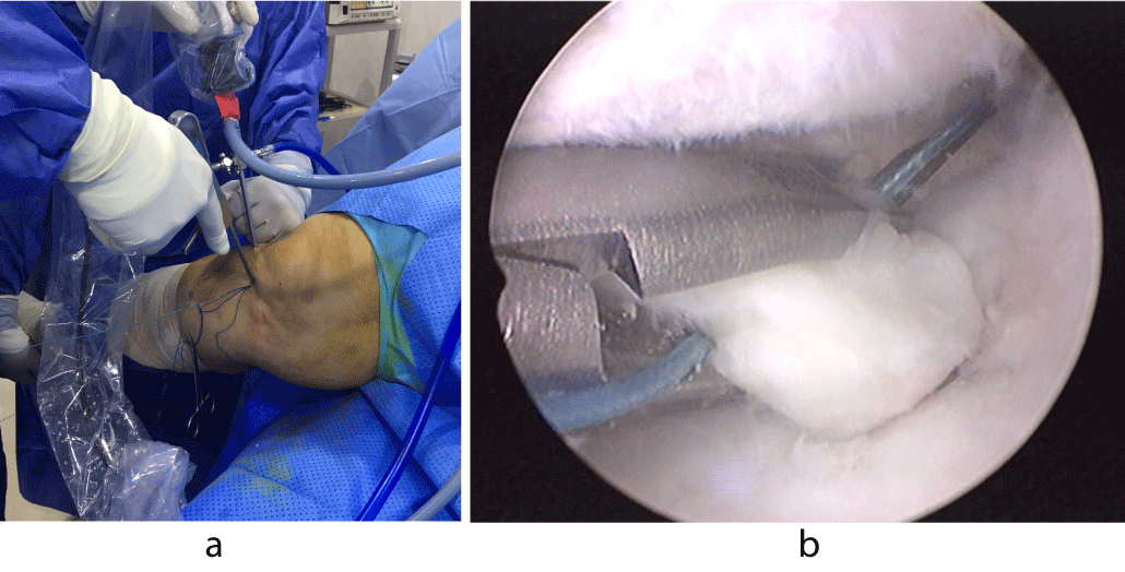
The patient is placed in the supine position on the operating table. After induction of spinal or general anesthesia, a knee examination is performed to evaluate for any associated ligamentous injuries, and to assess for knee range of motion. A well-padded high-thigh tourniquet is placed on the operative leg and then a thigh side support is fixed to the table to provide fulcrum for valgus position to open up medial joint space. The foot of the patient is then lowered from the side of the table with flexing of the knee so that it is resting on the surgeon’s lap.
Operative steps
Standard anterolateral and anteromedial portals are made adjacent to the patellar tendon. The joint is filled with normal saline and visualized with a 30 arthroscopic camera (Smith & Nephew, Andover, MA). An arthroscopic shaver (Smith & Nephew) is introduced and any associated injuries are dealt with. In most of the cases, there will be a medial femoral condyle chondral lesion for which any unstable cartilage fragments are debrided gently with a shaver. The affected meniscal root should be examined to assess for tear pattern and its condition. Medial collateral ligament release is done in its posterior half using a pie-crusting technique with a needle just above the medial meniscus to open up the posteromedial joint space to make instruments maneuvers easier.
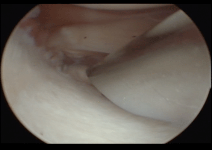
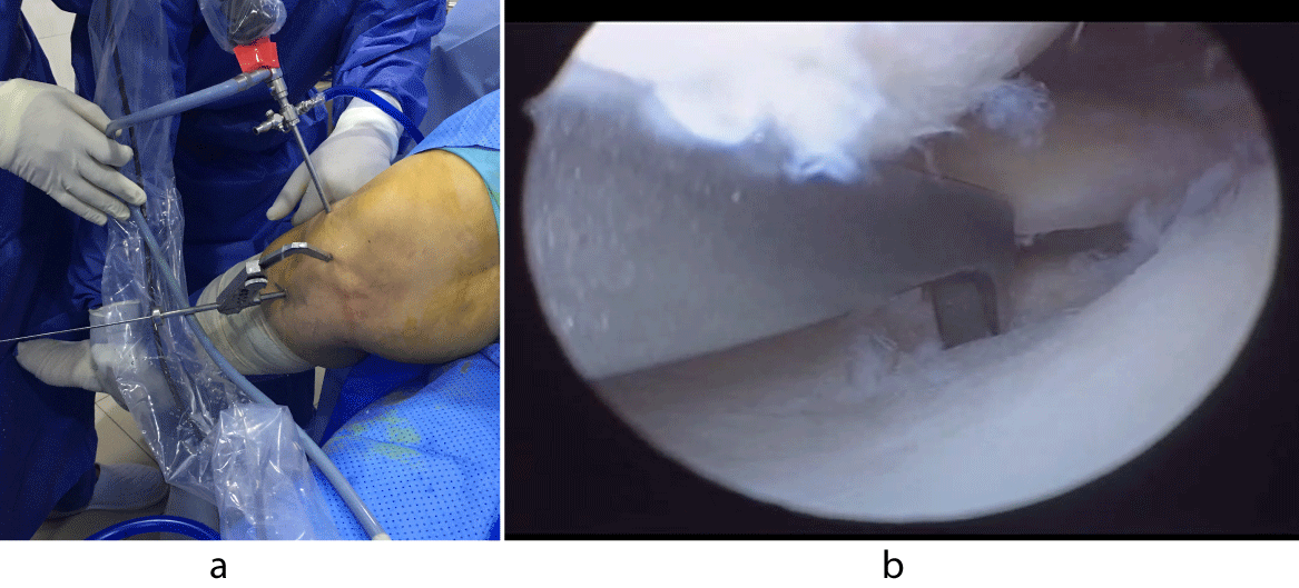
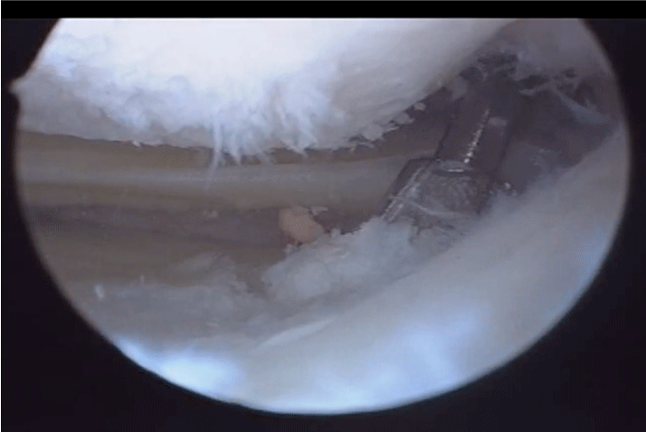
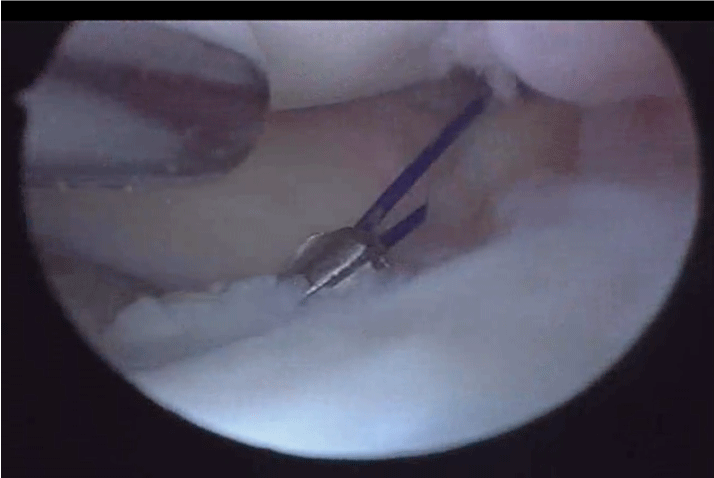
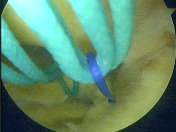
The site for the planned root repair on the tibial plateau cartilage should then be debrided to expose subchondral bone an arthroscopic burr or acrominizer (Smith & Nephew) (Figure 1). A grasper is used to pull on the torn meniscal root and to check for its mobility to the ideal location for performing the reattachment. An aiming device with a cannulated sleeve (Smith & Nephew), positioned at the site debrided for the root attachment is used to drill a transtibial tunnel with the drill pin just medial to the tibial tubercle (Figure 2a and Figure 2b). A 1 cm skin incision is made longitudinally at the entry point of the pin down to bone. A cannulated drill bit 4.5 mm (Smith & Nephew) is then used to ream the tunnel to the center of the meniscal root attachment site (Figure 3). The drill pin is removed while leaving the cannulated drill bit in place for passing the sutures (Figure 4). A finger of the assistant is put on the inlet of the cannulated drill pit to prevent fluid extravasation from the knee. A suture-passing device (Expresso, Mitek) is used to pass a simple polyfilament non absorbable No.2 suture (Orthocord, Mitek) through the far posterior portion of the detached medial meniscus root, approximately 5 mm medial to its lateral edge, passing from the tibial to the femoral side (Figure 5a and Figure 5b). The suture loop is pulled out through the anteromedial portal while the device is removed if it has a suture retrieving mechanism or it can be retrieved with a ring forceps or a grasper. It is important to confirm that there are no soft tissue bridges in the arthroscopic portal entangling the passing sutures. Then the ends of suture are passed through the its loop and then pulled from the same portal so as to make a locking loop stitch around the meniscus.
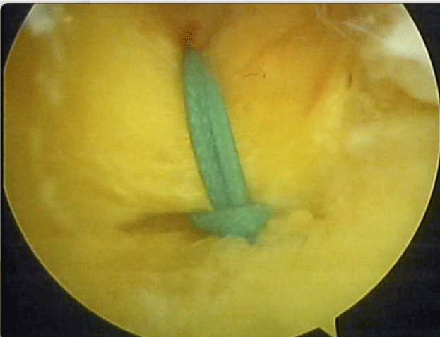
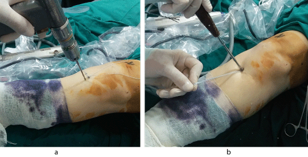
Then, a monofilament nonabsorbable No.2 fashioned into a loop is passed through the cannulated drill pit into the knee and retrieved from the anteromedial portal with a ring forceps or a grasper then after removal of the cannulated drill pit, the monofilament suture is used to shuttle the polyfilament suture from the anteromedial portal down through the tibial tunnel (Figure 6) The sutures are then secured and tied over a post with 4 mm cancellous screw (36-40 mm long) on the anteromedial tibia while the posterior root is visualized arthroscopically (Figure 7) to confirm a secure fixation in a reduced position (Figures 8a and Figures 8b).
Postoperative rehabilitation
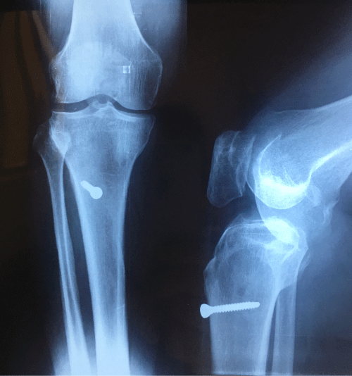
Post-operative X-ray of the knee is done to confirm position of the screw (Figure 9) Patients are allowed for partial weight-bearing on crutches for 6 weeks in an immobilizer or a hinged knee brace. Physical therapy could be started as soon as possible after surgery, which should include quadriceps drill strengthening exercises to ascertain full knee extension but flexion range of motion exercises is postponed for 2 weeks. After 2 weeks, knee flexion is started in a safe zone of 0 to 45 degrees for 2 weeks and is increased to 90 degrees for the next 2 weeks. Advancement to full weight-bearing begins gradually at 6-8 weeks. Deep leg presses and squats greater than 90 degrees of knee flexion should be avoided for at least 3 months after surgery.
Discussion
Pagnani, et al. [8] first described meniscal root tears, and during the last decade the functional importance of meniscal root and its integrity became more evident. An injury to the meniscal attachment, especially on the medial side, can lead to meniscal extrusion which increases stress to the cartilage, by decreasing the contact surface. This causes impairment of hoop stress function and its forces dissipation with accelerated articular degeneration [9]. Meniscal root tears (MRTs) are defined as radial tears within 1 cm of the meniscus insertion, or avulsions at the insertion of the meniscus [10]. Extrusion of the medial meniscus has been observed in both partial and total meniscal root tears, with extrusion > 3 mm being associated with increased cartilage degeneration and osteophyte formation [11]. The posterior root of the medial meniscus inserts 9.6 mm posterior and 0.7 mm lateral to the apex of the medial tibial eminence, in a position 3.5 mm lateral to the inflection point of the medial plateau articular cartilage and 8.2 mm anterior to the posterior cruciate ligament (PCL). It serves as an anchor point for the meniscus. In addition to this direct insertion, radially oriented fibers have been described on the posterior aspect of the posterior root of the medial meniscus. These fibers are referred to as “shiny white fibers” because of their arthroscopic appearance [12]. The etiology of posterior medial meniscus root tears is often degenerative, as seen in middle- aged women [13], but it can also be seen rarely in an acute setting in association with multiple ligament knee injuries [14]. Up to one-fifth of medial meniscal tears are found in the posterior root [13]. Patients with a lateral meniscus root tear were more likely to have an ACL tear, while patients with a medial meniscus root tear were more likely to have concomitant chondral defects [15].
Medial meniscus posterior root tear (MMPRT) is the most common lesion, with a prevalence of 10 to 21% of meniscal surgeries [16]. Because of its reduced mobility and significant loads, the posterior root of the medial meniscus is more liable to injury than the posterior lateral root [17]. Increased incidence of MMPRT has been described in those part of the world, where kneeling and squatting are common daily activities [16]. Increased BMI, varus mechanical axis, female sex, low activity level, together with some unknown intrinsic factors, have been associated with an increased risk of MMPRT [13]. Female gender was associated with worse final subjective outcome scores and high rates of subsequent total knee arthroplasty. In addition, 88% of females were considered failures at follow-up as compared to 60% of males.
Elevated BMI also resulted in a higher rate of failure and progression to subsequent arthroplasty [18]. Choi, et al. [19] showed that higher amount of extrusion was associated with worsening cartilage degeneration. The critical point of meniscus subluxation was defined as an index of 0.38 corresponding to a 44% chance of cartilage degradation to grade 3-4 at 2 years. Costa, et al. and Lerer, et al. [11,17] evaluated the effect of meniscus extrusion on cartilage degeneration and reported an extrusion critical point of 3 mm on mid-coronal imaging. Lee, et al. [20] determined that the amount of meniscus extrusion played a bigger role in subsequent arthritis development than the location of the tear. Forty-two to 64% of knees with medial meniscus extrusion had associated posterior root tears on MRI. [11,17] Some authors showed that the 3 mm threshold had high specificity (98%), but low sensitivity (54%), and introduced the meniscal length/transverse ratio (L/T ratio) as another index for MMPRTs [21].
The mean L/T ratio was 13% with meniscal root tears and 5% in the control groups. An extrusion ratio threshold was determined at 10% (79% sensitivity, 86% specificity) [22]. In a cadaveric study analyzing loading and kinematics in intact knees, knees with a posterior root tear of the medial meniscus, knees with a repaired posterior root, and knees with a total meniscectomy, Allaire, et al. [23] found that a root tear resulted in increased tibiofemoral contact pressure equal to that following a total meniscectomy, whereas repair restored contact pressure to that of the uninjured knee. Marzo and Gurske-DePerio [11] confirmed these findings in a cadaveric study of contact pressure in intact, posteromedial root-injured, and root-repaired cadaveric knees. Chung, et al. [24] compared the results of posterior medial root repair and partial medial root meniscectomy, in patients with Kellgren-Lawrence grade 0 to 2, at a minimum of 5-years postoperatively. They concluded that osteoarthritic changes were decelerated as compared with partial meniscectomy. This finding is also supported by Kim, et al. [25] who investigated medial meniscal root tears that were treated with repair or meniscectomy. At second-look MRI and radiography, the repair group had significantly less joint space narrowing than the meniscectomy group, indicating a slower progression of knee osteoarthritis. Another study investigated the results of 2 medial meniscal root repair techniques. At a minimum of 2 years after arthroscopy, radiographic evaluation revealed no significant change in Kellgren-Lawrence grade from preoperative assessment to postoperative assessment for either group [26]. Overall, the evidence available in the present literature supports the efficacy of meniscal root repair with significant improvement in patient outcomes and delaying of the future knee joint degeneration. Unfortunately, repair of root tears can be technically demanding and it is difficult to determine accurate suture points [7].
Sometimes the meniscus is too degenerative and weak and may tear up with suture passage. Passing the suture through the posterior horn away from the edge may create excessive tension on the meniscal body, whereas placing the suture too close to the edge may result in suture cutout and loss of fixation; this may be considered as an additional factor for failure of meniscal root repair [27]. While meniscal root repair demonstrates the ability to slow the progression of arthritic changes compared with meniscectomy, the procedure does not completely prevent the development of future degenerative disease. In the systematic review by Feucht, et al. [5] the data revealed a predominantly female population (83%) with a mean age of 55.3 years; demonstrated no progression of cartilage degeneration in 84% and 82% of patients who were evaluated with conventional radiographs and MRI, respectively; and demonstrated decreased meniscal extrusion in 56% of patients.
Healing, assessed with a combination of MRI and second-look arthroscopy, was reported as complete in 62% of patients, partial in 34%, and failed in 3%. All studies demonstrated improvements in subjective and functional scores at a mean of 30.2 months, with the mean Lysholm score increasing from 52.4 preoperatively to 85.9 postoperatively. Lee, et al. [28] reported a 5% rate of reoperation, most commonly performed because of recurrent symptoms related to a complete or partial retear of the meniscal root. Different surgical meniscal root refixation techniques have been described [29] aiming to regain the meniscus ring so that to maintain the circular hoop stress. To avoid overloading and to restore the intraarticular pressure distribution, an anatomical fixation should be performed [30].
Two competing techniques are mainly used which are anchor repair and transtibial refixation. Despite higher load-to-failure stability of the anchor fixation techniques, some disadvantages of the anchor techniques have to be considered. It is a difficult procedure with sophisticated steps and suture passage that requires an additional posterior approach. The direct visualization might be difficult, and the anchor positioning might fail or be tedious [31]. Kim, et al. [27] recommended that the sutures be passed through the red-red zone of the meniscus and meniscocapsular junction located 7 mm medial to the edge of the tear. The method had at least 2 advantages. First, the red-red zone and meniscocapsular junction tissues are not torn easily when the suture material is pulled out at the proximal tibia for tying. Second, because both are highly vascular and some tissues are inserted through the tibial tunnel when the suture material is pulled out, the potential for healing is high. One of the goals of medial meniscal root repair surgery is to slow or ultimately halt the progression of medial compartment arthritis. Overall, the evidence found within the several late research literatures supports the efficacy of meniscal root repair and patient outcomes have been reported to be significantly improved with possible delay of future joint degeneration. The technical steps are simple with a short learning curve for the arthroscopic surgeons.
Conclusion
Transtibial single-tunnel pull-out meniscal root repair with fixation over screw post provided secured, easy and reproducible method to repair these difficult types of meniscal tear.
Conflicts of Interest
The author reports that he has no conflicts of interest in the authorship and publication of this article.
References
- Strauss EJ, Day MS, Ryan M, et al. (2016) Evaluation, treatment, and outcomes of meniscal root tears: A critical analysis review. JBJS reviews 4: e4.
- Moatshe G, Chahla J, Slette E, et al. (2016) Posterior meniscal root injuries. A comprehensive review from anatomy to surgical treatment. Acta Orthopaedica 87: 452-458.
- La Prade CM, Jansson KS, Dorman G, et al. (2014) Altered tibiofemoral contact mechanics due to lateral meniscus posterior horn root avulsions and radial tears can be restored with in situ pull-out suture repairs. J Bone Joint Surg Am 96: 471-479.
- Padalecki JR, Jansson KS, Smith SD, et al. (2014) Biomechanical consequences of a complete radial tear adjacent to the medial meniscus posterior root attachment site: In-situ pullout repair restores derangement of joint mechanics. Am J Sports Med 42: 699-707.
- Feucht MJ, Kuhle J, Bode G, et al. (2015) Arthroscopic transtibial pullout repair for posterior medial meniscus root tears: A systematic review of clinical, radiographic, and second-look arthroscopic results. Arthroscopy 31: 1808-1816.
- Ozkoc G, Circi E, Gonc U, et al. (2008) Radial tears in the root of the posterior horn of the medial meniscus. Knee Surg Sports Traumatol Arthrosc 16: 849-854.
- Bhatia S, La Prade CM, Ellman MB, et al. (2014) Meniscal root tears: Significance, diagnosis, and treatment. Am J Sports Med 42: 3016-3030.
- Pagnani MJ, Cooper DE, Warren RF (1991) Extrusion of the medial meniscus. Arthroscopy 7: 297-300.
- Hein CN, Deperio JG, Ehrensberger MT, et al. (2011) Effects of medial meniscal posterior horn avulsion and repair on meniscal displacement. Knee 18:189-192.
- Marzo JM, Gurske DePerio J (2009) Effects of medial meniscus posterior horn avulsion and repair on tibiofemoral contact area and peak contact pressure with clinical implications. Am J Sports Med 37: 124-129.
- Lerer DB, Umans HR, Hu MX, et al. (2004) The role of meniscal root pathology and radial meniscal tear in medial meniscal extrusion. Skeletal Radiol 33: 569-574.
- Johannsen AM, Civitarese DM, Padalecki JR, et al. (2012) Qualitative and quantitative anatomic analysis of the posterior root attachments of the medial and lateral menisci. Am J Sports Med 40: 2342-2347.
- Hwang BY, Kim SJ, Lee SW, et al. (2012) Risk factors for medial meniscus posterior root tear. Am J Sports Med 40: 1606-1610.
- Ra HJ, Ha JK, Jang HS, et al. (2015) Traumatic posterior root tear of the medial meniscus in patients with severe medial instability of the knee. Knee Surg Sports Traumatol Arthrosc 23: 3121-3126.
- Matheny LM, Ockuly AC, Steadman JR, et al. (2015) Posterior meniscus root tears: associated pathologies to assist as diagnostic tools. Knee Surg Sports Traumatol Arthrosc 23: 3127-3131.
- Bin SI, Kim JM, Shin SJ (2004) Radial tears of the posterior horn of the medial meniscus. Arthroscopy 20: 373-378.
- Costa CR, Morrison WB, Carrino JA (2004) Medial meniscus extrusion on knee MRI: Is extent associated with severity of degeneration or type of tear?. Am J Roentgenol 183: 17-23.
- Aaron JK, Nick RJ, Michael JS, et al. (2018) Partial meniscectomy provides no benefit for symptomatic degenerative medial meniscus posterior root tears. Knee Surg Sports Traumatol Arthrosc 26: 1117-1122.
- Choi YR, Kim JH, Chung JH, et al. (2014) The association between meniscal subluxation and cartilage degeneration. Eur J Orthop Surg Trauma 24: 79-84.
- Lee DH, Lee BS, Kim JM, et al. (2011) Predictors of degenerative medial meniscus extrusion: Radial component and knee osteoarthritis. Knee Surg Sports Traumatol Arthrosc 19: 222-229.
- Park HJ, Kim SS, Lee SY, et al. (2012) Medial meniscal root tears and meniscal extrusion transverse length ratios on MRI. Brit J Radiol 85: e1032-1037.
- Mohankumar R, Palisch A, Khan W, et al. (2014) Meniscal ossicle: Post-traumatic origin and association with posterior meniscal root tears. Am J Roentgenol 203: 1040-1046.
- Allaire R, Muriuki M, Gilbertson L, et al. (2008) Biomechanical consequences of a tear of the posterior root of the medial meniscus. Similar to total meniscectomy. J Bone Joint Surg Am 90: 1922-1931.
- Chung KS, Ha JK, Yeom CH, et al. (2015) Comparison of clinical and radiologic results between partial meniscectomy and refixation of medial meniscus posterior root tears: A mini- mum 5-year follow-up. Arthroscopy 31: 1941-1950.
- Kim SB, Ha JK, Lee SW, et al. (2011) Medial meniscus root tear refixation: Comparison of clinical, radiologic, and arthroscopic findings with medial meniscectomy. Arthroscopy 27: 346-354.
- Kim JH, Chung JH, Lee DH, et al. (2011) Arthroscopic suture anchor repair versus pullout suture repair in posterior root tear of the medial meniscus: A prospective comparison study. Arthroscopy 27: 1644-1653.
- Kim YM, Joo YB, Noh CK, et al. (2016) The optimal suture site for the repair of posterior horn root tears: Biomechanical evaluation of pullout strength in porcine menisci. Knee Surg Relat Res 28: 147-152.
- Lee JH, Lim YJ, Kim KB, et al. (2009) Arthroscopic pullout suture repair of posterior root tear of the medial meniscus: Radiographic and clinical results with a 2-year follow-up. Arthroscopy 25: 951-958.
- Feucht MJ, Grande E, Brunhuber J, et al. (2015) Biomechanical evaluation of different suture materials for arthroscopic transtibial pull-out repair of posterior meniscus root tears. Knee Surg Sports Traumatol Arthrosc 23: 132-139.
- Starke C, Kopf S, Grobel KH, et al. (2010) The effect of a nonanatomic repair of the meniscal horn attachment on meniscal tension: A biomechanical study. Arthroscopy 26: 358-365.
- Feucht MJ, Grande E, Brunhuber J, et al. (2014) Biomechanical comparison between suture anchor and transtibial pull-out repair for posterior medial meniscus root tears. Am J Sports Med 42: 187-193.
Corresponding Author
Ahmad M Wagih, Department of Orthopaedic Surgery, the National Institute of Neuromotor System, Imbabah, Cairo, Egypt
Copyright
© 2022 Wagih AM. This is an open-access article distributed under the terms of the Creative Commons Attribution License, which permits unrestricted use, distribution, and reproduction in any medium, provided the original author and source are credited.