Arthroscopically-Assisted Technique for Anatomic Reconstruction of Chronic Acromio-Clavicular Joint Dislocations Using an Autologous Graft with AC Joint Augmentation Using Acromial Tunnels - Short Term Results
Abstract
AC joint dislocation is a common lesion after a direct impact to the lateral shoulder or a fall over an outstretched arm. There are countless techniques available for the treatment of this type of lesion and if left untreated, this type of lesion can lead to persistent pain, loss of strength, and even shoulder dyskinesia in some cases. When treating this lesion in the chronic stage, a number of additional problems must be considered, such as the need for additional biological support. In the present study we describe an arthroscopically assisted technique for the treatment of chronic AC joint dislocations. We also describe the clinical and radiological results in the short term. A cohort of 20 male patients with an average age of 40.8 was studied. All the patients underwent the same technique and the clinical and radiological results at 1, 3 and 6 months were evaluated. This arthroscopically-assisted technique for anatomic reconstruction of chronic acromioclavicular joint dislocations using an autologous graft with AC joint augmentation using acromial tunnels appears to be a reliable technique with excellent clinical and radiological outcomes in the short term. A longer follow-up period is needed to prove these results in the long term.
Introduction
Acromioclavicular (AC) dislocations usually present as the result of a fall that produces trauma to the lateral aspect of the shoulder. It brings about a variable separation of the acromioclavicular joint depending on the degree of damage to the capsule, the acromioclavicular ligaments, as well as the coracoclavicular (CC) ligaments. Rockwood I and II lesions are normally treated non-surgically. Surgical treatment is recommended for grade IV, V and VI lesions according to Rockwood classification; however, there is still a controversy regarding the treatment of type III lesions [1].
In the treatment of acute lesions, the usual approach is to reduce and stabilize the clavicle and expect healing of the CC ligaments to occur. In chronic cases, however, this healing is not expected, and reconstruction of the ligaments is necessary. Techniques of reconstruction using autologous, cadaveric, and synthetic grafts have been proposed. However, studies comparing the benefits and outcomes of each type of graft are still lacking [2]. Another aspect to be considered, in the chronic setting, is the late development of AC joint osteoarthritis, even after surgical repair of the AC joint. In this context, some studies reported incidences as high as 20% [3,4]. In the present study we propose a novel technique for the treatment of AC joint dislocations in the subacute (> 3 weeks) and chronic setting (>6 weeks). The authors present the clinical and radiologic results obtained using an arthroscopic coracoclavicular ligament reconstruction technique using an autologous semitendinosus graft. This technique adds a Mumford and augmentation technique to prevent late-onset arthrosis as well as providing additional horizontal stability.
Methods
In the present study a population of 20 patients with chronic AC joint dislocation were evaluated. Only patients with a grade III or higher AC joint dislocation according to the Rockwood classification were included in this study. The radiologic results were evaluated by two different observers at 1, 3 and 6 months post operatively, regarding loss or reduction or eventual complications seen on x ray (i.e. osteolysis, bone migration, fractures of the clavicle acromion or coracoid). The clinical results consisted of analyzing pain using visual analogue scale (VAS), and range of motion (ROM) values obtained by the patients. The disabilities of the arm, shoulder and hand (DASH), American shoulder and elbow surgeons (ASES) shoulder and University of California at Los Angeles (UCLA) shoulder scores were also evaluated at 6 months post operatively.
Surgical Technique
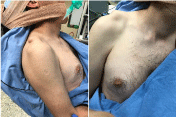
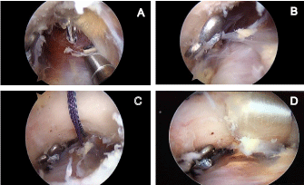
The authors describe the anatomic technique of reconstruction of both the CC ligaments and superior AC ligament with autologous semitendinosus graft. Under general anesthesia, with the patient in the beach chair position, the shoulder and arm are draped free (Figure 1). The semitendinosus graft is harvested and prepared by the standard technique. Shoulder arthroscopy is then performed using the standard technique using a 30° arthroscope. Posterior, anterior and anterolateral portals are used. Using the posterior portal as a viewing portal, associated lesions, if present, are diagnosed and treated (i.e. SLAP lesions or rotator cuff tears) and the general status of the glenohumeral joint is assessed. After a complete examination is performed, we change the viewing portal to anterolateral and the anterior portal as the working portal. Medial progression and dissection are performed along the superior border of the subscapularis, in order to find the inferior surface of the coracoid. The inferior margin of the coracoid process is then cleared of any soft tissue using a radiofrequency device, and then its inferior surface is abraded with a bone shaver. The musculocutaneous nerve lies distally on the medial edge of coracoid and care must be taken not to damage it.
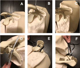
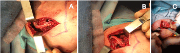
A longitudinal incision is then made along the distal end of the clavicle. A point approximately 35 mm medial (the conoid ligament is inserted 45 mm medial and the trapezoid ligament 25 mm medial to the AC joint) [5] to AC joint is identified and a 4-5 mm drill hole is made, using the orientation drill guide. At this point, the drilling of the coracoid should be checked arthroscopically and the positioning of the drillhole in the coracoid evaluated - ideally it should be centered in the mediolateral aspect of the bone, and as close as possible to its base (Figure 2). Next, a loop-ended Nitinol wire is inserted into the drill from superior to inferior. After the wire becomes visible on the inferior surface of the drill, it should be retrieved through the anterior portal using a suture retrieval instrument. The Endobutton system can be deployed in this moment according to its surgical technique. Before inserting the second button (the clavicle button), it is important to place the graft in the propper position. To do so, two Nitinol wires are used as shuttles (one from the posterior clavicle to the medial coracoid and the other from the anterior clavicle to the lateral coracoid); each of these shuttles will transport one edge of the graft, hence, forming a loop around the coracoid and clavicle (Figure 3).
Once the previous steps are completed, a small resection of the distal end of the clavicle (about 3~5 mm) is performed, as well as excision of the AC joint meniscus. This will prevent the development of late AC joint pain. The clavicle is reduced and the Endobutton system tightened. During the reduction maneuver it is important that the assistant makes a vertical upward force from the elbow in order to elevate the shoulder and facilitate reduction. Afterwards the graft is also tightened with a knot to itself (this knot is then secured with a no. 2 high-strength nonabsorbable suture). The remaining tops of the tendon are then passed along the AC joint and reinserted into the acromion (by drilling 2 tunnels in the acromion) (Figure 4).
Results
In this study, a population of 20 patients was evaluated. All of the patients were male. The median age was 40.8 (range 34-53). All patients were satisfied with the result and, when questioned, all answered that would underwent the surgery again. The mean pain value (using the VAS) was 0.66 (range 0-2). When evaluated for ROM the mean values obtained were 160° for forward flexion; 150° for abduction and 80° for IR. All the patients were able to internally rotate until the hand reached L1 level. Concerning the scores, the median ASES shoulder score obtained was 86.67; the modified UCLA obtained was 27 and the average Quick DASH value was 4.5. All of the previous described results were evaluated only at 6 months post operatively When analyzing the radiological results, we found no loss of reduction or osteolysis at 1 and 3 months post operatively. At 6 months post op we found a discrete loss of reduction of an average 3 mm (range 2-5 mm).
Discussion
There are innumerous techniques described in the literature for the treatment of AC joint dislocation. However, if we narrow the search to surgical treatment of chronic AC joint dislocations, the number of papers we find dramatically decreases. The technique we present seems to be an interesting option to consider when treating this type of lesion. Although, in the title, we describe to this technique as an arthroscopic technique, it has in fact an open part (the AC joint augmentation). However, we consider that a 3~4 cm incision is not very aggressive in terms of exposure and allows us to have better control of the reduction, while facilitating meniscal excision, excision of the distal clavicle, augmentation and graft fixation.
The technique we describe is very similar to other techniques already described, but, has the advantage of addressing the late development of AC joint pain. In the research we performed Chernchujit, et al. [6] described a very similar technique. The only minor difference is that in our technique, we don't do an interposition of the AC joint with the graft. Instead, we bridge the AC joint above it (as shown in Figure 4) We believe that this technique allows us to restore vertical stability (provided by the enbobutton + the loop) and horizontal stability (provided by the augmentation of the AC joint) while preventing the late-onset development of AC joint osteoarthritis or pain (through resection of the distal clavicle and meniscal excision).
As for the clinical results, we find them to be extremely positive with a complete ROM and a very low mean VAS value. The fact that all patients would repeat the procedure demonstrates this. In terms of radiological results, although we found a mean reduction loss of 3 mm after 6 months, this reduction loss was clinically negligible. One can speculate whether this value can increase over time. We believe that complete healing and integration of the graft has occurred at this point, so no further loss of reduction is expected. However, only time will tell if this is the case. Some studies describe post operative complications such as osteolysis of the distal clavicle, ossification of the CC interspace and infection [7]. One of the limitations of this study is the short follow-up period. A longer follow-up period is needed to obtain more conclusive long-term data on this technique (in terms of clinical and radiological outcomes).
Conclusion
AC joint dislocation is a well-known pathology. Over the years, a wide variety of techniques have been described for its treatment in the acute phase. However, for the chronic phase of the pathology, the number of available techniques decreases drastically. The authors present an arthroscopically-assisted technique for the treatment of this pathology in the chronic phase which simultaneously addresses the problem of late-onset AC joint pain. This technique presents positive clinical and radiological results in the short term. Further studies and a longer follow-up are needed in order to assess its effectiveness in the long term.
Conflicts of Interest
The authors have no conflicts of interest to disclose.
References
- Ceccarelli E, Bondi R, Alviti F, et al. (2008) Treatment of acute grade III acromioclavicular dislocation: A lack of evidence. J Orthop Traumatol 9: 105-108.
- Fauci F, Merolla G, Paladini P, et al. (2013) Surgical treatment of chronic acromioclavicular dislocation with biologic graft vs synthetic ligament: A prospective randomized comparative study. J Orthop Traumatol 14: 283-290.
- Bergfeld JA, Andrish JT, Clancy WG, et al. (1978) Evaluation of the acromioclavicular joint fol lowi ng first-and second-degree sprains. Am J Sports Med 6: 153-159.
- Menge TJ, Boykin RE, Bushnell BD, et al. (2014) Acromioclavicular osteoarthritis: A common cause of shoulder pain. South Med J 107: 324-329.
- Rios CG, Arciero RA, Mazzocca AD (2007) Anatomy of the clavicle and coracoid process for reconstruction of the coracoclavicular ligaments. Am J Sports Med 35: 811-817.
- Chernchujit B, Parate P (2017) Surgical technique for arthroscopy-assisted anatomical reconstruction of acromioclavicular and coracoclavicular ligaments using autologous hamstring graft in chronic acromioclavicular joint dislocations. Arthrosc Tech 6: e641-e648.
- Choi NH, Lim SM, Lee SY, et al. (2017) Loss of reduction and complications of coracoclavicular ligament reconstruction with autogenous tendon graft in acute acromioclavicular dislocations. J Shoulder Elbow Surg 26: 692-698.
Corresponding Author
André Miguel Carvalho, Department of Orthopedics and Traumatology, Hospital Distrital da Figueira da Foz, Coimbra, Rua Hospital 3094-001, Portugal, Tel: +351-913-316-902
Copyright
© 2021 Carvalho AM, et al. This is an open-access article distributed under the terms of the Creative Commons Attribution License, which permits unrestricted use, distribution, and reproduction in any medium, provided the original author and source are credited.