Acute Arterial Iliac-Femoral Thrombosis During Doble Lumbar Approach for Degenerative Spondylolisthesis
Abstract
Vascular complications during anterior lumbar approach are well known, however arterial thrombosis is extremely rare. We report a case of an acute left external iliac artery and common femoral artery thrombosis during a doble lumbar approach. The diagnosis was made in the immediate postoperative period and a iliofemoral thromboendarterectomy was performed by a cardiovascular surgeon. The patient developed a left leg compartment syndrome due to a reperfusion injury which was treated in the same surgical stage through a wide fasciotomy. After appropriate treatment the patient had complete recovery without sequelae.
Keywords
Artery, Thrombosis, Lumbar, Spondylolisthesis, Compartment síndrome
Introduction
Anterior lumbar approach (ALIF) is a well-know option for the treatment of different lumbar conditions, with a low intraoperative complication rate, among them, vascular damage is the most concerning one. Vascular injury is more frequent in vein structures, arterial injury is a rare entity and can be fatal due to hypovolemic shock, compartment syndrome and rhabdomyolysis [1]. We report a case of a 64-year-old male patient who suffered a compartment syndrome secondary to an acute left external iliac and common femoral artery thrombosis during the anterior stage of a double lumbar approach surgery for degenerative spondylolisthesis. Immediate postoperative treatment through thrombectomy followed by extended fasciotomy was performed with a satisfactory outcome.
Case Presentation
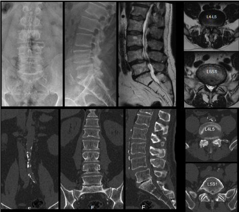
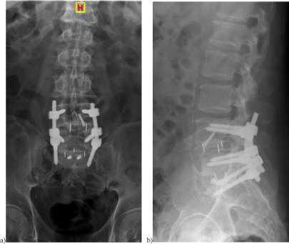
A 64-year-old patient without history of vascular disease underwent a double lumbar approach due to spinal stenosis secondary to L4-L5 spondylolisthesis (Figure 1). After decubitus positioning, anterior lumbar stage was performed first through a left paramedian retroperitoneal approach in collaboration with an access general surgeon. Vascular structures were properly identified and slightly movilized with three retractors in order to access to lumbosacral region. A peek cage with autologous bone and hidroxiapatita was introduced at level L5-S1 after complete discectomy (small size kili cage of 9 mm high with 6º of lordosis and 2 screws fixation to sacrum). After this, structures were retracted medially according to original oblique technique to access L4-L5 level [2], a peek cage was introduced at this level (medium size Kili cage of 9 mm high with 11º of lordosis). The procedure was performed without complications with an estimated bleeding of 250 ml, the operative time was 70 minutes for both L5-S1 and L4-L5 procedures.
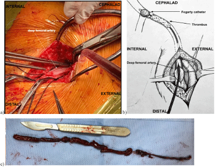
The patient was turned to prone position, and posterior approach was performed (L4-S1 lumbar decompression and fusion). The total procedure including anterior and posterior stage lasted 300 minutes, with an estimated bleeding of 520 ml. During the anesthesia wake-time period, lower temperature and mottled color on the left lower extremity along with decrease motion was noticed. After complete examination, femoral and posterior tibial pulses were not detected on the left side. Acute arterial thrombosis was suspected and confirmed with arterial doppler. Vascular surgeon was rapidly contacted, and an external iliac and femoral artery thrombectomy using the fogarty technique [3] was performed (Figure 2).
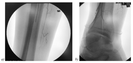
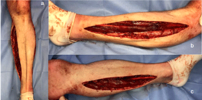
After this procedure, normal revascularization of the limb was detected and confirmed with intraoperative angiography (Figure 3), however, increased left lower extremity diameter and tension were noticed, intracompartmental pressure measurement was performed which was above normal (51 mmHg) requiring a two incision leg fasciotomy according to Mubarak's technique [4] and a vacuum assisted closure placement (Figure 4). The whole salvage procedure lasted 170 minutes. The patient was transferred to the coronary unit for close monitoring. The vacuum assisted closure was revised and changed every 48 hours and removed after 6 days. Lower limb controls were performed with Doppler during the 10 days postoperative, not additional or remanent vascular occlusions were detected. After 10 days, progressive rehabilitation started and the patient was discharged at day 15th. The antiaggregation protocol included/followed a dual antiplatelet therapy (DAPT) with 100 mg/day aspirin and 75 mg/day clopidogrel during fourth months [5-7]. Postoperative outcome was satisfactory without sequela. At fourth month follow up, the patient had complete function recovery and satisfactory X Ray (Figure 5).
Discussion
Anterior lumbar approach is a common and relatively safe technique for the treatment of different lumbar spinal condition including degenerative disc disease, spondylolisthesis, tumors, infections, and fractures. Vascular injury such as direct damage of iliac and iliolumbar vessels, bleeding from the median sacral vessels and postoperative deep vein thrombosis appear to be the most common complications with a reported incidence ranging from less than 1 % to almost 15 % [8-11].
Arterial thrombosis during anterior lumbar procedures are rare, they can lead to compartment syndrome, acute limb ischemia and fatal acidosis secondary to rhabdomyolysis [1,12]. They usually occur due to direct injury of the vascular structures. Even there are few reports in the literature, it is believed that prolonged retraction of the vessels with the valves during the approach has a clear role in the genesis of the arterial thrombus, and presumably secondary to arterial flow stasis, intimal tear or occult atherosclerotic plaques that might be precursors of thrombotic occlusion [9,13-15].
Fantini, et al. [16] described in a review several factors related to vascular injury during anterior lumbar surgery as anterior osteophyte formation, spondylolisthesis, current or previous osteomyelitis or discogenic infection, anterior migration of interbody device and previous anterior spinal surgery. Also smoke history is noticed to be a mayor risk for arterial complications following ALIF [1,12]. Another risk factors include obesity, long operation time, hypotensive anesthesia, history of thrombosis, and diabetes [17]. In this case, the patient had osteophyte formation and vascular calcification evident on preoperative X-rays and CT (Computed Tomography) (Figure 1). We believe that microtrauma/damage during vascular mobilization associated with aortic calcification may contribute to an arterial thrombosis. The rate of vascular injury associated with vessels calcification is reported in about 25% and anterior osteophyte formation about 17% [18].
As in previous reports [1], in our case there wasn't a direct manipulation of the greater vessels, but we believe that when approaching L4-L5 level, medial retraction of the common iliac artery during the exposure (more than 30 minutes) may have had an impact on the genesis of the arterial thrombosis. It is recommended to release traction on the major vessels at regular intervals of no longer than 20 min, based on the notion that prolonged traction will increase intimal tear and vasospasm [16]. In addition, is known that distal mobilization of the left iliac artery toward the femoral canal is preferred, as this can prevent stretching and intimal tears that may provoke thrombosis [19].
Left lower limb oxygen saturation (SaO2) monitoring has been recommended as an additional preventive measure using pulse oximetry in the toes of the left foot. Brau, et al. evaluated the incidence of vascular injury during anterior lumbar procedures and found that close to 60% of patients undergoing ALIF at L4-L5 level had reductions of somatosensory evoked potentials (SSEP) and SaO2 secondary to the retraction of the left iliac vessels; these changes return to normal signals after removal of the retractors [20].
Sometimes, the initial symptoms include leg pain and numbness of the lateral shank, which can confuse as a result of lumbar nerve root irritation from surgery [14], also surgeons should be aware and have a suspect when there are only sensory symptoms, especially in cases of previous abdominal surgery and when approaching L4-L5 level, this could be symptoms of progressive thrombotic occlusion of left common iliac artery [21]. Electrophysiological monitoring would be a good practice to take into account in anterior approaches in order to notice any disturbance in the vessels [20,22]. In this case, after thrombectomy and normal revascularization of the limb, an acute leg compartment syndrome was diagnosed, it is known that the reestablishment of blood flow in this situation could lead to severe reperfusion syndrome, mainly due to release of several muscle elements such as myoglobin, potassium and free radicals. These can lead to acute renal failure [23].
We believe that high suspicion and early recognition are important tools in those cases, in addition, a multidisciplinary team is essential to deal with this uncommon but severe complication. Fortunately, in the presented case the diagnosis and resolution of this complication was made promptly, since we noticed the problem in the early postoperative course. In this case we performed the anterior approach in collaboration with a general surgeon. However, Quraishi, et al. [10] remark that with adequate training and judgment, anterior access surgery to the lumbar spine can be performed safely by spinal surgeons without direct "access surgeon" support, but they should be available if required, and one should employ particular care with surgical exposures at the L4-L5 level. In addition, a systematic review including 8028 patients studied the rate of complications following anterior lumbar approach with and without an "access surgeon", and showed that anterior approaches with and without an access surgeon are both safe approaches [24].
Conclusion
Vascular injury during anterior lumbar approach are relatively common complications. We described an rare complication of a leg compartment syndrome secondary to an acute arterial iliac-femoral thrombosis during doble lumbar approach for degenerative spondylolisthesis. Postoperative outcomes were safistactory after early recognition and treatment of this condition. We encourage surgeons to be aware of this uncommon but severe complication and promote the development of intensive preoperative surgical risk assessments, especially when thinking in approaching L4-L5 level trough anterior retroperitoneal access.
References
- Kulkarni SS, Lowery GL, Ross RE, et al. (2003) Arterial complications following anterior lumbar interbody fusion: Report of eight cases. Eur Spine J 12: 48-54.
- Mayer HM (1997) A new microsurgical technique for minimally invasive anterior lumbar interbody fusion. Spine 22: 691-699.
- Fogarty TJ, Cranley JJ (1965) Catheter technic for arterial embolectomy. Ann Surg 161: 325-330.
- Mubarak SJ, Owen CA (1977) Double-incision fasciotomy of the leg for decompression in compartment syndromes. J Bone Joint Surg Am 59: 184-187.
- Hess CN, Hiatt WR (2018) Antithrombotic therapy for peripheral artery disease in 2018. JAMA 319: 2329-2330.
- Bötticher G, Gäbel G, Weiss N, et al. (2012) Antithrombotic therapy after peripheral vascular treatment: What is evidence-based? Zentralbl Chir 137: 425-429.
- Alonso Coello P, Bellmunt S, McGorrian C, et al. (2012) Antithrombotic therapy in peripheral artery disease: Antithrombotic therapy and prevention of thrombosis, 9th ed: american college of chest physicians evidence-based clinical practice guidelines. Chest 141: e669S-e690S.
- Sasso RC, Best NM, Mummaneni PV, et al. (2005) Analysis of operative complications in a series of 471 anterior lumbar interbody fusion procedures. Spine. 30: 670-674.
- Inamasu J, Guiot BH (2006) Vascular injury and complication in neurosurgical spine surgery. Acta Neurochir (Wien) 148: 375-387.
- Quraishi NA, Konig M, Booker SJ, et al. (2013) Access related complications in anterior lumbar surgery performed by spinal surgeons. Eur Spine J 1: S16-S20.
- Jarrett CD, Heller JG, Tsai L (2009) Anterior exposure of the lumbar spine with and without an "access surgeon": morbidity analysis of 265 consecutive cases. J Spinal Disord Tech 22: 559-564.
- Huang JH, Lee CH, Tsai TC, et al. (2008) Perioperative thrombotic occlusion of left external iliac artery during anterior lumbar interbody fusion. Arch Orthop Trauma Surg 128: 1107-1110.
- Marsicano J, Mirovsky Y, Remer S, et al. (1994) Thrombotic occlusion of the left common iliac artery after an anterior retroperitoneal approach to the lumbar spine. Spine 19: 357-359.
- Hackenberg L, Liljenqvist U, Halm H, et al. (2001) Occlusion of the left common iliac artery and consecutive thromboembolism of the left popliteal artery following anterior lumbar interbody fusion. J Spinal Disord 14: 365-368.
- Watkins R (1992) Anterior lumbar interbody fusion surgical complications. Clin Orthop Relat Res 284: 47-53.
- Fantini GA, Pappou IP, Girardi FP, et al. (2007) Major vascular injury during anterior lumbar spinal surgery: Incidence, risk factors, and management. Spine 32: 2751-2758.
- Chang YS, Guyer RD, Ohnmeiss DD, et al. (2003) Case report: Intraoperative left common iliac occlusion in a scheduled 360-degree spinal fusion. Spine 28: E316-E319.
- Rothenfluh DA, Koenig M, Stokes OM, et al. (2014) Access-related complications in anterior lumbar surgery in patients over 60 years of age. Eur Spine J 1: S86-S92.
- Brau SA, Delamarter RB, Schiffman ML, et al. (2004) Vascular injury during anterior lumbar surgery. Spine J 4: 409-412.
- Brau SA, Spoonamore MJ, Snyder L, et al. (2003) Nerve monitoring changes related to iliac artery compression during anterior lumbar spine surgery. Spine J 3: 351-355.
- Khazim R, Boos N, Webb JK (1998) Progressive thrombotic occlusion of the left common iliac artery after anterior lumbar interbody fusion. Eur Spine J. 7: 239-241.
- Nair MN, Ramakrishna R, Slimp J, et al. (2010) Left iliac artery injury during anterior lumbar spine surgery diagnosed by intraoperative neurophysiological monitoring. Eur Spine J 2: S203-S205.
- Coban YK (2014) Rhabdomyolysis, compartment syndrome and thermal injury. World J Crit Care Med 3: 1-7.
- Phan K, Xu J, Scherman DB, et al. (2017) Anterior Lumbar Interbody Fusion With and Without an "Access Surgeon": A Systematic Review and Meta-analysis. Spine 42: E592-601.
Corresponding Author
Martín M Estefan, Institute of Orthopedics "Carlos E. Ottolenghi," Hospital Italiano de Buenos Aires, Buenos Aires, Argentina.
Copyright
© 2021 Estefan MM, et al. This is an open-access article distributed under the terms of the Creative Commons Attribution License, which permits unrestricted use, distribution, and reproduction in any medium, provided the original author and source are credited.


