Decompression at the Suprascapular Notch: Arthroscopic Landmarks and Surgical Technique
Abstract
Background: To investigate effect of arthroscopic treatment of suprascapular nerve compression syndrome caused by SLAP injury combined with with paralabral cyst.
Methods: Three patients with SLAP injury and suprascapular nerve entrapment syndrome were treated from January 2017 to January 2018. There were 2 males and 1 female, aged from 28 to 33 years. All patients underwent arthroscopic suprascapular nerve release, repair of SLAP injury and cystectomy. The symptoms and muscle strength recovery were observed.
Results: The pain of patient's neck and shoulder disappeared after one week operation. Shoulder abduction, external rotation and abduction muscle strength were restored to grade 5. At 1, 6, 12 and 24 months follow-up after operation, there was no recurrence of symptoms and significant improvement of muscle atrophy.
Conclusions: This study highlight the interesting nature of compression of the suprascapular nerve, as well as arthroscopic treatment of suprascapular nerve compression syndrome caused by SLAP injury combined with paralabral cyst is effective.
Keywords
SLAP injury, Suprascapular Nerve, Paralabral Cyst, Arthroscopy
Abbreviations
SLAP: Superior Labrum Anterior and Posterior
Background
Suprascapular nerve entrapment syndrome is a group of symptoms and caused by the entrapment of the suprascapular nerve at the suprascapular notch [1]. It accounts for about 2% of all cases of shoulder pain. The suprascapular nerve is running laterally beneath trapezius and omohyoid, it enters the supraspinous fossa through the suprascapular notch and passed spinoglenoid notch to the infraspinous fossa, below the transverse scapular ligament. Then the nerve gives off branches to the surrounding muscles, such as supraspinatus and infraspinatus muscle [2].
Some reasons causing compression of the suprascapular nerve have become increasingly recognized in the recent literature, fracture, overhead sports, anatomic variations or spinoglenoid cysts have all been implicated [3]. Scapular notch entrapment is mainly caused by ossification of suprascapular transverse ligament and spinoglenoid cysts tearing of labium. Non-operative therapy has had limited success where spinoglenoid cysts cause suprascapular nerve dysfunction and surgical intervention is usually required [4].
There are significant advantages of an arthroscopic technique over traditional open procedures for suprascapular nerve decompression. Radic, et al. [1] reported a case of suprascapular neuropathy caused by spinoglenoid cysts tearing of labium. We report a few cases of a spinoglenoid notchl cyst causing such compression, as well as the results of arthroscopic treatment aimed at repairing the glenoid labrum in conjunction with decompression of the suprascapular notch.
Methods
Patient demographics
We identified a total of 3 patients with SLAP injury and suprascapular nerve entrapment syndromes were treated from January 2016 to January 2017. There were 2 males and 1 female, aged from 21 to 36 years. Fall injury on level ground was the most common mechanism of injury, accounting for 2 (67%), sprain injury accounting for 1(33%) of 3. Average operation time was approximately 90 minutes. A follow-up period of 12 months was attained in 3 patients (Table 1).
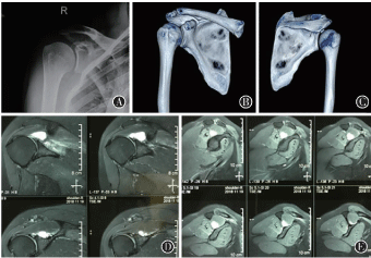
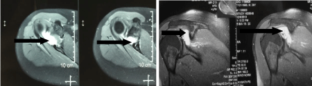
Right shoulder muscle atrophied and electromyography demonstrated marked wasting of both supra-and infraspinatus muscles. Abduction and external rotation were grade 4/5 in strength in two patients and another is grade 3/5, O'Brien, speed and neer test was positive. The patient showed full range of motion (ROM) with no instability of anterior or posterior. Clinical tests demonstrated normal subscapularis. Magnetic resonance imaging (MRI) of three patients revealed a large cyst arising from the postero glenoid labrum extending past the spinoglenoid notch into the suprascapular notch (Figure1, Figure 2 and Figure 3).
Surgical technique
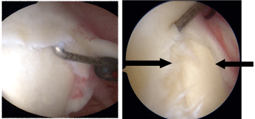
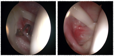
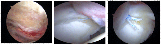
At arthroscopy, all chondral surfaces were found to be normal (Figure 4, Figure 5 and Figure 6). The cyst was decompressed by the use of shaver suction. The superior and posterior labral tear was repaired via two anchors placed to the biceps tendon (Figure 6). Then arthroscope got into the subcromial space via the lateral portal. Cleaning of subacromial synovium and hyperplastic bone, it is difficult and can be time consuming to dissection in this area. Importantly, the suprascapular artery is identified running superficial to the transverse scapular ligament. It is careful to protect this vessel to prevent post-operative bleeding. Lastly, the suprascapular nerve released.
Results
Post-operatively, the patient was placed in a shoulder joint abduction 30° brace for first day after operation with active ROM exercises allowing elevation to shoulder height. Thereafter, patients were took full ROM with physiotherapy exercise. Repeat EMG studies at 3 months post-operatively showed a good level of reinnervation of both suprascapular nerve and infraspinatus. MRI studies at 3 months post-operatively demonstrated complete disappear of the complex paralabral cyst. Clinical follow-up and review revealed improving strength of supra- and infraspinatus. The patient returned to work shortly after the 3 months review. Interestingly, in conjunction with his physiotherapist, the patient felt satisfied on the biceps and labrum during the catch criminal.
Discussion
The suprascapular nerve is a mixture of motor and sensory nerves, originating from the upper trunk of the brachial plexus [5]. The suprascapular nerve sends out the sensation of multiple fine sensory branches matching the shoulder joint and acromioclavicular joint through the scapular notch. The suprascapular nerve and the suprascapular arteriovenous vein bypass the scapular ganglia and suprascapular notch laterally, enter the subganglionic fossa in an arc manner, and send out muscular branches innervating the infraspinatus muscle and sending out multiple sensory fibers to dominate the glenohumeral joint capsule [6]. This is the anatomical basis of suprascapular nerve compression syndrome.
Shoulder joint is a very flexible joint, circular glenoid is a static stable structure of shoulder joint. The upper glenoid lip is closely connected with the biceps brachii tendon to form the labial complex of the biceps brachii tendon. Biomechanical studies have shown that the long head tendon of the biceps brachii and the upper glenoid labium complex play an important role in the stability of the glenohumeral joint. The injury of the anterior and posterior glenoid lip of the shoulder is called SLAP injury (Superior Labrum Anterior and Posterior), which mainly refers to the avulsion of the upper lip of the glenoid edge of the shoulder from anterior to backward, involving the attachment of the tendon of the long head of biceps brachii muscle [6,7]. In the literature, the incidence of SLAP injury was 4% of intra-shoulder diseases. Before arthroscopic application, the diagnosis and treatment of SLAP injury is very difficult. With the application of arthroscopy and the study of biomechanics, the recognition of SLAP damage is increasing. Due to the poor blood supply of the upper lip of the glenoid margin, and because of the biceps tendon glenoid complex has the function of stabilizing the anterior shoulder joint, it is found that the SLAP injury rarely heal themselves and often cause pain and dysfunction. Furthermore, the injury can cause the anterior instability of the shoulder joint in the biomechanical experiment [7]. With SLAP injury, the glenoid lip was lacerated, the joint effusion flowed out along with the suprascapular nerve and suprascapular notch, and the suprascapular nerve was encased to form a cyst, which resulted in compression of the suprascapular nerve.
Patients often have a direct or indirect history of shoulder trauma, neck and shoulder discomfort, distension and blunt pain. The diagnostic basis was as follows: (1) Pain in scapula, obvious tenderness, weakness of ipsilateral upper limb, (2) Atrophy of supraspinatus muscle and infraspinatus muscle, (3) Special signs: The muscle strength decreased most obviously at 30° of shoulder abduction, and generally decreased to grade, (4) Electromyography showed that the motor conduction velocity of suprascapular nerve was significantly slower, and positive spikes or fibrillatory potentials appeared in supraspinatus muscle and infraspinatus muscle [8].
Shoulder dysfunction and pain was mainly caused by spinoglenoid cyst. Classically, the cyst can cause infraspinatus weakness [1,3]. This study, including 3 cases, showed that large cyst tracking the suprascapular notch can result in both infra- and supraspinatus weakness. This contrasts with the notion that proximal suprascapular nerve entrapment are generally caused by local anatomical variations of the suprascapular notch [9]. Narrow V-shaped notches, variations to the superior transverse ligament, including calcification, muscle tear, or hypertrophied ligaments, have all been identified.
Conservative therapy for suprascapular nerve lesions associated with SLAP injury with paralabral cyst is often unsuccessful [9]. It is our opinion that, when shoulder joint grow weakness or muscle atrophy, a series of conservative therapy consisting of rest, physiotherapy and stretching may be pursued. In the setting of muscle atrophy and weakness, such conservative approach is unlikely to be successful. A few studies have advocated an operative approach in the setting of compression [8,9]. There is still some controversy regarding the exact treatment for each issue. These include open excision of the cyst, arthroscopic evacuation of the cyst via an intra-articular approach or simple treatment of the associated labral tear.
Lafosse, et al. [10] first reported the successful cases of nerve decompression with shoulder arthroscopy in suprascapular nerve compression surgery. For patients with scapular neck cyst, the cyst should be excised, the suprascapular nerve should be protected, combined with preoperative imaging results, the relationship between cyst and surrounding tissue should be carefully identified, and the cyst should be fully removed. If necessary, all parts of the glenohumeral joint can be completely exposed. After removal of the cyst, suturing the side of the subganglionic muscle, Radic, et al. [1] reported a case of proximal suprascapular neuropathy caused by laceration of the glenoid lip. Arthroscopic glenoid repair and neurolysis. Cyst decompression through the probe puncture. Repair of tear of upper glenoid lip entered under the anterior passage of arthroscope and repaired with anchor. The posterior glenoid tear was repaired by a posterior, lateral approach with anchor. Then the cyst was explored and sucked, and the suprascapular nerve was decompressed. Intraoperative protection of suprascapular arteriovenous, to prevent postoperative bleeding. Excellent recovery was made in this case soon after arthroscopic treatment. In order to improve the effect of suprascapular nerve decompression, more reasonable surgical methods were adopted according to the specific manifestations and clinical symptoms of the patients.
The mechanism of injury is very interesting. Our thought is the repetitive stress was a significant contributor to the development of the SLAP tearing and subsequent cyst formation. Previous studies have showed repetitive microtrauma to the suprascapular nerve resulting in conduction shoulder impairment.
Conclusion
This study proved the clinical symptom whereby a SLAP injury with resulting cyst formation has caused a suprascapular neuropathy. We found the mechanism of injury interesting in that the patient's occupation, possibly contributed to the site of development of the labrum tearing. In keeping with other previous study, a fully arthroscopic approach to these injuries can result in better outcomes.
Declarations
Ethics approval and consent to participate
The study was approved by the institutional review board of Tianjin First Center Hospital and all authors and patients have read and approved the final manuscript.
Consent for publication
All authors have read and approved the final manuscript.
Availability of data and materials
All data generated or analyzed during this study are included in this published article.
Competing Interests
The authors declare that they have no competing interests.
Funding
The author(s) received no financial support for the research, authorship, and/or publication of this article.
Authors' Contributions
KS carried out the studies and drafted the manuscript. YJ L conceived of the study and participated in its design and coordination, and helped to draft the manuscript. All authors read and approved the final manuscript.
Acknowledgements
The authors would like to thank Tianjin First Center Hospital for providing the database.
References
- Radic R, Wallace A (2016) Arthroscopic release and labral repair for bifocal compression of the suprascapular nerve. Shoulder Elbow 8: 32-36.
- Knudsen ML, Hibbard JC, Nuckley DJ, et al. (2016) Anatomic landmarks for arthroscopic suprascapular nerve decompression. Knee Surg Sports Traumatol Arthrosc 24: 1900-1906.
- Naidoo N, Lazarus L, De Gama BZ, et al. (2014) The variant course of the suprascapular artery. Folia Morphol 73: 206-209.
- Hahn M, Muller A, Valderrabano V, et al. (2014) Der Karpaltunnel der Schulter/the carpal tunnel of the shoulder: Die arthroskopische dekompression des N. suprascapularis. Sports Orthopaed Traumatol 30: 215-219.
- Gupta S, Manjuladevi M, Vasudeva Upadhyaya KS, et al. (2016) Effects of irrigation fluid in shoulder arthroscopy. Indian J Anaesthes 60: 194-198.
- Said HG, Abdelkawi AF, Fetih TN, et al. (2017) A Shortcut to arthroscopic suprascapular nerve decompression at the suprascapular notch: Arthroscopic landmarks and surgical technique. Arthroscopy Techniques 6: 1709-1713.
- Yamakado K (2015) Quantification of the learning curve for arthroscopic suprascapular nerve decompression: An evaluation of 300 cases. Arthroscopy 31: 191-196.
- Chen AL, Ong BC, Rose DJ (2003) Arthroscopic management of spinoglenoid cysts associated with SLAP lesions and suprascapular neuropathy. Arthroscopy 19: E15-E21.
- Cummins CA, Messer TM, Nuber GW (2000) Suprascapular nerve entrapment. J Bone Joint Surg Am 82: 415-424.
- Lafosse L, Piper K, Lanz U (2011) Arthroscopic suprascapular nerve release: indications and technique. J Shoulder Elbow Surg 20: S9-S13.
Corresponding Author
Yijin Li, MD, Tianjin First Center Hospital, Fukang Road No. 24, Nankai District, Tianjin, 300192, China.
Copyright
© 2021 Sun K, et al. This is an open-access article distributed under the terms of the Creative Commons Attribution License, which permits unrestricted use, distribution, and reproduction in any medium, provided the original author and source are credited.