What Should Be the Surgical Position Choice in A Patient Having Proximal Humeral Fracture with Developed Hypotension?
Abstract
Proximal humerus fractures with increasing prevalence in the geriatric age group are treated in a wide range from nonoperative treatment to arthroplasty and the treatment issue is still controversial. Developments in proximal humeral locking plate technology have increased the plate osteosynthesis option. Although surgical position in shoulder arthroscopy is as controversial as fracture treatment, osteosynthesis with locking plate is performed in the beach chair and in the supine position. In our clinical experience, the osteosynthesis with a locking plate was performed in beach chair position. Due to the hypotension developing after anesthesia, lateral decubitus position was used in the presented case and the surgical procedure of the patient was completed safely. In this case, it was aimed to operate the presence of hypotension that may cause cerebral perfusion failure in such a way that does not cause cerebral desaturation for surgical safety of the patient.
Keywords
Lateral decubitus position, Proximal humerus fracture, Plate fixation, Hypotansion
Introduction
Proximal humerus fracture constitutes 4-5% of all fractures and it is the most frequent fracture type seen in the elderly after hip and distal radius fractures [1-3]. The purpose of the treatment of proximal humerus fractures is the restoration of a painless shoulder having a satisfactory function. This requires an understanding of the injury including information about the patient's age, expectations, medical condition, quality and current fixation techniques and limitations. Many different techniques consisting of osteosynthesis and prosthesis options have been identified for the fixation of proximal humerus fractures [4-7]. Surgical procedures are carried out in the beach chair and supine positions. In the case subject of the article, the hypotension developing after anesthesia was attributed to the Bezold-Jarisch reflex defined in 1867 and resulting in apnea, bradycardia and hypotension by stimulation of chemical and mechanical receptors [8]. It is known that this reflex can develop in orthostatic position and hypovolemia during neuroaxial and general anesthesia depending on pain and anxiety [9]. The surgical procedure on the developing hypotension was performed in the lateral decubitus position.
A distance is remained for lateral decubitus position especially in arthroscopic procedures that can be switched to open surgery in order not to have approach and orientation problems [10]. When the literature was examined, no study was found indicating that osteosynthesis of proximal humerus fracture with locking plate was performed in lateral decubitus position. Considering the presence of systemic hypertension (HT), renal stent history and smoking, which may cause cerebral perfusion failure, it is aimed to operate the patient in a way that will not cause cerebral desaturation for surgical safety. For the safety of surgical procedures of patients with proximal humerus fractures having a clinical picture that may cause cerebral perfusion failure, it may be beneficial to keep lateral decubitus position in mind as an alternative.
Case Report
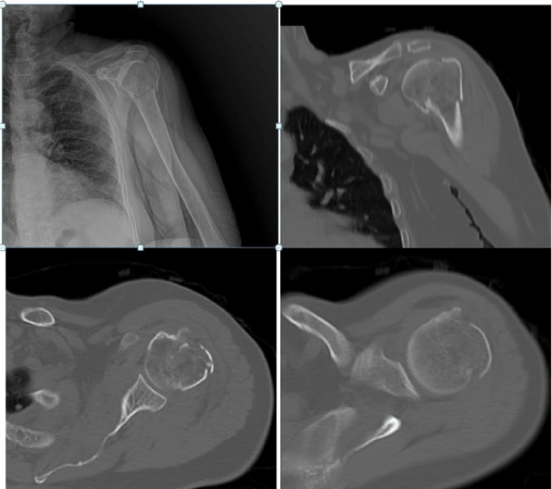
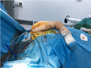
A 78 year-old female patient applied to the emergency room of our hospital due to the complaint of pain in the left shoulder after falling from a height of approximately 1 meter. In the examination and tests, there was a 3-part fracture in the left proximal humerus in which the surgical neck and tuberculum majus were broken according to Neer classification (Figure 1). There was no neurovascular deficit. In the patient's history, there was HT, history of renal stenting, and hemorrhoids. In addition, there were an undiagnosed preoperative hyperglycemia and smoking history. She was receiving verapamil treatment for HT. In her preoperative evaluation, it was assessed that her effort capacity was normal, hemodynamics was stable, and her ejection fraction was 55%. The feature was not found in the laboratory results and PA chest x-ray. It was monetarized with standard electrocardiography (ECG), heart rate (HR), noninvasive blood pressure (BP) and peripheral oxygen saturation (SPO2). No abnormalities were observed in the monitorization results. Anesthesia induction was performed with 2mg/kg propofol, 1 mg/kg lidocaine, 1 mcg/kg fentanyl, 20 mg ketamine and 0.6 mg/kg esmerone. After the induction, the patient with stabile hemodynamics was intubated without any problem once with the tube no: 7.0. For maintaining anesthesia, 2% sevoflurane was initiated. Since the patient who was brought to the sitting position for the operation was administered with atropine, ephedrine and noradrenaline. She was returned back to the supine position since her hypotension and bradycardia could not be fixed with supportive therapy. 1ml/kg of fluid replacement was given. The patient, whose hemodynamics quickly stabilized in the supine position, was brought back to the sitting position. However, since the deep hypotension and bradycardia were repeated, it was decided to perform the operation of the patient in lateral decubitus position in order not to disturb the cerebral vascular nutrition (Figure 2).
Surgical Procedure
We used scapular and sacral supports on the back, sternal and pubic supports on the front side and gel pad supports to protect the bone protrusion and superficial nerves in order to hold the patient in this position. Right arm flexion was increased in order to allow the surgeon to work more comfortably. The surgical procedure was performed using the standard deltopectoral approach. In order to facilitate the incision and approach, the table was tilted at partial distances to the right and left at certain stages of the surgery. For the reduction and determination of the displaced tuberculum majus, no: 2 polyethylene multifilament sutures were applied to the bone tendon junction. For the reduction of the humeral surgical neck fracture, manual traction was applied in arm abduction, the medial force effect of the pectoralis major muscle was eliminated with a folded surgical drape placed between the arm and the trunk. After the reduction of fracture lines, fixation was achieved with a locking proximal humerus plate after temporary fixation with multiple k wires.
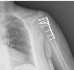
The sutures passing through the bone tendon junction were passed through suture holes in the plate proximal, knotted and the osteosynthesis was completed. Follow-ups of perioperative ECG, SPO2 blood pressure values were stable. The operation lasted for 75 minutes. After the muscle relaxant was antagonized, she was extubated without any problem. No hemodynamic problem was experienced in the postoperative period. For the radiological imaging, C-arm fluoroscopy was rotated under the surgical table and positioned so that the shoulder can be seen anteriorly. Proximal humerus Anteroposterior/Lateral images were obtained by moving the arm. A shoulder arm sling was used until the skin sutures were taken. Pendulum and assisted joint range of motion were then started. Strengthening exercises started in the 6th week. In the outpatient clinical follow-ups, it was found that bone healing and joint range of motion were complete (Figure 3).
Discussion
The best part of orthopedic and traumatology surgery is that a surgical procedure can be performed with different instruments, different techniques and in different positions. Proximal humerus fractures are among the most common fractures in human body with a peak incidence in elderly population. Its treatment is still a matter of discussion and it covers a wide range of options such as non-operative treatment, osteosynthesis and arthroplasty [6,7,11-14]. Depending also on surgical procedure, implants vary from k-wire to arthroplasty material. Open reduction internal fixation (ORİF) indications have expanded for some fracture types and in patients particularly having osteoporotic bone following the developments in locking plate technology in proximal humerus fractures [15]. Positions in shoulder surgery are beach chair, supine and lateral decubitus positions depending on the procedure, surgical experience and operating room facilities. When examining the hypotension that developed in the patient, the course without any complication for about 15 minutes following the intubation and the induction of anesthesia, sudden development of unresponsive bradycardia and hypotension to inotropes and atropine after placing the patient in the sitting position, a quick response to support treatment performed in the supine position but the inability to stabilize hemodynamics in the sitting position suggested us Bezold-Jarisch reflex. Hypotension, bradycardia, and asystole seen in sitting position depending on Bezold-Jarisch reflex quickly respond to the intervention when s/he is placed in the supine position and the findings can improve quickly [8,9]. In the osteosynthesis with locking plate in displaced and unstable proximal humerus fractures, we routinely prefer beach chair position. When the literature is examined, it is seen that patients are operated in beach chair or supine positions [16-18]. However, we did not find any study in the literature preferring lateral decubitus position. Optimal positioning according to the clinical condition of patient is necessary for a successful surgical procedure.
In the presented case, since TA values dropped after anesthesia and the desired increase could not be obtained at the desired time despite inotropic agents in the case, we changed the surgical plan as the lateral decubitus position. The possibility of cerebral oxygen desaturation during the beach chair position and hypotension of the patient lay on contribution to this change. Complications in patients after shoulder surgery in beach chair position vary from cranial nerve damage [19] to vision loss [20] and cerebral infarction [21-23]. Although the exact pathogenesis of these events has not been fully explained, it has been assumed that the beach chair position may be responsible for these complications. Moerman, et al. showed that cerebral oxygen desaturation occurred during the beach chair position by using Near-infrared spectroscopy [24]. Although there is a short-term orientation problem in the lateral decubitus position, the fact that it does not impair cerebral perfusion can be seen as a great advantage in this regard.
Conclusion
Although it is rare for a particular surgical procedure is performed only in a certain position, surgical positions can have various complications along with their pros and cons. Although a partial surgical orientation problem was experienced, lateral decubitus position enabled us to perform the surgical procedure safely in the patient who had proximal humerus fracture and developed hypotension. In addition, axial traction to the humerus for fracture reduction can be achieved more effectively manually or by using a traction system. In addition, the elimination of the medial force effect of the pectoralis major muscle with the folded surgical drape placed between the arm and the trunk, and easy reduction are other important advantages.
Conflicts of Interest and Source of Funding
The authors no conflict of interest.
The expenses of all materials used in this study were undertaken by the researcher.
Study conception and design: HA
Data collection: HA
Drafting of the article: HA, SE
Data analysis and interpretation: HA, SE
Critical revision of the article: HA
References
- Hallberg I, Rosenqvist AM, Kartous L, et al. (2004) Health-related quality of life after osteoporotic fractures. Osteoporos Int 15: 834-841.
- Kwon YW, Zuckerman JD (2005) Outcome after treatment of proximal humeral fractures with humeral head replacement. Instr Course Lect 54: 363-369.
- Lübbeke A, Stern R, Grab B, et al. (2005) Upper extremity fractures in the elderly: Consequences on utilization of rehabilitation care. Aging Clin Exp Res 17: 276-280.
- Park MC, Murthi AM, Roth NS, et al. (2003) Two-part and three-part fractures of the proximal humerus treated with suture fixation. J Orthop Trauma 17: 319-325.
- Robinson CM, Page RS, Hill RM, et al. (2003) Primary hemiarthroplasty for treatment of proximal humeral fractures. J Bone Joint Surg 85: 1215-1223.
- Lanting B, MacDermid J, Drosdowech D, et al. (2008) Proximal humeral fractures: A systematic review of treatment modalities. J Shoulder Elbow Surg 17: 42-54.
- Liew AS, Johnson JA, Patterson SD, et al. (2000) Effect of screw placement on fixation in the humeral head. J Shoulder Elbow Surg 9: 423-426.
- Von Bezold AV, Hirt L (1867) Uber die physiologischen wirkungen des essigsauren veratrins. Untersuchungen aus dem Physiologischen Laboratorium Wurzburg 1: 75-156.
- Jihyun S, Woo Jong S, Jae Hang S (2013) A cardiovascular collapse occurred in the beach chair position for shoulder arthroscopy under general anesthesia. Korean J Anesthesiol 64: 265-267.
- Arrington ED, Parada SA, Marchant BG (2012) Beach chair and lateral decubitus setup- pros and cons. In: Provencher MT, Romeo AA, (3rdedn), Shoulder Instability: A Comprehensive Approach, Philadelphia, PA: Elsevier Sounders 33-42.
- Wijgman AJ, Roolker W, Patt TW, et al. (2002) Open reduction and internal fixation of three and four-part fractures of the proximal part of the humerus. J Bone Joint Surg Am 84: 1919-1925.
- Misra A, Kapur R, Maffulli N (2001) Complex proximal humeral fractures in adults-a systematic review of management. Injury 32: 363-372.
- Niemeyer P, Hauschild O, Strohm PC, et al. (2004) Fracture treatment in the elderly. Acta Chir Orthop Traumatol Cech 71: 329-338.
- Shrader MW, Sanchez Sotelo J, Sperling JW, et al. (2005) Understanding proximal humerus fractures: Image analysis, classification, and treatment. J Shoulder Elbow Surg 14: 497-505.
- Agudelo J, Schürmann M, Stahel P, et al. (2007) Analysis of efficacy and failure in proximal humerus fractures treated with locking plates. J Orthop Trauma 21: 676-681.
- Konrad G, Bayer J, Hepp P, et al. (2009) Open Reduction and internal fixation of proximal humeral fractures with use of the locking proximal humerus plate surgical technique. J Bone Joint Surg Am 91: 1320-1328.
- Geiger EV, Maier M, Kelm A, et al. (2010) Functional outcome and complications following PHILOS plate fixation in proximal humeral fractures. Acta Orthop Traumatol Turc 44: 1-6.
- Chin Hsien Wu, Ching Hou Ma, James Jih Hsi Yeh, et al. (2011) Locked plating for proximal humeral fractures: Differences between the deltopectoral and deltoid-splitting approaches. J Trauma 71: 1364-1370.
- Boisseau N, Rabarijaona H, Grimaud D, et al. (2002) Tapia's syndrome following shoulder surgery. Br J Anaesth 88: 869-870.
- Bhatti MT, Enneking FK (2003) Visual loss and ophthalmoplegia after shoulder surgery. Anesth Analg 96: 899-902.
- Pohl A, Cullen DJ (2005) Cerebral ischemia during shoulder surgery in the upright position: A case series. J Clin Anesth 17: 463-469.
- Verbrugge SJ, Klimek M, Klein J (2004) A cerebral watershed infarction after general anaesthesia in a patient with increased anticardiolipin antibody level. Anaesthesist 53: 341-346.
- Dippmann C, Winge S, Nielsen HB (2010) Severe cerebral desaturation during shoulder arthroscopy in the beach-chair position. Arthroscopy 26: S148-S150.
- Moerman AT, De Hert SG, Jacobs TF, et al. (2012) Cerebral oxygen desaturation during beach chair position. Eur J Anaesthesiol 29: 82-87.
Corresponding Author
Harun Altinayak, PhD, MD, Health Sciences University, Samsun Training and Research Hospital, Department of Orthopaedics and Traumatology, Samsun, Turkey, Tel: 905-374-624-182.
Copyright
© 2021 Altinayak H, et al. This is an open-access article distributed under the terms of the Creative Commons Attribution License, which permits unrestricted use, distribution, and reproduction in any medium, provided the original author and source are credited.