Minimally Invasive Plate Osteosynthesis (MIPO) for Treatment of Comminuted Mid-Distal Third Humeral Shaft Fractures through the Anterior Approach
Abstract
This study was conducted to evaluate the results of management of comminuted fractures of the mid-distal third shaft of the humerus with indirect reduction and internal fixation with plate and screws through minimally invasive technique using the anterior approach. Twenty-three comminuted fractures of the mid-distal third humeral shaft in 23 adult patients were managed using the minimally invasive plate osteosynthesis. Fractures were classified according to the AO classification system, healing was assessed by plain radiographs. Functional outcomes were assessed using the American Shoulder and Elbow Surgeons score. All fractures united within 15 weeks (mean 12.8 weeks). One case had an iatrogenic radial nerve palsy that was regained 2 months later. No limitation of the shoulders and elbows movement on the injured side when compared to the uninjured one. No complications related to the implant; no implant failure, screw loosening or screw breakage. The alignment was restored to that of the contralateral non-affected side in all but 3 cases; these cases had varus deformities not more than 10 degrees without affecting shoulder or elbow function. No infection was reported in this series. Minimally invasive plate osteosynthesis is a safe and effective way for management of comminuted mid-distal third humeral shaft fractures, with early fracture healing, rapid recovery of shoulder and elbow function and cosmetically acceptable scar.
Keywords
Mini invasive, Fracture shaft humerus, Biological techniques, MIPO
Introduction
Humeral shaft fracture is a common entity encountered in orthopedic surgery, it represents about 1-3% of all fractures, 5-10% of all long bone fractures and 20% of all humeral fractures [1,2].
Although conservative treatment of these fractures results in satisfactory clinical and functional outcomes, it may result in varus deformity and limitation of shoulder and elbow motion in some patients [3].
The indications for operative treatment of these fractures increased widely with time in order to achieve healing with adequate function in the shortest possible time [2].
In spite of the presence of several techniques for operative management of humeral shaft fractures, yet, there is no agreement over the optimal method. However, plating remains the standard technique for management of this fracture [4].
Management of comminuted fractures of the shaft of the humerus is a challenge due to the high morbidity associated with plating by the traditional anterolateral or posterior approach while interlocking nails cannot control rotation sufficiently in unstable and distal humeral shaft fractures [5-7].
The concept currently known as Minimally Invasive Plate Osteosynthesis (MIPO) is based on the relative stability of the fracture with minimal damage to the surrounding soft tissues [8].
Minimally invasive treatment modalities with low morbidity, rapid patient recovery and earlier return to work and activities of daily living are favored to other modalities [9].
Anterior approach to the surface of the humerus is recommended to avoid injury of the nearby neurovascular structures [3].
The aim of this study was to report the results of application of the minimally invasive technique with bridging plates in the treatment of comminuted mid-distal third humeral shaft fractures through the anterior approach assessing the time of fracture union and the nearby joint function.
Material and Methods
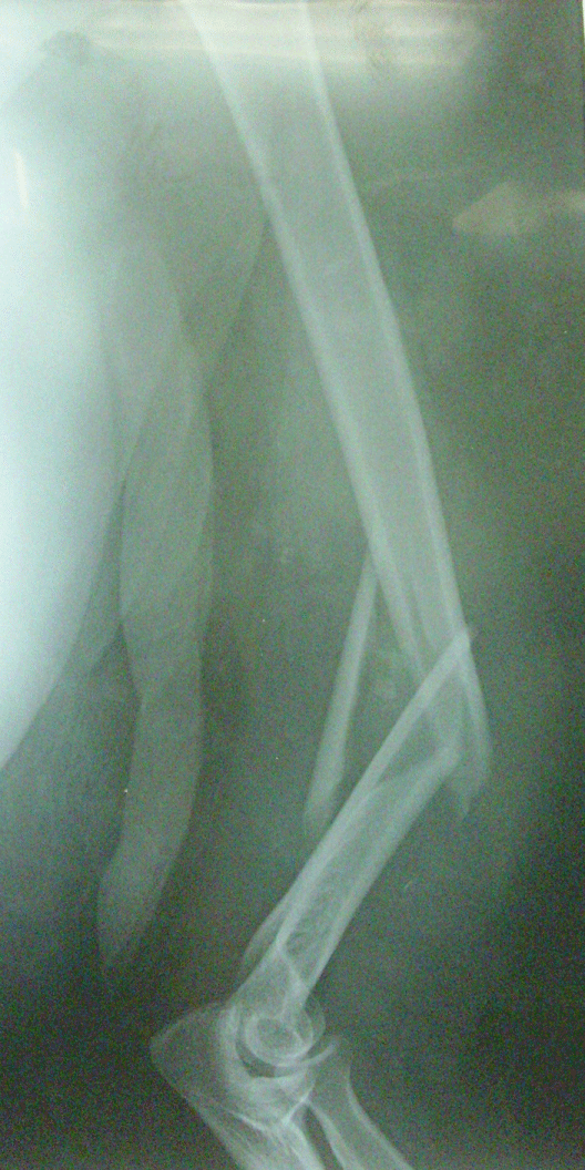
This prospective study was approved by institutional review board of University and consent was obtained from all patients. Between May 2010 and October 2013, twenty-three comminuted fractures of the mid-distal third humeral shaft in twenty-three adult patients were managed in University hospitals using the MIPO technique with indirect reduction method through the anterior approach. Fractures were classified according to the AO classification system, there were 16 cases type 12B and 7 cases type 12C [10].
Adult patients with comminuted mid-distal third humeral shaft fractures were included in the study, all fractures in the study were comminuted and required fixation to restore alignment. Patients with proximal third fractures, open fractures; associated radial nerve palsy and children were excluded from the study.
Technique
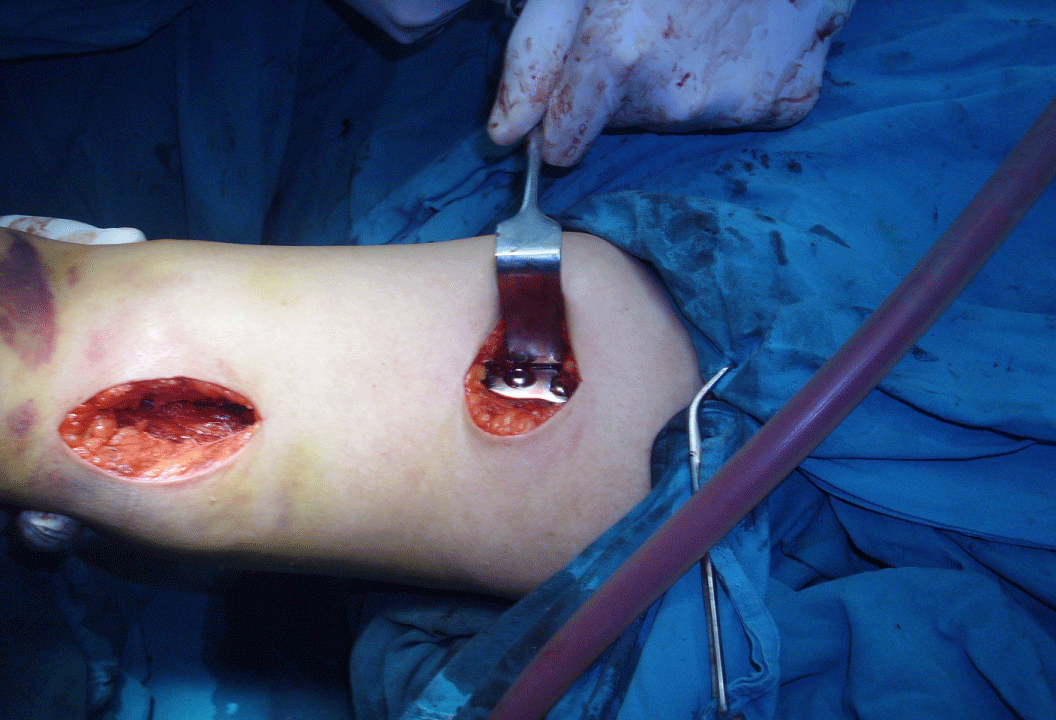
Surgery is performed with the patient in a supine position on a radiolucent operating table, and the injured arm is abducted 60° under image intensifier control. The approach includes 2 skin incisions: The proximal one is a part of the deltopectoral approach, and the distal one is a 4-5 cm long incision on the anterior side proximal to the elbow crease over the biceps tendon taking care to preserve the sensory branch of the musculocutaneous nerve (Figure 1.1 and Figure 2.1). The biceps muscle is then retracted, and the brachialis muscle is split by blunt dissection. Tunneling of the ventral side of the humeral shaft using a nine to twelve holes Locking Compression Plate (LCP) pushed gently from proximal to distal while being in close contact to the bone. Tunneling is done smoothly with only slight resistance by the deltoid insertion.
The technical tips and tricks should be followed to obtain the best reduction with this indirect reduction technique and to avoid iatrogenic radial nerve palsy:
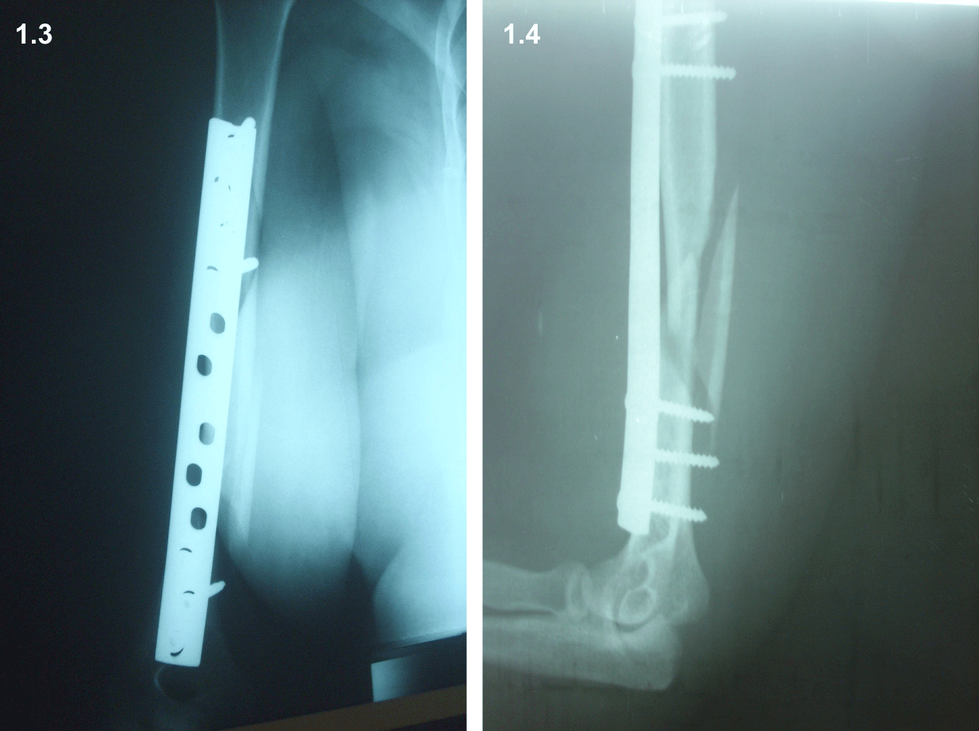
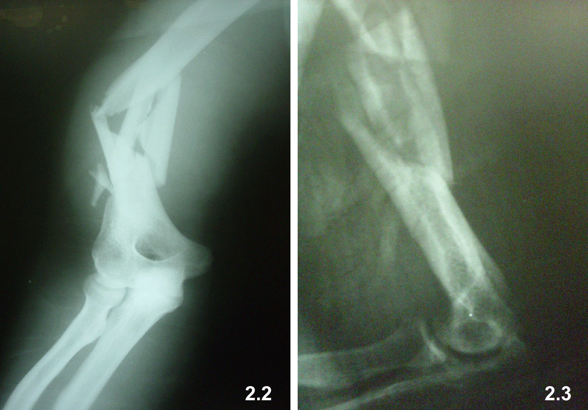
The forearm should be in a supine position to bring the radial nerve away from bone, arm abduction 60°. The Locking Compression Plate (LCP) is fixed provisionally on the anterior surface of the humerus to the distal main fragment with the most distal screw is the first to be inserted without tightening as this allows better manipulation, and the fracture is reduced by manipulation under image control for proper adjustment of length, alignment and rotation, then plate is fixed to the proximal main fragment and the screws are tightened and another two or more additional screws are inserted in each fragment [5-13] (Figure 1.2, Figure 1.3, Figure 1.4, Figure 2.2 and Figure 2.3).
The authors used the AO guides for obtaining control on alignment and rotation depending on the difference of cortical thickness and diameter difference of proximal and distal main fragments that appears on x rays in cases of rotation.
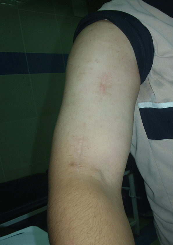
The incisions are then sutured with simple separated stitches without drains; postoperative immobilization with humeral brace till union is achieved radiologically by callus formation in 3 of 4 cortices in anteroposterior and lateral views.
Early rehabilitation in the form of shoulder pendulum exercises and active and passive wrist and elbow movements were started by the first week post-surgery.
Fracture healing was assessed by plain radiograph. Functional outcomes were assessed using the American Shoulder and Elbow Surgeons score [14].
Patients were followed up weekly for two weeks and monthly thereafter till union then every 3 months till one year and yearly after that till the last follow up. Radiographs and clinical examinations were done in each visit to evaluate fracture healing and shoulder and elbow function.
Radiological assessment to measure alignment through drawing the anatomical axis of the proximal and distal fragments and measuring the angle in between them and compare it to that of the contralateral side. Range of Motion (ROM) was measured by goniometer on the fractured and healthy sides during follow up visits.
Statistical Analysis
The data of patients were statistically assessed using the Statistical Package for Social Sciences (SPSS) version 19 for windows. Independent sample t-test and paired sample test were used to define relations between functional outcomes at 3 months, at 6 months and at the last follow up, the relation between varus complication and the final follow up score and the effect of AO classification on the final results. Probability values of less than 0.05 were considered significant.
Results
Twenty-three patients with 23 humeral fractures were included in the study, there were sixteen males (69.6%) and seven females (30.4%), with the age of patients ranged from 19 to 61 years (mean 36.2 years). Thirteen patients were affected on the right side and 10 on the left side.
All fractures were isolated fracture humerus except for 2 cases with associated forearm fractures.
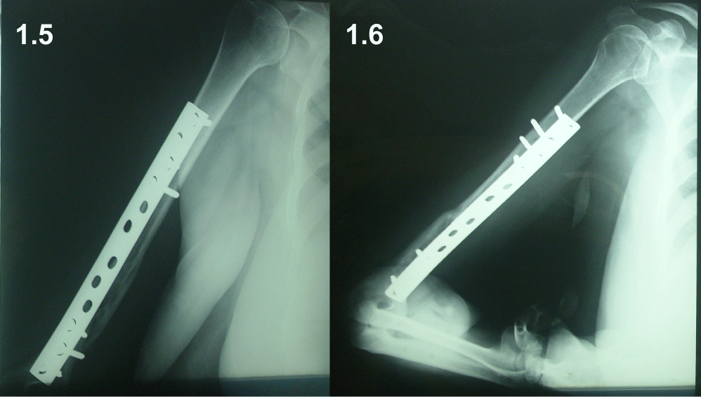
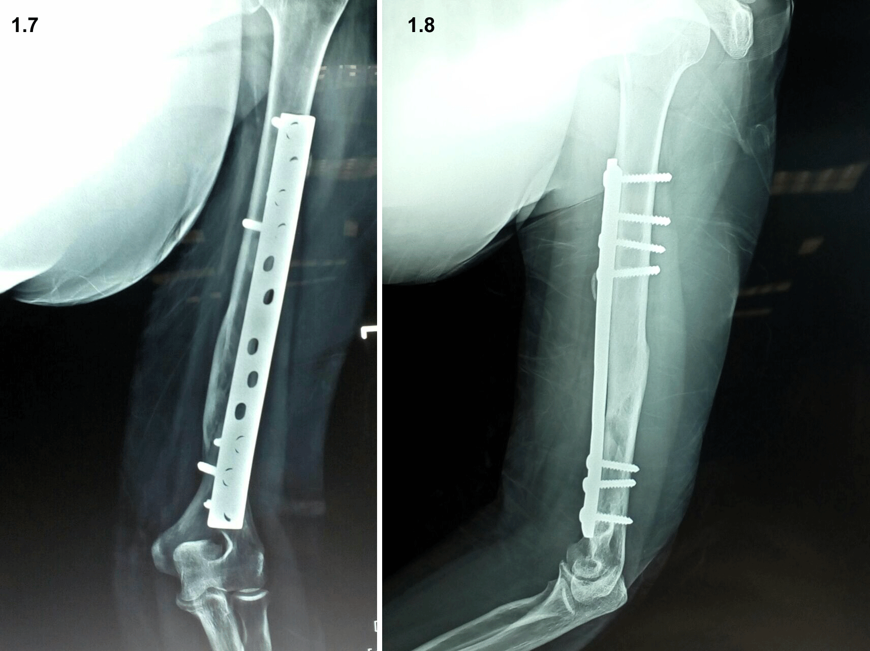
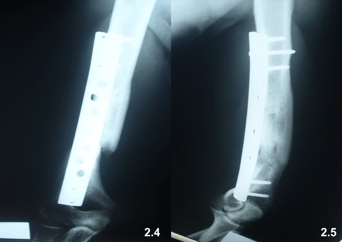
All cases were followed to a mean period of 34.2 months (range 14-73 months). All fractures united within 15 weeks (mean 12.6 weeks) (Figure 1.5, Figure 1.6, Figure 1.7, Figure 1.8, Figure 2.4 and Figure 2.5).
One case had an iatrogenic radial nerve concussion that was regained 2 months later. 2 cases with associated forearm fractures in which the forearm bones were fixed with plates and screws without effect on elbow mobility.
No limitations of the shoulders and elbows movements were observed on the operated when compared to the uninjured side. No complications related to the implant; no implant failure, screw loosening or screw breakage. The alignment as measured on radiographs was restored to that of the contralateral non-affected side in all cases except for 3 cases with varus deformity not more than 10 degrees which has no statistical effect on the results P = 0.243. No infection was reported in this series.
American Shoulder and Elbow Surgeons Score (ASES) were done at 3 months, 6 months, and 12 months and at the last follow up. At 3 months the mean ASES was 94.3 (range 90-100), at 6 months it was 97.5 (range 95-100), at 12 months it was 97.8 (range 96-100), after one year there were no further improvement in the outcome, the mean ASES was 97.8 (range 96-100), by the last follow up. There was a significant improve in the in the score at 6 months (p < 0.001), and at 12 months (P < 0.001). The fracture type according to AO doesn't significantly affect the final outcome as measured by the ASES p = 0.726 or the union time (P > 0.05) (Table 1).
Discussion
Absolute stability of fixation offers solid union of fractures. However, direct precise reduction and absolute stability are achieved on the expense of extensive soft tissue stripping and disruption of periosteal blood supply [15].
The concept of biological osteosynthesis preserves the regional blood supply enhancing fracture union. Minimally Invasive Plate Osteosynthesis (MIPO) follows the rules of biological osteosynthesis and has become a widely accepted method of fixation [12]. The use of MIPO prevents large surgical approach, extensive soft tissue stripping, and disruption of periosteal blood supply and iatrogenic nerve injuries. MIPO shortened the time for union and the period of rehabilitation [16].
An, et al. in their comparative study for treating mid-distal hu�meral shaft fractures with ORIF and MIPO, they found 31.3% Iatrogenic radial nerve injury with the ORIF while no case had this complication in the MIPO group; the mean union time was shorter in the MIPO group with similar satisfactory functional out-comes in both groups [17].
Both IMN and minimally invasive plating osteosynthesis are valid treatment options with nearly similar good results for mid-distal humeral shaft fractures. IMN required a longer rehabilitation time for shoulder and elbow functions as compared to minimally invasive plating osteosynthesis. Exploration for the ra�dial nerves is not required with minimally invasive plating osteosynthesis using an anteriorly placed plate to treat humeral shaft fractures, which is not the case with IMN as the nerve may be entrapped at the fracture site and put in danger while the nail is being inserted [11].
Many humeral shaft fractures are managed successfully by conservative means, there is a wide range of acceptance as the large range of shoulder motion can compensate for malunion with little or no functional impairment. When surgery is indicated Minimally Invasive Plate Osteosynthesis (MIPO) and intramedullary nailing are considered the treatments of choice as biologic fixation procedures [8,11,18].
Many authors agree that humeral plating is the method of choice for fixation of humeral shaft fractures, plating has a high union rate, and doesn't interfere with nearby joint function. MIPO unlike traditional plating offers an approach that limits surgical soft tissue trauma at the fracture site. The minimal disruption of the periosteum associated with MIPO enhances bone healing and limits nonunion [4,17,19,20].
The MIPO is a relatively flexible fixation as full reduction and compression of the fractured fragments is not accomplished, in flexible fixation Healing occurs indirectly by formation of callus which is faster and more effective than direct healings in absolute hard fixations [21].
Approaching the humerus through its anterior surface does not interfere with nerves or major blood vessels and allows safe percutaneous plating to bridge comminuted fractures [5,12].
Two points of concern about the MIPO technique; varus malunion and the potential risk of iatrogenic neurovascular injury, however, considerable varus deformity did not occur in many studies with MIPO. Furthermore, with mild varus angulation shoulder and elbow functions are preserved due to wide range of motion [3].
The neurovascular injury can be avoided by adhering strictly to operative technique; the forearm must be positioned in supination; Pronation brings the radial nerve closer to plate. Taking in mind the danger zone for musculocutaneous and radial nerves; Apivatthakakul, et al. in their cadaveric study found a danger zone for radial nerve to be located between 42% and 59% of the humeral length from the lateral epicondyle, hence the safe zone for application of the screws in MIPO is roughly in the distal and proximal 40% of humeral shaft [12,21,22].
Take care to be in the proper intermuscular plain and the plate advanced gently in close contact to bone over the anterior surface in a proximal to distal direction to protect deltoid insertion [18].
In this study all fractures united within 15 weeks (mean 12.6 weeks). One case had an iatrogenic radial nerve palsy that was regained within 3 months. No limitation of the shoulders and elbows movement was observed on the operated when compared to the uninjured side. No complications related to the implant; no implant failure, screw loosening or screw breakage. The alignment as measured on radiographs was restored to that of the contralateral non-affected side in all cases except for 3 cases with varus deformity not more than 10 degrees. No infection was reported in this series.
The functional outcomes assessed by the American Shoulder and Elbow Surgeons score. Functional Scores are equivalent in the affected side to that of the contralateral non-affected one which is consistent with the findings previously reported [17,20]. Many studies were done to assess the validity of the ASES score and concluded it is a repeatable, a reliable and a valid outcome tool [13,23].
In this study there was a significant improve in the in the score at 6 months (P < 0.001), and at 12 months (P < 0.001). After one year there were no further improvement in the outcome, by the last follow up the mean ASES was 97.8 (range 96-100).
MIPO technique for the treatment of diaphyseal fractures of the humerus resulted in satisfactory results in many series [5,12,16,21].
Malhan, et al. studied the results of MIPO using a locking compression plate and found that all fractures united after 14 weeks except for 2 cases with delayed union, and only one patient had iatrogenic radial nerve palsy [24].
Esmailiejah, et al. found better results with MIPO when compared to open reduction and plating as regard to the time of surgery and iatrogenic radial nerve injury (3% versus 12%) and the rate of infection (0% versus 6%), patients managed with the MIPO technique had also shorter time for union and earlier return to their previous level of activities [3].
MIPO is preferred to conventional plating due to limited incision which offers better cosmotic appearance and less risk of infection, limited possibility of iatrogenic radial nerve injury due to anterior approach and insertion of the plate away from the radial nerve, biological character of the technique preserving the periosteum and fracture hematoma which increase the potential for and accelerates fracture healing [3]. Conventional plating technique has increased risk of radial nerve injury, non-union and infection [5,12].
An, et al. compared the results of managing humeral shaft fractures with conventional plating and MIPO and found high incidence of iatrogenic radial nerve injury in the ORIF group while none were found in the MIPO group. They also found accelerated union time in the MIPO with similar satisfactory Functional outcomes [17].
Shin, et al. reported satisfactory results without iatrogenic radial nerve injury with the MIPO technique. Oh, et al. in their comparative study found that the primary union rate is higher in their patients treated with MIPO than those treated with open reduction and internal fixation whereas; the functional outcomes were the same [25,26].
In 2004, Livani and Belangero found that MIPO is an applicable and safe method and had better results when compared to the traditional plating in providing a shorter recovery time, more rapid fracture healing and early return of function in shoulder and elbow [5].
Although both Intramedullary Nailing (IMN) and MIPO have similar good results; IMN delay shoulder and elbow functional recovery when compared with MIPO, and the operative time for MIPO is shorter. Antegrade nailing may be problematic for the shoulder function; retrograde nail introduced to avoid shoulder dysfunction increases the risk of iatrogenic humeral fractures [3,11].
The radial nerve is not dissected when performing MIPO. The radial nerve along the spiral groove is away from sites of screws insertion; hence it is away from danger whereas, during IMN, the danger zone for the radial nerve is wide [26].
With MIPO the mean distance of the closest part of the radial nerve from the plate is 3.2 mm. The plate is placed at the anterior surface of the humerus under the brachial muscle which protects the radial nerve from injury [12].
Ji, et al. reported one case of iatrogenic radial nerve palsy out of 23 cases with MIPO although they used the less safe lateral approach. Livani, et al. had no radial nerve palsy in their study on 35 cases of mid-distal humeral shaft fractures managed with MIPO. Which is consistent with the results of Zhiquan, et al. and Pospula, et al. in their series [5,13,27,28].
The authors and others advocate this technique for distal third humeral fractures with radial nerve palsy after exploration of the radial nerve through a separate incision at the junction of the middle and distal thirds of the arm. The radial nerve is exposed between the brachialis and brachioradialis muscles till its exit through the lateral intramuscular septum [29].
Although the aim of the study was achieved but this study was limited by the small number of cases, a study including a larger number will give more convenient results. This study addressed only the results of MIPO and not a comparative study to assess the results of other methods of fixation.
Conclusion
Minimally invasive plate osteosynthesis is a safe and effective way for management of comminuted mid-distal third humeral shaft fractures, with early fracture healing, rapid recovery of shoulder and elbow function and cosmetically acceptable scar.
Compliance with Ethical Standards
Conflict of interest
The authors declare that they have no conflict of interest.
References
- Tytherleigh Strong G, Walls N, McQueen M (1998) The epidemiology of humeral shaft fractures. J Bone Joint Surg Br 80: 249-253.
- Sharma V, Awasthi B, Mehta S, et al. (2010) Evaluation of results of different treatment modalities in the management of diaphyseal fractures of the humerus. Internet J Orthop Surg 16: 1531-1568.
- Esmailiejah AA, Abbasian MR, Safdari F, et al. (2015) Treatment of humeral shaft fractures: Minimally invasive plate osteosynthesis versus open reduction and internal fixation. Trauma Mon 20: e26271.
- Mckee MD, Larsson S (2010) Humeral shaft fractures. In: Bucholz RW, Heckman JD, Court Brown CM, Tornetta P Rockwood and Green's Fractures. 1000-1038.
- Livani B, Belangero WD (2004) Bridging plate osteosynthesis of humeral shaft fractures. Injury 35: 587-595.
- Rommens P, Kuechle R, Bord T, et al. (2008) Humeral nailing revisited. Injury 39: 1319-1328.
- Changulani M, Jain U, Keswani T (2007) Comparison of the use of the humerus intramedullary nail and dynamic compression plate for the management of diaphyseal fractures of the humerus. A randomised controlled study. Int Orthop 31: 391-395.
- Zogaib RK, Morgan S, Belangero PS, et al. (2014) Minimal invasive ostheosintesis for treatment of diaphiseal transverse humeral shaft fractures. Acta Ortop Bras 22: 94-98.
- Garnavos C, Lasanianos N, Kanakaris NK, et al. (2009) A new modular nail for the diaphyseal fractures of the humerus. Injury 40: 604-610.
- Müller ME, Koch P, Najarian S, et al. (1990) The comprehensive classification of fractures of the long bones. Berlin Heidelberg, Springer, New York.
- Lian K, Wang L, Lin D, et al. (2013) Minimally invasive plating osteosynthesis for mid-distal third humeral shaft fractures. Orthopedics 36: e1025-e1032.
- Apivatthakakul T, Arpornchayanon O, Bavornratanavech S (2005) Minimally invasive plate osteosynthesis (MIPO) of the humeral shaft fracture. Is it possible? A cadaveric study and preliminary report. Injury 36: 530-538.
- Zhiquan A, Bingfang Z, Yeming W, et al. (2007) Minimally invasive plating osteosynthesis (MIPO) of middle and distal third humeral shaft fractures. J Orthop Trauma 21: 628-633.
- Michener LA, McClure PW, Sennett BJ (2002) American shoulder and elbow surgeons standardized shoulder assessment form, patient self-report section: Reliability, validity, and responsiveness. J Shoulder Elbow Surg 11: 587-594.
- Li M, Zhang X, Liu X, et al. (2012) The recent development of MIPO in long bone fractures. Open Journal of Orthopedics 2: 159-165.
- Livani B, Belangero W, Andrade K, et al. (2009) Is MIPO in humeral shaft fractures really safe? Postoperative ultrasonographic evaluation. Int Orthop 33: 1719-1723.
- Huri G, Bicer OS, Ozturk H, et al. (2014) Functional outcomes of minimal invasive percutaneous plate osteosynthesis (MIPPO) in humerus shaft fractures: A clinical study. Acta Orthop Traumatol Turc 48: 406-412.
- An Z, Zeng B, He X, et al. (2010) Plating osteosynthesis of mid-distal humeral shaft fractures: minimally invasive versus conventional open reduction technique. Int Orthop 34: 131-135.
- Bhandari M, Devereaux P, Mckee DM, et al. (2006) Compression plating versus intramedullary nailing of humeral shaft fractures- a meta-analysis. Acta Orthop 77: 279-284.
- Niall D, O'Mahony J, McElwain J (2004) Plating of humeral shaft fractures- has the pendulum swung back? Injury 35: 580-586.
- Aksu N, Karaca S, Kara AN, et al. (2012) Minimally invasive plate osteosynthesis (MIPO) in diaphyseal humerus and proximal humerus fractures. Acta Orthop Traumatol Turc 46: 154-160.
- Apivatthakakul T, Patiyasikan S, Luevitoonvechkit S (2010) Danger zone for locking screw placement in minimally invasive plate osteosynthesis (MIPO) of humeral shaft fractures: A cadaveric study. Injury 41: 169-172.
- Yahia A, Guermazi M, Khmekhem M, et al. (2011) Translation into Arabic and validation of the ASES index in assessment of shoulder disabilities. Annals of Physical and Rehabilitation Medicine 54: 59-72.
- Malhan S, Thomas S, Srivastav S, et al. (2012) Minimally invasive plate osteosynthesis using a locking compression plate for diaphyseal humeral fractures. J Orthop Surg 20: 292-296.
- Shin SJ, Sohn HS, Do NH (2012) Minimally invasive plate osteosynthesis of humeral shaft fractures: A technique to aid fracture reduction and minimize complications. Journal of Orthopaedic Trauma 26: 585-589.
- Oh CW, Byun YS, Oh JK, et al. (2012) Plating of humeral shaft fractures: comparison of standard conventional plating versus minimally invasive plating. Orthopaedics & Traumatology: Surgery & Research 98: 54-60.
- Pospula W, Abu Noor T (2006) Percutaneous fixation of comminuted fractures of the humerus: Initial experience at Al Razi hospital, Kuwait. Med Princ Pract 15: 423-426.
- Ji F, Tong D, Tang H, et al. (2009) Minimally invasive percutaneous plate osteosynthesis (MIPPO) technique applied in the treatment of humeral shaft distal fractures through a lateral approach. Int Orthop 33: 543-547.
- Livani B, Belangero WD, Castro de Medeiros R (2006) Fractures of the distal third of the humerus with palsy of the radial nerve: Management using minimally-invasive percutaneous plate osteosynthesis. The Journal of Bone and Joint Surgery British 88: 1625-1628.
Corresponding Author
Hatem SA Elgohary, Associate Professor, Department of Orthopedic Surgery, Kafr elsheikh University, Egypt.
Copyright
© 2017 Elgohary HSA, et al. This is an open-access article distributed under the terms of the Creative Commons Attribution License, which permits unrestricted use, distribution, and reproduction in any medium, provided the original author and source are credited.