Massive Patellar Tendon Ossification: Excision and Simultaneous Total Knee Replacement
Abstract
Tendon ossification has a multifactorial aetiology. We present a case of a massive ossification within the substance of patellar tendon. A 79-years-old male who had a previous history of patellar tendon rupture and its surgical repair. He gradually developed a large bony mass within the patellar tendon and progressive symptoms of arthritis in the knee joint. The patient underwent a total knee replacement along with simultaneous excision of the entire bony lump and primary repair of the patellar tendon. The knee was mobilised gradually after surgery and achieved satisfactory range of motion, function and alleviation of symptoms without any complication.
Keywords
Tendon ossification, Knee replacement, Patellar tendon, Arthritic symptoms, Osteoarthritis
The aetiology of intratendinous ossification remains debatable, however it is considered to be multifactorial [1]. The major contributory factors are trauma and previous surgery but many other intrinsic and extrinsic factors have been described, which include age-related degeneration, peritendinitis, seronegative arthropathies, renal failure, gout, diabetes and occupational problems [2,3]. We present an unusual case of a patient who underwent total knee replacement for arthritic symptoms and simultaneous excision of a massive and symptomatic ossification of the patellar tendon.
Case History
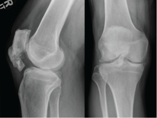
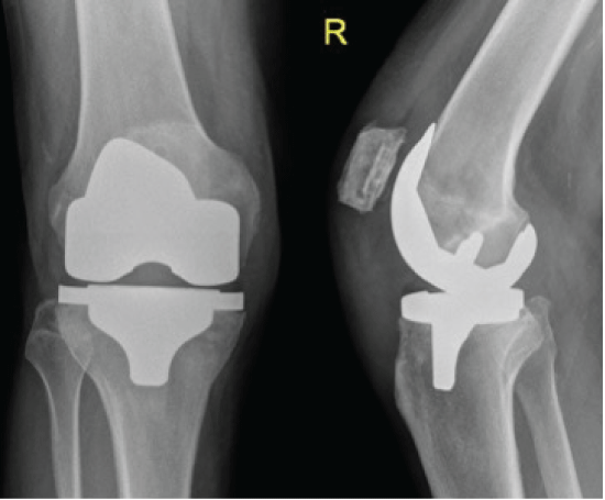
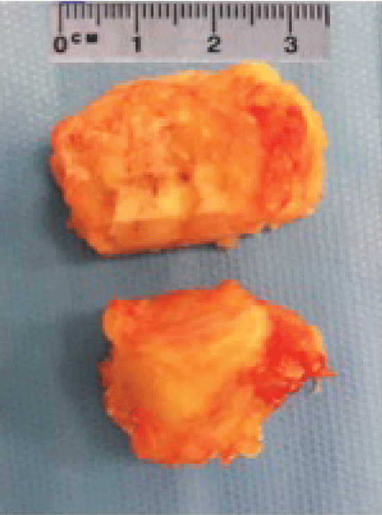
A 79-years-old male who was referred to an elective knee clinic for assessment of his arthritic symptoms in his right knee. He sustained an injury to his right knee at the age of 30 years when he was involved in a car accident. He underwent surgery and was thought to have patellar tendon repair, however no documentation was available regarding the details of surgery. Overall he had regained satisfactory functional recovery after that surgery but reported an intermittent discomfort, difficulty in kneeling down and inability to fully extend the knee afterwards. During the recent years his knee pain gradually worsened. He described medial as well as anterior knee pain. His examination showed a well healed slightly curved transverse scar at the infrapatellar region. He had a palpable and prominent bony lump occupying almost the entire length of the patellar tendon. Movements ranged from 10 to 90 degrees with associated pain medially and anteriorly. Plain radiographs of his knee confirmed the presence of degenerative changes (narrowing of medial joint space, osteophytes, subchondral sclerosis) and a large ossified mass within the patellar tendon (Figure 1). At the time of referral to our clinic, his symptoms were considered to be a combination of degenerative changes and the presence of the large ossified lump. He had exhausted and failed the non-operative treatment options and was offered surgery in view of the severity of his symptoms. He underwent total knee replacement using fixed-bearing cruciate retaining implants along with patellar resurfacing. The surgery was performed in a clean air theatre, using a pneumatic tourniquet through a midline skin incision and medial parapatellar approach. The insertion of the implants was not fraught with any difficulty however the large lump of the ossified patellar tendon led to maltracking of the patella. It occupied the middle two-thirds of the width of the patellar tendon, proximal two-thirds of its height extending from the lower pole of the patella and full thickness in anteroposterior dimensions. Intraoperatively it was decided to excise the ossified mass in its entire dimensions. It was shelled out of the tendon avoiding damage to the peripheral parts of the tendon or its attachments proximally and distally (Figure 2). The tendon was repaired longitudinally using an absorbable multifilament suture using a continuous suture technique. Patellar tracking was found satisfactory after excision of the ossified lump. Postoperatively the knee was mobilised from 0 to 60 degrees using a range of motion brace for a period of six weeks in order to protect the tendon repair, however the weight bearing was not restricted in view of the longitudinal repair of the tendon. The patient was given prophylactic antibiotics and prophylaxis against venous thromboembolism. There were no intra- or post-operative complications. Plain radiographs confirmed a satisfactory excision of the ossified mass and the position of the implants (Figure 3). At six weeks review the patient was allowed free range of motion out of the brace and underwent physiotherapy for quadriceps strengthening. On review at five months he had a pain free range of motion from 5 to 100 degrees and had returned to full level of normal activities including playing golf.
Discussion
Patellar tendon ossification is a rare condition. Different theories have been suggested for development of ossification within the tendon substance. Tamam, et al. hypothesised that persistent lowering of tissue oxygenation tension causes transformation of the tendon in to areas of fibrocartilage in which chondrocytes mediate the deposition of calcium at multiple foci developing in to a bony mass [4]. Sasaki, et al. reported that intratendinous ossification or calcification may be a result of degenerative changes in the collagen [3], however, Richards, et al. did not find any evidence of degenerative or inflammatory changes in their reported study on this subject [5]. In the literature, ossification of the patellar tendon has been reported mainly after knee injuries, as a consequence of patellectomy, after intramedullary nailing of the tibia and after Anterior Cruciate Ligament (ACL) reconstruction using bone-patellar tendon-bone graft [6-10]. Our case was different in a way that the patient underwent total knee replacement, simultaneous excision of the large ossified lump and primary longitudinal repair of the patellar tendon. To our knowledge such a case has not been described in the literature. This was technically challenging in view of the extensive dimensions of the lump, its dissection and excision from the tendon substance and repair of the residual tendon. Restriction of the range of movements using a brace and gradual physiotherapy rehabilitation led to satisfactory outcome and alleviation of patient's symptoms which were a combination of degenerative changes and the presence of a sizable bony lump.
References
- Majeed H, Deall C, Mann A, et al. (2015) Multiple intratendinous ossified deposits of the Achilles tendon: Case report of an unusual pattern of ossification. Foot Ankle Surg 21: e33-e35.
- Cortbaoui C, Matta J, Elkattah R (2013) Could ossification of the achilles tendon have a hereditary component? Case Rep Orthop 2013: 539740.
- Sasaki D, Hatori M, Kotajima S, et al. (2005) Ossification of the Achilles tendon-a case report. Scott Med J 50: 174-175.
- Tamam C, Yildirim D, Tamam M, et al. (2011) Bilateral Achilles tendon ossification: diagnosis with ultrasonography and Single Photon Emission Computed Tomography/Computed Tomography. Case report. Med Ultrason 13: 320-322.
- Richards PJ, Braid JC, Carmont MR, et al. (2008) Achilles tendon ossification: pathology, imaging and aetiology. Disabil Rehabil 30: 1651-1665.
- Bruijn JD, Sanders RJ, Jansen BR (1993) Ossification in the patellar tendon and patella alta following sports injuries in children. Complications of sleeve fractures after conservative treatment. Arch Orthop Trauma Surg 112: 157-158.
- Cakici H, Hapa O, Ozturan K, et al. (2010) Patellar tendon ossification after partial patellectomy: a case report. J Med Case Rep 4: 47.
- Camillieri G, Di Sanzo V, Ferretti M, et al. (2013) Patellar tendon ossification after anterior cruciate ligament reconstruction using bone-patellar tendon-bone autograft. BMC Musculoskelet Disord 14: 164.
- Gosselin RA, Belzer JP, Contreras DM (1993) Heterotopic ossification of the patellar tendon following intramedullary nailing of the tibia: report on two cases. J Trauma 34: 161-163.
- Howell RD, Park JH, Egol KA (2011) Late symptomatic heterotopic ossification of the patellar tendon after medial parapatellar intramedullary nailing of the tibia. Orthopedics 34: 226.
Corresponding Author
Haroon Majeed, MBBS, MRCS, MSc (Tr & Orth), FEBOT, FRCS (Tr & Orth), Trauma and Orthopaedic Surgeon, The Royal Stoke University Hospital, Newcastle Road, Stoke-on-Trent England, ST4 6QB, UK, Tel: +44-7973-824289.
Copyright
© 2017 Majeed H, et al. This is an open-access article distributed under the terms of the Creative Commons Attribution License, which permits unrestricted use, distribution, and reproduction in any medium, provided the original author and source are credited.


