Necrotizing Fasciitis following Laparoscopic Appendicectomy: Case Report of Unpredict Surgical Site Complication
Abstract
Necrotizing fasciitis represents a deep surgical site infection, which compromises the patient muscular tissues and fascia's. Therefore, it manifests with rapidly advancing polymicrobial (gram positive, negative and anaerobic microorganisms) soft tissue infection and secondary necrosis. In most of cases, the patient evolves with systemic inflammatory response and organ dysfunction if resuscitative measures and aggressive surgical debridement could not be expedited. The necrotizing fasciitis is more common after laparotomy for contaminated or infected abdominal procedures. The complication is less frequent during minimally invasive surgery (laparoscopic approach) and there are few reports addressing the issue. So, it is presented a rare case of a patient with necrotizing fasciitis following laparoscopic appendicectomy. The report call attention to the successfully management of this condition, with appropriate surgical intervention, intensive support and antimicrobial treatment.
Keywords
Necrotizing fasciitis, Laparoscopy, Appendicitis, Appendicectomy
Background
Acute appendicitis is one of the most common causes of acute abdomen and one of the most frequently performed surgical emergencies procedures worldwide. Its incidence is approximately 1 case per 1000 inhabitants, generally occurring between the second and third decade of life and indicating a higher prevalence in males [1]. The appendix can be graded as to different levels of appendicitis based upon its visual appearance during laparoscopy (Table 1) [2].
Necrotizing fasciitis (NF) is an uncommon soft tissue infection, usually caused by toxin producing virulent bacteria. It is characterized by wide spread facial necrosis, involving superficial, deep fascia's and subcutaneous adipose tissue with relative sparing of skin and underlying musculature. Often associated with severe and rapidly fatal systemic toxicity, if not promptly recognized and treated [3].
The incidence of postoperative complications for laparoscopic appendicectomy, including wound infection, is significantly less than for the open procedure, because the inflamed appendix is extracted within the trocar and did not touch the abdominal wall [4].
Therefore, since NF is a very rare complication of appendicectomy, especially if performed by laparoscopy, there are many reports of NF after laparotomy appendicectomy and several laparoscopic surgeries, but few studies have reported the occurrence after laparoscopic appendicectomy.
Case Report
A 52-year-old male patient, hypertensive and diabetic, presented with typical symptoms and signs consistent with acute appendicitis, underwent a laparoscopic appendicectomy for grade V appendicitis and were given intravenous antibiotics (500 mg metronidazole and 2 g ceftriaxone). A three port technique was used: An umbilical 12 mm optical port, a 5 mm suprapubic port and a 12 mm optical left iliac fossa (LIF) port for specimen retrieval.
On the first postoperative day, the patient was afebrile and tolerating liquid fluids. By the fifth postoperative day, he was well enough for transfer to the base ward. On the eighth postoperative day the patient was discharged home healthy.
19 days after the procedure, the patient has come back to the hospital with abdominal pain and swelling in the right iliac fossa (RIF), evolving with infection of the surgical site, being treated with drainage and 500 mg amoxicillin and 125 mg clavulanate, with apparent resolution, being discharged home past 4 days.
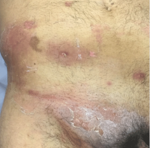
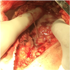
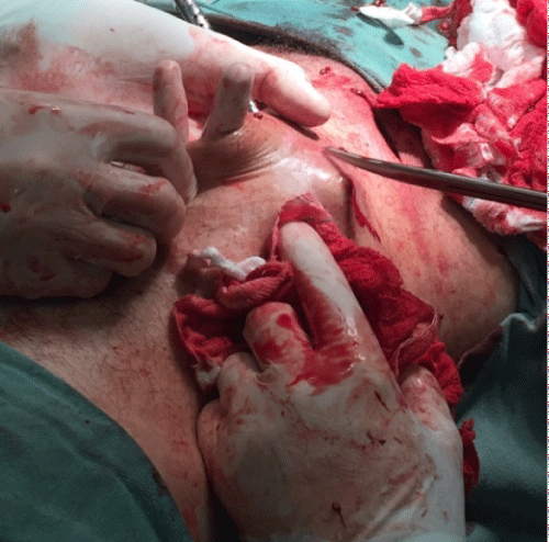
In the late postoperative period (45 days after the procedure) the patient presented severe sepsis and copious purulent drainage at surgical sites, with local edema and erythema (Figure 1). The abdomen was visualized by an exploratory laparotomy and there was no evidence of intra-abdominal sepsis. Findings were consistent with NF with a loss of sheen and texture of the fascia and pus extending along the fascial planes. An abscess drainage and a fasciotomy was performed with extensive debridement of necrotic tissues (Figure 2 and Figure 3) and escharotomy in the abdominal wall (Figure 4). The surgical site was filled with silver sulfadiazine compresses without primary closure.
Patient was referred to intensive care unit where wide spectrum antibiotics (meropenem and vancomycin) were administered following microbiological advice. The wound swabs taken at operation grew Klebsiella pneumoniae, classifying the infection as Type I based on microbiology. It was analyzed the use of hyperbaric oxygen therapy, however, according to this case, there was no need in the treatment. The patient in question presented a significant improvement after the interventions, and was discharged 14 days after fasciotomy.
Outcome and Follow-Up
After discharged home, the patient was reviewed at 2 months at which time the wound was fully epithelialized and he did not present any associated symptoms.
Discussion
NF is an uncommon, rapidly progressive, and highly lethal disease with a poor prognosis unless promptly treated. The incidence of NF in the United States of America (USA) is estimated to be around 500-1500 cases per year [5].
The classic initial presentation of NF includes severe pain that appears disproportionate to physical findings, tenderness, erythema, tense edema, crepitus and cellulitis. It usually presents systemic signs of sepsis as fever with toxic appearance, altered mental state, tachycardia and tachypnea. At the first revisit, there were only common abdominal pain and swelling in the RIF, without another symptom that points to the diagnosis of NF, suggesting a superficial infection [6].
Fournier's gangrene is a variant of NF which involves the genitalia and perineum. Although the redness on the patient right groin, the NF was presented only on the abdominal wall, with sparing of the genital and perineal regions [7].
The spreading speed of the necrosis is an important point to be noticed. The infection can advance as fast as 1 inch per hour with little overlying skin change. Delay in source control for patients with NF has been repeatedly associated with a greater mortality. Therefore, the NF is an emergency and the surgical intervention should be immediately [8].
Even with surgical treatment, NF has a mortality rate in USA of approximately 25%, can even reach higher in some studies [9]. On the other hand, in a specific study in Brazil the mortality general rate was about 20.9% [10]. The main causes of death are septic shock, disseminated intravascular coagulation and/or multiple organ dysfunction syndrome [11]. This case documents the successful management of this condition following laparoscopic appendicectomy.
Laparoscopic appendicectomy for acute appendicitis has been shown to be of benefit reducing postoperative recovery time and complication rate, including wound infections, then open appendicectomy [12].
Laparoscopic appendectomy can be indicated as surgical treatment of appendicitis in such several circumstances (Table 2). Since laparoscopic equipment and skills are available, laparoscopic appendicectomy should be chosen instead laparotomy appendicectomy, offering clear advantages in older patients and patients with comorbidities [13]. In this case, the appendicitis has occurred in a 52-year-old male patient with hypertension and diabetes.
According to microbiology, the Streptococcus species seem to be the most common single causative organism, followed by Staphylococcus aureus, Enterococcus species, Lactobacillus, and Corynebacterium species. Already, the Klebsiella pneumoniae and Escherichia coli were the commonest facultative anaerobic bacteria isolated, followed by anaerobic cocci. However, many cases of NF are polymicrobial with wound cultures yielding a combination of gram-positive cocci, gram-negative rods, and anaerobes [14].
The treatment of patients with NF summarize in early diagnosis and differentiation between necrotizing and non-necrotizing soft-tissue infection, early launch of wide spectrum antibacterial coverage, surgical intervention for abscess drainage and debridement of necrotizing soft tissue infections and identification of the pathogen with adjustment of antimicrobial coverage [15].
Necrotizing soft-tissue infection is one of 12 reimbursable indications for the use of hyperbaric oxygen by The Undersea and Hyperbaric Medical Society. Its physiologic effects at the tissue level include increased killing ability of leukocytes, reducing the tissue edema, killing of certain anaerobes, increasing collagen formation and stimulating the fibroblast growth [16].
The first and most important treatment of NF is surgical, requiring immediate exploration and debridement of all necrotic tissue and fasciotomy. Early diagnosis with appropriate surgical intervention, intensive support and antimicrobial treatment can lead to therapeutic success.
References
-
Bhangu A, SøreideK, Di Saverio S, et al. (2015) Acute appendicitis: Modern understanding of pathogenesis, diagnosis, and management. Lancet 386: 1278-1287.
-
Gomes CA, Nunes TA, Fonseca Chebli JM, et al. (2012) Laparoscopy grading system of acute appendicitis: New insight for future trials. Surg Laparosc Endosc Percutan Tech 22: 463-466.
-
Puvanendran R, Huey JC, Pasupathy S (2009) Necrotizing fasciitis. Can Fam Physician 55: 981-987.
-
B Wei, CL Qi, Chen TF, et al. (2011) Laparoscopic versus open appendectomy for acute appendicitis: A metaanalysis. Surg Endosc 25: 1199-1208.
-
Anaya DA, Dellinger EP (2007) Necrotizing soft-tissue infection: Diagnosis and management. Clin Infect Dis 44: 705-710.
-
Sartelli M, Malagoni MA, May AK, et al. (2014) World Society of Emergency Surgery (WSES) guidelines for management of skin and soft tissue infections. World J Emerg Surg 9: 57.
-
Thwaini A, A Khan, A Malik, et al. (2006) Fournier's gangrene and its emergency management. Postgrad Med J 82: 516-519.
-
Sarani B, Strong M, Pascual J, et al. (2008) Necrotizing fasciitis: Current concepts and review of the literature. J Am Coll Surg 208: 279-288.
-
Shimizu T, Tokuda Y (2010) Necrotizing fasciitis. Intern Med 49: 1051-1057.
-
Wang JM, Lim HK (2014) Necrotizing fasciitis: Eight-year experience and literature review. Braz J Infect Dis 18: 137-143.
-
Mishra SP, Singh S, Gupta SK (2013) Necrotizing soft tissue infections: Surgeon's prospective. Int J Inflam 2013: 609628.
-
Li X, Zhang J, Sang L, et al. (2010) Laparoscopic versus conventional appendectomy - a meta-analysis of randomized controlled trials. BMC Gastroenterol 10: 129.
-
Di Saverio, Birindelli A, Kelly MD, et al. (2016) WSES Jerusalem guidelines for diagnosis and treatment of acute appendicitis. World J Emerg Surg 11: 34.
-
Lancerotto L, Tocco I, Salmaso R, et al. (2012) Necrotizing fasciitis: Classification, diagnosis, and management. J Trauma Acute Care Surg 72: 560-566.
-
Wallace HA, Perera TB (2018) Necrotizing Fasciitis. StatPearls.
-
Green RJ, Dafoe DC, Raffin TA (1996) Necrotizing Fasciitis. Chest 110: 219-229.
Corresponding Author
Carlos Augusto Gomes, MD, PhD, TCBC, Faculdade de Ciências Médicas e da Saúde de Juiz de Fora - Suprema Alameda Salvaterra, Surgery Department, School of Medicine, Hospital Universitário Terezinha de Jesus N° 200, Bairro Salvaterra, CEP 36.033-003 - Juiz de Fora (MG), Brazil.
Copyright
© 2018 Tavares RL, et al. This is an open-access article distributed under the terms of the Creative Commons Attribution License, which permits unrestricted use, distribution, and reproduction in any medium, provided the original author and source are credited.