Postoperative Surgicel Mimicking Pelvic Abscess Causing Mechanical Bowel Obstruction following Abdominal Hysterectomy
Abstract
Oxidized regenerated cellulose (Surgicel), a sterile knitted fabric that causes thrombus formation due to its physical properties, is frequently used for intraoperative hemostasis. Surgicel is bioabsorbable and can be left in the surgical bed. On postoperative CT scans, the appearance of the retained oxidized cellulose can often mimic an abscess leading to dilemma in management. We report the appearance of Surgicel in the pelvis, on postoperative CT scan in a 46-year-old woman who underwent total abdominal hysterectomy for abnormal uterine bleeding. This case stresses the importance of familiarity with common appearance of surgicel on postoperative imaging so that an erroneous diagnosis of an abscess with unnecessary intervention can be avoided.
Introduction
Surgicel (Ethicon, Johnson & Johnson) is a plant based, absorbable, local hemostatic agent composed of oxidized regenerated cellulose. It is locally applied in a variety of surgical procedures where complete hemostasis is difficult to attain. Biodegradation of surgicel usually begins within 24-48 hours and is completed in as early as 7-14 days [1]. There are previous reports where, surgicel gave a picture of postoperative abscess on CT scans causing diagnostic dilemma [2-5]. We present the CT appearance of Surgicel used during abdominal hysterectomy in a woman who developed postoperative abdominal pain and distension due to bowel obstruction. To our knowledge, this is the first case of mechanical bowel obstruction reported following use of surgicel in pelvic surgery.
Case Report
A 46-year-old para 6 with history of one cesarean section was followed up in Gynecology clinic with abnormal uterine bleeding associated with fibroid uterus. She was a known diabetic and hypertensive with past surgical history of excision of presacral schwannoma 8 years before and craniotomy with clipping of left middle cerebral artery bifurcation aneurysm 3 years before. She underwent total abdominal hysterectomy with bilateral salpingo-oophorectomy under general anesthesia as the endometrial sampling showed complex hyperplasia with atypia. Uterus was irregularly enlarged to 16 weeks size, densely adherent to anterior abdominal wall, urinary bladder was pulled up and adherent to anterior surface of uterus. After hysterectomy, a large surgicel (12 × 6 cm) was placed at the bladder base for control of venous oozing from the raw area. Blood loss during surgery was 500 ml. She received prophylactic Inj. Cefuroxime and Metronidazole and Cefuroxime was continued in the postoperative period. Immediate postoperative period was uneventful.
Patient developed abdominal distension with nausea and vomiting on 2nd postoperative day; diagnosed as paralytic ileus and managed with naso gastric tube, I.V fluids and potassium replacement. Even though she was passing flatus and had scanty bowel movement; her abdominal distension, nausea and vomiting was increasing. She also developed low grade fever with mild elevation of C-Reactive protein and White cell count. Urine culture and blood culture were negative.
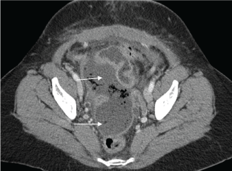
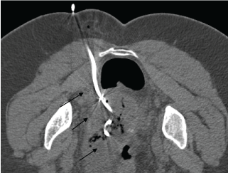
Ultrasound pelvis showed a heterogenous, avascular mass of 8 × 5 cm in the right lower abdomen. Computed Tomography (CT) scan of pelvis on 6th postoperative day showed a well-defined hypodense area with rim enhancement and internal pockets of air, anterior to the rectum extending to the right side of the abdomen (Figure 1). It measured 12 × 6 cm suggestive of collection/abscess. Small bowel loops were dilated with collapsed large bowel. The initial impression was pus collection in the pelvis extending to the right side of abdomen causing mechanical obstruction of ileum. Even though there was a diagnostic confusion of retained surgicel with hematoma mimicking an abscess; in view of her persistent symptoms decided to proceed with aspiration. She underwent CT guided drainage of the pelvic collection with pigtail catheter placement through transgluteal approach (Figure 2). Only 10 ml of serosanguinous fluid was obtained despite the appearance of big hematoma on CT scan. Pigtail catheter was kept in for 48 hours with only minimal drainage. Culture of the aspirate did not show any growth. Histopathology of the surgical specimen showed multiple leiomyoma, adenomyosis, endometrial polyp and complex hyperplasia of endometrium.
Patient's symptoms improved and discharged home on 10th postoperative day. On follow up two weeks after discharge, she was clinically well and ultrasound scan showed complete resolution of the mass.
Discussion
Surgicel is a sterile, bioabsorbable, thrombogenic agent used intraoperatively to assist hemostasis. It has been commonly used in gynecologic and pelvic surgeries; both open and laparoscopic. It is a gauze-like material placed directly onto the tissue to assist in capillary, venous and small arterial bleeding when other conventional methods of hemostasis are not practical or effective [3]. When saturated with blood, Surgicel forms a gelatinous mass which acts as a matrix for fibrin deposition and promote formation of platelet plug helping in hemostasis [4]. During the degradation process, foci of gas are trapped within the gauze resulting in multiple imaging patterns, which can be misinterpreted [4].
On ultrasound examination, surgicel appears as a highly echogenic mass with posterior reverberation artifact, similar to the finding expected in an abscess caused by gas forming organisms [6]. CT findings of retained surgicel is described more frequently as CT is the better diagnostic imaging tool in postoperative patients presenting with abdominal pain or fever. Behbehani and Tulandi [5] had reported the CT appearance of intrapelvic surgicel after gynecologic surgery, but there was no ultrasound correlation in those cases.
On CT scan, retained oxidized cellulose most commonly appears as a collection of gas at the surgical site, imitating the appearance of an abscess [2]. Differentiating features of retained cellulose from an abscess include a unifocal collection of gas with a linear or punctate pattern without any air-fluid level. This pattern of air collection is caused by the air trapped within the spaces of the hemostatic gauze. Postoperative abscess typically shows air-fluid levels and scattered air bubbles [7]. As CT imaging can be often confusing, Magnetic Resonance Imaging (MRI) has been suggested as an alternative diagnostic modality in differentiating between retained oxidized cellulose and an abscess [6].
On MRI, surgicel has a short T2 relaxation time leading to a hypointense mass on T2-weighted images, which might be helpful in differentiating it from an abscess, which is typically T2 hyperintense [8].
Although the literature from Ethicon states that the absorption time of surgicel is 7-14 days, there are cases where, it persisted for 4-8 weeks mimicking an abscess [6,9]. Such patients can be managed conservatively if imaging characteristics support postoperative appearance of surgicel and clinical symptoms improve. This can prevent unnecessary work-up or procedures and prevent additional costs [10].
In our case, patient showed features of paralytic ileus that did not respond to conservative management. Though CT scan was suggestive of a large pelvic abscess causing mechanical bowel obstruction, drainage did not reveal any abscess. Radiologists were not informed about the use of surgicel during surgery, even though it was recorded in the operative notes. It is essential to notify the interpreting radiologist whenever a local hemostatic agent is used during surgery. Knowledge of imaging characteristics in addition to patient's clinical symptoms will help to differentiate between the two conditions.
Conclusion
When oxidized regenerated cellulose (Surgicel) is left in situ for hemostasis, foci of gas trapped within the gauze may mimic an abscess on CT imaging. While using hemostatic agents the smallest amount required should be used. Proper documentation of the use of surgicel in the operative notes and notifying the Radiologist prior to imaging can prevent misinterpretation and unnecessary intervention.
References
- (2012) SURGICEL* Absorbable Hemostat Full Prescribing Information, Ethicon, Inc. SGC-048-12.
- Young ST, Paulson EK, McCann RL, et al. (1993) Appearance of oxidized cellulose (Surgicel) on postoperative CT scans: similarity to postoperative abscess. Am J Roentgenol 160: 275-277.
- Arnold AC, Sodickson A (2008) Postoperative Surgicel mimicking abscesses following cholecystectomy and liver biopsy. Emerg Radiol 15: 183-185.
- Tam T, Harkins G, Dykes T, et al. (2014) Oxidised regenerated cellulose resembling vaginal cuff abscess. JSLS 18: 353-356.
- Behbehani S, Tulandi T (2013) Oxidized regenerated cellulose imitating pelvic abscess. Obstet Gynecol 121: 447-449.
- Melamed JW, Paulson EK, Kliewer MA (1995) Sonographic appearance of oxidized cellulose (Surgicel): pitfall in the diagnosis of postoperative abscess. J Ultrasound Med 14: 27-30.
- Mausner EV, Yitta S, Slywotzky CM, et al. (2011) Commonly encountered foreign bodies and devices in the female pelvis: MDCT appearances. AJR Am J Roentgenol 196: 461-470.
- Oto A, Remer EM, O'Malley CM, et al. (1999) MR characteristics of oxidized cellulose (Surgicel). Am J Roentgenol 172: 1481-1484.
- Ibrahim MF, Aps C, Young CP (2002) A foreign body reaction to Surgicel mimicking an abscess following cardiac surgery. Eur J Cardiothorac Surg 22: 489-490.
- Zhang F, Bondie MJ, Ventrelli SM, et al. (2015) Intraovarian oxidized cellulose (Surgicel) mimicking acute ovarian pathology after recent pelvic surgery. Radiology Case Rep 10: 39-41.
Corresponding Author
Mariam Mathew, Department of Obstetrics & Gynecology, Qaboos University Hospital, Muscat, Oman.
Copyright
© 2018 Mathew M, et al. This is an open-access article distributed under the terms of the Creative Commons Attribution License, which permits unrestricted use, distribution, and reproduction in any medium, provided the original author and source are credited.


