Effects of ELF-EMF Treatment on Depression
Clinical Image
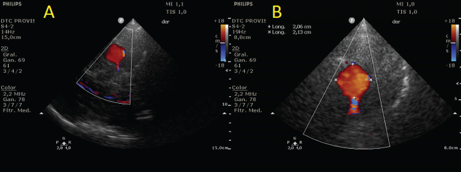
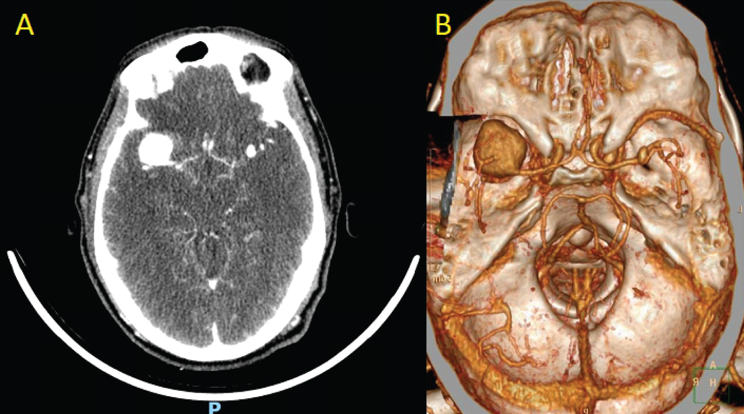
A 50-year-old male with no relevant clinical history presented sudden headache accompanied by nausea, which evolved with facial paresis and drowsiness with a computed tomography (CT) showing bilateral subarachnoid bleeding, being admitted to the intensive care unit. We performed a transcranial doppler ultrasonography (TCD) that showed the presence of a rounded image at M2 portion of the right middle cerebral artery (rMCA) that measured 20 mm × 21 mm with a positive color Doppler signal Figure 1. CT angiography with 3D reconstruction showed an aneurysmal formation of 20 mm in the projection of the rMCA and another small one at the level of the left middle cerebral artery Figure 2. An angiography of intracerebral vessels confirmed a giant aneurysm of 22 mm × 25 mm at the rMCA, requiring stent placement and embolization of the aneurysm with coils. During the hospital stay, he presented vasospasm of the rMCA with a good response to treatment, evolving clinically stable, with subsequent discharge. This case demonstrates that TCD can be useful for the diagnosis of cerebral aneurysms.
Conflicts of Interest
The author declares that he has no conflict of interest.
Corresponding Author
Issac Cheong, Intensive Care Unit, Department of Critical Care Medicine, Sanatorio De los Arcos, Juan B. Justo 909, CABA, Buenos Aires, Argentina.
Copyright
© 2022 Cheong I. This is an open-access article distributed under the terms of the Creative Commons Attribution License, which permits unrestricted use, distribution, and reproduction in any medium, provided the original author and source are credited.


