Cutaneous Metastatic Renal Cell Carcinoma to the Skin Presenting as a Zosteriform Eruption
Abstract
Cutaneous metastasis of internal malignancy is an uncommon but not rare phenomenon. Renal cell carcinoma is a visceral malignancy that rarely metastasizes to the skin and typically presents as a solitary red, purple nodule when it does. We describe a case of metastatic renal cell carcinoma to the skin presenting as a zosteriform eruption. Clinicians should have a low threshold to biopsy atypical lesions or rashes in patients with a history of malignancy and in patients that are immunosuppressed. Dermatologists are in a unique position to detect cutaneous metastasis in this population.
Keywords
Cancer, Renal cell carcinoma, Metastasis, Cutaneous, Herpes zoster
Abbreviations
PET: Positron Emission Test; RCC: Renal Cell Carcinoma; PAX2: Paired-box 2; PAX8: Paired-box 8
Introduction
Cutaneous metastasis of internal malignancy is an uncommon occurrence with an incidence between 0.6% to 10% [1,2]. The most common internal malignancy with cutaneous metastasis in females is breast cancer and in males is lung cancer [3]. A particular malignancy that rarely presents with skin metastasis is renal cell carcinoma. The total incidence of metastasis is 30%, but only 8% of these cases present on the skin [4]. Cutaneous metastasis of renal cell carcinoma typically presents as rapidly growing solitary red-purple nodules on the head and neck region [5-11]. Several morphological appearances have also been reported in literature with clinical findings similar to pyogenic granuloma and abscess [7,12]. We describe the case of a cutaneous metastasis of renal cell carcinoma masquerading as a zosteriform eruption.
Methods
Literature search was performed with PubMed (National Library of Medicine) using key terms including cutaneous metastasis, renal cell carcinoma, herpes zoster, and immunosuppression. Over 300 articles including cases reports and clinical reviews were found and further narrowed by hand-selection based on presenting information.
Case Presentation
A 74-year-old man presented to our dermatology office for evaluation of an erythematous pruritic rash located on the right superior flank that developed three weeks prior. The patient denied any other skin conditions prior to the eruption. His medical history was significant for metastatic renal cell carcinoma. His previous PET scans demonstrated metastasis to the spleen, right lung, abdomen, and axillary and cervical lymph nodes. He had currently finished his third chemotherapy regimen and was scheduled to start nivolumab within the next several weeks.
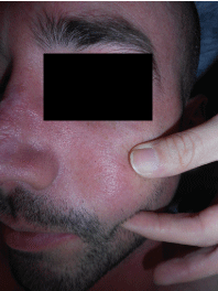
Physical examination revealed a well-developed, well-nourished Caucasian male in no acute distress. Skin findings were significant for several erythematous papules coalescing into plaques and erythematous macules coalescing into patches located on the right superior flank (Figure 1). Based on these findings, the differential consisted of herpes zoster, herpes simplex, drug eruption, and cutaneous metastasis of renal cell carcinoma.
A punch biopsy was performed and taken from the large patch over the right superior flank. Histopathological examination demonstrated mild to moderate irregular epidermal hyperplasia. The papillary and mid-reticular dermis revealed several variably sized islands of cells with ample clear cytoplasm with hemorrhage throughout the islands (Figure 2). The cells demonstrated variably hyperchromatic nuclei as well. Moderate lymphocytic infiltrate was also noted surrounding the tumoral islands. Immunohistochemistry was performed for PAX2, PAX8, and RCC, which were all positive (Figure 3). Viral culture was negative for herpes simplex type 1, herpes simplex type 2, and herpes zoster. These histopathological findings confirmed a diagnosis of cutaneous metastasis of the patient's renal cell carcinoma. The patient's oncologist was notified of the results and the patient was referred for further workup and treatment.
Discussion
Cutaneous metastasis occurs in approximately 0.6% - 10.4% of all patients with cancer and comprises up to 2% of all skin tumors [1,2]. Prevalence of cutaneous metastasis varies based on population. In females, the highest prevalence occurs in breast cancer followed by colorectal cancer. In males, the highest prevalence occurs in lung cancer followed by colorectal cancer [3]. The most common site for cutaneous metastasis is on the trunk although it can occur in any site [9]. In about 20% of cases, cutaneous metastasis is the first sign of an internal malignancy [1]. This highlights the importance of a thorough dermatologic evaluation to rule out malignancy.
To the best of our knowledge, this is the first case of metastatic cutaneous renal cell carcinoma to present as a zosteriform eruption. Zosteriform eruption pattern has been described as a unilateral and belt or girdle-like distribution along a dermatome [13-16]. Renal cell carcinoma comprises a total of 2% of all malignancies and 85% of renal cancers. Renal cell carcinoma occurs more often in males over the age of 60 and has a poor prognosis [17,18]. Risk factors for renal cell carcinoma include smoking, obesity, and hypertension [19]. There are also several autosomal dominant familial disorders which can increase the risk of renal cell carcinoma. One such disorder is von Hippel-Lindau disease, which can present with other dermatologic manifestations such as café au lait spots and vascular malformations on the head and neck [20]. Classically, renal cell carcinoma presents late with a triad of hematuria, flank pain, and an abdominal mass [21].
Over 30% of patients with renal cell carcinoma initially present with metastatic disease with common sites including the lung, soft tissue, and bone. Approximately 8% of metastasis occur in the skin4. These metastases typically present as black or purple solitary nodules [2]. Microscopically, renal cell carcinoma presents with several different histologic features and subtypes. The most common type is termed clear cell carcinoma which demonstrates a high cytoplasmic lipid and glycogen content. This results in the cytoplasm appearing clear after histological processing [20]. Additionally, clear cell carcinoma often presents with prominent hemorrhage and hemosiderin deposition [22]. The differential diagnosis of these tumors includes pyogenic granuloma, Kaposi sarcoma, hemangioma, and other vascular pathologies [2]. Definitive diagnosis of renal cell cancer may require immunohistological evaluation with the use of several specific markers including PAX2, PAX8, and RCC-Ma [23].
Patients with internal malignancy on immunosuppressive therapy can also present with unusual skin findings. This case presented a cutaneous metastasis resembling a zosteriform eruption upon physical examination which raised our suspicion of a possible generalized herpes zoster infection secondary to his immunosuppressed status. Localized herpes zoster is one of several skin infections that can manifest in immunosuppressed patients with an incidence of 14.9% in patients with solid cancers [24]. Typically, these generalized infections will present more severely with widespread distribution lasting several months. Additional dermatologic findings seen in patients with immunosuppressed status include candidiasis, herpes simplex, pyoderma gangrenosum, and erythema nodosum [25].
Conclusions
Although rare, cutaneous metastasis can occur and sometimes appears as the first sign of internal malignancy [1]. We present a rare and unique case of cutaneous metastasis of renal cell carcinoma masquerading as a zosteriform eruption. This case highlights the need for clinicians to have a heightened level of awareness in evaluating patients that are immunosuppressed and/or have a history of malignancy. A low threshold for biopsy of atypical rashes should be employed in these patients as well. Dermatologists are in a unique position to detect metastatic disease in this subset of patients.
References
- Nashan D, Muller ML, Braun-Falco M, et al. (2018) Cutaneous metastasis of visceral tumours: A review. J Cancer Res Clin Oncol 135: 1-14.
- Alcaraz I, Cerroni L, Rutten A, et al. (2012) Cutaneous metastasis from internal malignancies: A clinicopathologic and immunohistochemical review. Am J Dermatopathol 34: 347-393.
- Brownstein MH, Helwig EB (1972) Patterns of cutaneous metastasis. Arch Dermatol 105: 862-868.
- Corgna E, Betti M, Gatta G, et al. (2007) Renal cancer. Crit Rev Oncol Hemat 64: 247-262.
- Kandemir NO, Barut F, Yilmaz K, et al. (2010) Renal cell carcinoma presenting with cutaneous metastasis: A case report. Case Rep Med 2010: 1-5.
- Gottlieb MD, Roland JT (1998) Paradoxical spread of renal cell carcinoma to the head and neck. Laryngoscope 108: 1301-1305.
- Porter NA, Anderson HL, Al-Dujaily S (2006) Renal cell carcinoma presenting as a solitary cutaneous facial metastasis: Case report and review of the literature. Int Semin Surg Oncol 3: 27-31.
- Carlesimo M, Abruzzese C, Narcisi A, et al. (2012) Renal cell carcinoma diagnosed by cutaneous metastasis: A case report. Cutis 90: 196-199.
- Fernandez-Rueda P, Ruiz-Lopez P, Ramierz-Negrin MA, et al. (2015) Cutaneous metastasis of renal cell carcinoma: A case report and review of the literature. Gac Med Mex 151: 497-501.
- Mahmoudi HR, Kamyab K, Daneshpazhooh M (2015) Cutaneous metastasis of renal cell carcinoma: A case report. Dermatol Online J 18: 12-14.
- Pan D, Niall O, Sharma H, et al. (2006) Isolated scalp nodule in patient with undiagnosed RCC. Sci World J 6: 2430-2432.
- Batres E, Knox JM, Wolf JE (1978) Metastatic renal cell carcinoma resembling a pyogenic granuloma. Arch Dermatol 114: 1082-1083.
- Hayderi L, Libon F, Nikkels-Tassoudji N, et al. (2015) Zosteriform dermatoses - A review. Glob Dermatol 2: 163-173.
- Arvin AM (1996) Varicella-zoster virus: Overview and clinical manifestations. Semin Dermatol 15: 4-7.
- Nikkels AF, Pierard GE (2002) Oral antivrials revisited in the treatment of herpes zoster: What do they accomplish? Am J Clin Dermatol 3: 591-598.
- Mueller NH, Gilden DH, Cohrs RJ, et al. (2008) Varicella zoster virus infection: Clinical features, molecular pathogenesis of disease, and latency. Neurol Clin 26: 675-697.
- Motzer RJ, Bander NH, Nanus DM (1996) Renal cell carcinoma. N Engl J Med 335: 865-875.
- Truong LD, Shen SS (2011) Immunohistochemical diagnosis of renal neoplasms. Arch Pathol Lab Med 135: 92-109.
- Cohen HT, McGovern FJ (2005) Renal cell carcinoma. N Engl J Med 353: 2477-2490.
- Ferzli PG, Millett CR, Newman MD, et al. (2008) The dermatologist's guide to hereditary syndromes with renal tumors. Cutis 81: 41-48.
- Rini BL, Campbell SC, Escudier B (2009) Renal cell carcinoma. Lancet 373: 1119-1132.
- Patterson JW (2016) Weedon's Skin Pathology. (4th edn), Churchill Livingston, UK, 1123-1124.
- Perna AG, Ostler DA, Ivan D, et al. (2007) Renal cell carcinoma marker (RCC-Ma) is specific for cutaneous metastasis of renal cell carcinoma. J Cutan Pathol 34: 381-385.
- Yenikomshian MA, Guignard AP, Haguinet F, et al. (2015) The Epidemiology of Herpes Zoster and Its Complications in Medicare Cancer Patients. BMC Infect Dis 15: 106-116.
- Hall, John C (2014) Skin Diseases in the Immunocompromised. (1st edn), Springer, UK, 45-55.
Corresponding Author
Neil F Fernandes, MD, University of Arizona College of Medicine, Phoenix; The Skin and Cancer Center of Arizona, 725 South Dobson Road, Suite 200, Chandler, AZ, 85224, USA, Tel: (480)-899-7546.
Copyright
© 2018 Menon H, et al. This is an open-access article distributed under the terms of the Creative Commons Attribution License, which permits unrestricted use, distribution, and reproduction in any medium, provided the original author and source are credited.