Multiple Myeloma with Skin and Breast Involvement: A Case Report
Abstract
Multiple myeloma; that constitutes 10% of all hematologic malignancies and approximately 1% of whole cancers, is a plasma cell dyscrasia. Extramedullary involvement is often seen in the upper skeletal system, besides it can be seen on the skin and breast, rarely. Extramedullary involvement is generally limited to a single organ. In this article, we present a 70-year-old woman with recurrent skin and breast involvement after multiple treatment with MM diagnosis.
Keywords
Multiple myeloma, Skin, Breast, Extramedullary involvement
Introduction
Multiple Myeloma (MM) is plasma cell malignancy and extramedullary involvement is seen in about 5% of cases [1]. Skin and breast involvement can be seen at the time of diagnosis in MM as well as at the time of treatment or relapse. Never the less, metastatic cutaneous and breast lesions may develop without bone marrow involvement, rarely [2-6]. We aimed to present a very rare case of a seventy-year-old female patient who have multiple myeloma with breast and skin involvement, independently from treatment.
Case Report
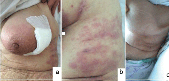
A 70-year-old woman applied with complaints of swelling, redness and pain in her left breast for one month. She pointed out that her complaints could not be relieved by antibiotics. On the dermatologic examination, an erythematous infiltrated plaque, which do not have warmth, on the left nipple and left outer quadrant of left breast and a large number of erythematous nodular lesions on the left midaxillary line were observed (Figure 1a and Figure 1b).
We was informed that she had been treated with melphalan, methylprednisolone and thalidomide (the MPT regime) for one year diagnosed with IgG lambda-type MM since two years and that bortezomib, dexamethasone and lenalidomide (the VRD regime) were administered for one more year, after evaluating as relapse. At the time of diagnosis, it was learned that there was extensive bone involvement but no organ involvement. However, the treatment has been interrupted for three months, because of the remission and the patient has been followed.
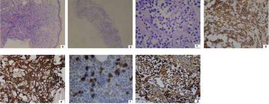
On the tomography examination, there were numerous well-defined hypoechoic heterogeneous opacities in the mammary. The patient was performed a true-cut biopsy from the breast tissue and deep punch biopsy from the skin, simultaneously. In the histopathological examination from the skin; atypical plasma cell infiltration starting from the deep dermis to the subcutaneous tissue were revealed (Figure 2a). Besides, diffuse, exenteric nucleus plasma cell infiltration was seen between normal ducts in the mammary on the histopathological examination of true-cut biopsy from breast (Figure 2b and Figure 2c). Immunohistochemical examination of skin and breast material revealed that plasma cells showed positivity staining with CD38, CD138 and lambda and negativity staining with kappa (Figure 2d, Figure 2e, Figure 2f and Figure 2g). Plasma cell ratio was detected as 1% in the bone marrow biopsy of the patient considered to be relapsed and serum and urine immunfixation electrophoresis studies did not reveal monoclonal gammapathia. It is suggested that she was in full remission for classical myeloma, because determining as hgb: 11 gr/dl, creatinine: 0.4 mg/dl and serum calcium: 8.4 mg/dl. Positron emission tomography determined left femoral head involvement.
In addition to solitary plasmacytoma, we were diagnosed the case as extramedullary MM with skin and breast involvement. Multidrug treatment, which consists of bortezomib, dexamethasone and cyclophosphamide, was started. In the third week of treatment, skin lesions with swelling and redness of the breast were seen to be regressed (Figure 1c). Two months after the end of the treatment, the patient's breast complaints recurred. The patient was ex due to septic shock after pneumonia.
Discussion
Multiple myeloma is a disease characterized by proliferation of malignant plasma cells and abnormal monoclonal protein synthesis called M protein. It constitutes approximately for 10% of hematologic malignancies and 1% of all cancers [7]. The average age of appearance is 65. Plasma cell diseases have four subtypes including; Classical multiple myeloma, Extramedullary Plasmacytomas (EMP), solitary plasmacytoma of bone and plasma cell leukemia [1,3]. Amonsgt diagnosis criteria of the disease are serum/urine monoclonal protein presence, hypercalcemia, renal failure, anemia and bone lesions. The high sedimentation rate, anemia, and albumin/globulin ratio reversal that are detected in laboratory tests help to predict disease [2,3].
In the pathogenesis of extramedullary involvement, it is thought that plasma cells, with reduced expression of adhesion molecules such as VLA-4, CD44 and CD56, form the appropriate microenvironment and are deposited as plasmatomas in tissues [4].
Although extramedullary MM cases [5,6,8-10] with skin or breast involvement are reported in the literature, there is only one case where both skin and breast involvement are seen together [5]. Skin lesions occur in 5-10% of MM cases. The most observed is many erythematous/violose colored nodules, papules and plaques on the body, extremity, and face [3,8]. The skin lesions of the disease are divided into two groups, specific and non-specific. Specific lesions, whether with hematological spread or bone extension, always develop due to plasma cell infiltration; besides nonspecific skin lesions are; amyloidosis, pyoderma gangrenosum, leukocystic vasculitis, necrobiotic xanthogranuloma, ichthyosiform dermatitis and alopecia [8]. Cutaneous/subcutaneous nodules containing atypical plasma cells or diffuse interstitial infiltration are observed in histopathological examination of cutaneous lesions [8-10]. In our case, interstitial atypical plasma cell infiltration was detected in the cutaneous and subcutaneous area. Immunohistochemical examination showed intense positive staining with malignant plasma cell markers CD38, CD138 and lambda.
Immunoglobulin (Ig) G type monoclonal protein is most frequently detected in MM. Similarly, skin and breast involvement are most commonly seen in IgG type MM [9]. IgG type MM was detected in our case in accordance with the literature.
Skin involvement in extramedullary MM is usually detected late in the disease, but rarely, it can be detected at diagnosis, treatment and/or relapse term of the disease [8,10]. MM accompanied by skin involvement was associated with poor prognosis [3]. Metastatic skin involvement without bone involvement is rare in MM [3]. In our case, bone and skin involvement were seen together.
Although EMP frequently shows thoracic involvement, there are few case reports with nodular lesions of the deep or mammary glands [5,6,8,11]. In EMP cases with breast involvement, mammography and tomography findings have been observed nonspecific [5,6]. Examination of the tomography of our case revealed numerous well-defined hypoechoic heterogeneous opacities in the mammary. In EMP, breast involvement is associated with poor prognosis [5,6,11]. Extramedullary MM has no standard treatment regimen.
In our case treatment; although the potent combination chemotherapy bortezomib, dexamethasone, and cyclophosphamide had been applied, patient died in two month period.
As a result, MM patients may manifest with skin and/or organ involvement at baseline. In a particularly elderly patient who admitted to the dermatology polyclinic, MM preliminary diagnosis should be considered if there is a high rate of sedimentation, bone pain and anemia with deeply erythematous nodular and/or plaque-like lesions.
References
- Usmani SZ, Heuck C, Mitchell A, et al. (2012) Extramedullary disease portends poor prognosis in multiple myeloma and is over-represented in high risk disease even in the era of novel agents. Heamatologica 97: 1761-1767.
- Guvenc B, Canataroglu A, Gumurdulu Y, et al. (2001) Multiple Myeloma with skin involvement. J Eur Acad Dermatol Venerol 15: 328-329.
- Reuena L, Kutzner H, Palmedo G, et al. (2003) Cutaneous involvement in multiple myeloma: A clinicopathologic, immunohistochemical and cytogeneticstudy of 8 cases. Arch Dermatol 139: 475-486.
- Blade J, Fernandez de Larrea C, Rosinol L (2012) Extramedullary involvement in multiple myeloma. Haematologica 97: 1618-1619.
- Bouzguenda R, Khanfir A, Toumi N, et al. (2013) Multiple myeloma presenting as bilateral breast lumps in pregnant woman. Int J Hematol 98: 487-490.
- Pasquini E, Rinaldi P, Nicolini M, et al. (2000) Breast involvement in immunolymphoproliferative disorders: report of two cases of multiple myeloma of the breast. Ann Oncol 11: 1353-1359.
- Becker N (2011) Epidemiology of multiple myeloma. Recent Results Cancer Res 183: 25-35.
- Kosari F, Saffar H, Aghaei S (2016) Cutaneous Involvement in Plasma Cell Myeloma: Report of a Case. Acta Med Iran 54: 817-819.
- Bayer-Carner IB, Smoller BR (2003) The spectrum of cutaneous disease in multiple myeloma. J Am Acad Dermatol 48: 497-507.
- Behera B, Pattnaik M, Sahu B, et al. (2016) Cutaneous Manifestations of Multiple Myeloma. Indian J Dermatol 61: 668-671.
- Bauer C, Peigne V, Gisselbrecht (2008) Unusual presentation of myeloma in an elderly woman: breast and cutaneous involvement. M Eur J Intern Med Mar 19: 150-151.
Corresponding Author
Suzan Demir Pektas, M.D, Assistant Professor, Department of Dermatology, Mugla Sitki Kocman University, 48000, Mugla, Turkey, Tel: +90-252-211-5219, Fax: +90-312-3116768.
Copyright
© 2017 Pektas SD, et al. This is an open-access article distributed under the terms of the Creative Commons Attribution License, which permits unrestricted use, distribution, and reproduction in any medium, provided the original author and source are credited.