The History of Cerebrospinal Fluid Analysis in Multiple Sclerosis: A Great Development over the Last Centuries
Abstract
Cerebrospinal Fluid (CSF) analysis acquired a great role in the 1990's when applied to Multiple Sclerosis (MS) diagnostic work-up. The "watery fluid" by Andreas Vesalius remained unstudied till the introduction of Lumbar Puncture (LP) at the eighteenth century. Technical developments exploded in the 1990's: When new applications to the CSF analysis opened to the hypothesis of MS as the model of intrathecal synthesis of immunoglobulins. Over the last decades liquoral findings remained of interest in MS not only for supporting diagnosis but also for their possible role as markers for disease prognosis.
Keywords
Lumbar puncture, History of neurology, Multiple sclerosis
Text
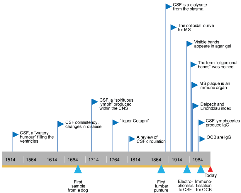
The existence of the Cerebrospinal Fluid (CSF) dated back to Hippocrates (460-375 before Christ, Greece), in relation to the drainage in hydrocephalus. Despite this early report, brain anatomy was described during the sixteenth and seventeenth centuries, including the first descriptions of a "watery humour" and "spirituous lymph" filling the ventricles written by Andreas Vesalius (1514-1564, Belgium), and Emanuel Swedenborg (1688-1772, Sweden) [1]. The lumbar puncture was introduced contemporarily by Walter Wynter (1860-1945, UK) and Heinrich Quincke (1842-1922, Germany), but this techniques remained applied for one century only to the diagnosis of meningitides, and the treatment of hydrocephalus [2,3]. Then, in 1912, William Mestrezat (1883-1928, France) described CSF chemical composition as a dialysate in osmotic equilibrium with plasma [4]. The innovation came with Karl Friedrich Lange (1883-1953, Germany), who studied qualitatively CSF proteins with "colloidal gold" test according to flocculation. The resulting curves were affected by presence of CSF gamma globulin, but not by albumin, alpha or beta globulins, and had a specific shape appearances in neurosyphilis and MS, that were called "dementia paralytica formula" [5]. Thirty years later, Elvin Kabat (1914-2000, USA) applied the electrophoretic techniques to CSF confirming the presence of increased gamma globulins in neurosyphilis and MS, thus opening to the hypothesis of the production of Immunoglobulins (Ig) within the CNS [6]. Furthermore, he established the normal ranges for CSF albumin and Ig [7,8]. Electrophoresis had a great development in the middle of the nineteen century with the agar technique by Denise Karcher. In 1960 her co-worker Armand Lowental (1919-2001, The Netherland) applied electrophoresis to CFS assessing qualitatively abnormal proteins in MS. Increased gamma globulins were divided into fractions with different mobility in agar gel that appeared as visible bands [9,10]. The term Oligoclonal Bands (OCB) was coined by Christian Laterre (1933-1998, Belgium) [11,12]. In 1972, the focusing technique substituted agar gel: Proteins separated according to their isoelectric point and OCB were detected with higher sensitivity [13]. While Lowental was working at electrophoresis, Wallace Tourtellotte (1933, USA) focused on the MS demyelinating plaque in the Central Nervous System (CNS) as an immune organ, able to synthesize oligoclonal Ig type G (IgG) detectable in CSF and not in serum. In 1970 Tourtellotte proposed a formula to measure permeability across the Blood-Brain Barrier (BBB) using the albumin ratio [14,15].
Few years later, investigators tried to determine the range of intrathecal synthesis comparing concentrations of IgG and albumin in CSF and serum. Historically this ratio, or IgG index, was called "Delpech and Lichtblau" index [16]. Despite this index was elevated in the 70% of MS patients, OCB were selected as the most reliable marker for MS by Hans Link (Sweden) in 1987. He also identified OCB in the gamma region as IgG, and recommended to compare bands patterns in CSF and serum to evidence intrathecal synthesis [17]. Moreover, Link recently confirmed the high sensitivity and specificity of OCB determined by isoeletric focusing for MS, detectable in more than 95% of the patients [18]. Support to this finding was given by Magnhild Sandberg-Wollheim (1937, Sweden) who studied in vitro MS CSF lymphocytes, discovering they produced increased IgG that migrated in the gamma region too (as in vivo IgG) with isoelectric focusing [19,20]. CSF analysis detecting intrathecal synthesis become then the gold standard test in suspected MS. A time-line of the historical development of CSF analysis is presented in Figure 1. Nowadays liquoral findings are not straightly recommended to diagnose MS (except for the progressive forms), and CSF gained a role in the search for biomarkers related to disease prognosis. The idea of predicting disability and neurodegeneration, instead of defining the among of inflammation the early phases, pointed out again the role of liquoral analysis. Fox example, neurofilaments' levels, especially of the light subunit, resulted elevated in the progressive forms: This finding suggested a biomarker that could reflect the axonal damage since the time of MS diagnostic work-up [21]. The need of monitoring and predicting MS prognosis allowed CSF to remain a key part of the MS history over time.
In conclusion, the interest in CSF increased rapidly both in clinical practice and in research when LP was introduced to collect samples in humans. Analytical techniques developed rapidly in the twentieth century, and have been immediately applied to CSF analysis. MS diagnostic work-up has been significantly enriched with liquoral indexes and OCB detection. Moreover the extensive research around CSF anticipated, generated, and strengthened the crucial hypothesis of MS as an autoimmune disease mediated by clonally expanded lymphocytes.
Acknowledgement
I would like to thank Professor GG who first suggested me to write this essay few years ago: he continues to be a great teacher at every talk.
Domizia Vecchio has no disclosures for this manuscript.
References
- Hajdu SI (2003) A note from history: Discovery of the cerebrospinal fluid. Ann Clin Lab Sci 33: 334-336.
- Quincke H (1891) Die Lumbalpunction des Hydrocephalus. Berl Klin Wochenschr 28: 929-933.
- Wynter WE (1981) Four cases of tubercular meningitis in which paracentesis of the theca vertebralis was performed for the relief of fluid pressure. Lancet 137: 981-982.
- Mestrezat W (1912) Le liquide céphalo-rachidien normal etiopathologique, valeur clinique de l'examen chimique. Maloine, Paris.
- Lange C, Harris AH (1945) Interpretation of finding in the cerebrospinal fluid. Arch NeurPsych 53: 116-124.
- Kabat EA, Moore DH, Landow H (1942) An electrophoretical study of the protein components in cerebrospinal fluid and their relationships to the serum proteins. J Clin Invest 21: 571-577.
- Kabat EA, Freedman DA (1950) A study of the crystalline albumin, gamma globulin and total protein in the cerebrospinal fluid of 100 cases of multiple sclerosis and in other diseases. Am J Med Sci 219: 55-64.
- Yahr MD, Goldensohn SS, Kabat EA (1954) Further studies on the gamma globulin content of cerebrospinal fluid in multiple sclerosis and other neurological diseases. Annals of the New York Academy of Sciences 58: 613-624.
- Karcher D, Van Sande M, Lowenthal A (1959) Micro-electrophoresis in agar gel of proteins of the cerebrospinal fluid and central nervous system. J Neurochem 4: 135-140.
- Lowenthal A, van Sande M, Karcher D (1960) The differential diagnosis of neurological diseases by fractionating electrophoretically the CSF gamma-globulins. J Neurochem 6: 51-56.
- Holmøy T (2009) The discovery of oligoclonal bands: A 50-year anniversary. Eur Neurol 62: 311-315.
- Sindic CJ, Monteyne P, Laterre EC (1994) The intrathecal synthesis of virus-specific oligoclonal IgG in multiple sclerosis. J Neuroimmunol 54: 75-80.
- Delmotte P (1972) Comparative results of agar electrophoresis and electrofocalization examination of gamma globulins of the cerebrospinal fluid. Acta Neurol Belg 72: 226-234.
- Tourtellotte W, Ma BI (1978) Multiple sclerosis. The blood‐brain‐barrier and the measurement of de novo central nervous system IgG synthesis. Neurology 28: 76-83.
- Tourtellotte W, Potvin AR, Fleming JO, et al. (1980) Multiple sclerosis: Measurement and validation of central nervous system IgG synthesis rate. Neurology 30: 240-244.
- Delpech B, Lichtblau E (1972) Etude quantitative des immunoglobulines G et de l'albumine du liquide cephalo rachidien. Clinica Chimica Acta 37: 15-23.
- Link H (1967) Qualitative changes in immunoglobulin G in multiple sclerosis-cerebrospinal fluid. Acta Neurol Scand 43: 180+.
- Link H (1987) Contribution of CSF studies to diagnosis of multiple sclerosis. Ital J Neurol Sci 6: 57-69.
- Link H, Huang YM (2006) Oligoclonal bands in multiple sclerosis cerebrospinal fluid: An update on methodology and clinical usefulness. J Neuroimmunol 180: 17-28.
- Sandberg-Wollheim M, Zettervall O, Müller R (1969) In vitro synthesis of IgG by cells from the cerebrospinal fluid in a patient with multiple sclerosis. Clin Exp Immunol 4: 401-405.
- Giovannoni G (2006) Multiple sclerosis cerebrospinal fluid biomarkers. Dis Marker 22: 187-196.
Corresponding Author
Domizia Vecchio, MD, Department of Neurology, University of Piemonte Orientale, Corso Mazzini, 18, 28100, Novara, Italy.
Copyright
© 2017 Vecchio D. This is an open-access article distributed under the terms of the Creative Commons Attribution License, which permits unrestricted use, distribution, and reproduction in any medium, provided the original author and source are credited.


