Biochemical Analysis and Antimicrobial Activity of Ethanolic and Aqueous Leaf Extracts of Acalypha wilkesiana var morea Müll.Arg. (Copperleaf)
Abstract
There has been a renewed interest in the use of medicinal plants for the treatment of various ailments in the world. The biochemical composition and antimicrobial effects of crude leaf extracts of Acalypha wilkesiana var morea were studied. The leaves of the plant were collected, dried, pulverized and extracted with ethanol and distilled water. The respective crude extracts were concentrated using a rotary evaporator and biochemical screening was performed using standard methodologies. The antimicrobial sensitivity screening was done using cup diffusion method. The biochemical constituents found in the plant included tannins, flavonoids, phenols, alkaloids, anthocyanins, acids and reducing sugars. Proteins, amino acids and anthraquinones were completely absent. The antimicrobial experiment revealed that the extracts had inhibitory effects. The zones of inhibition for ethanolic extract at concentration 500 mg/ml were: 23.33 ± 0.9 mm (Staphylococcus aureus), 21.33 ± 1.5 mm (Escherichia coli), 26.33 ± 1.9 mm (Salmonella typhi), 16.33 ± 2.2 mm (Aspergillus niger). The antimicrobial sensitivity screening of the aqueous leaf extract had effects only on E. coli at the different concentrations used. The concentration of 500 mg/ml recorded the highest inhibition zone of 24.33 ± 0.7 mm while 62.5 mg/ml had the least, 14 ± 1.2 mm. The extracts will be effective in treatment of Staphylococcus aureus, Salmonella typhi, E coli based infections.
Keywords
Biochemical analysis, Antimicrobial activity, Cup diffusion, Leaf, Extracts, Acalypha wilkesiana
Introduction
Medicinal plants are plants which are used for therapeutic purposes and have enjoyed great popularity in the treatment of various diseases for many centuries. The discovery of these useful plants was as a result of man's inquisitive and inventive nature as well as necessity to feed [1]. Based on their physiological and pharmacological actions and uses, medicinal plants are classified as; Central Nervous System (CNS) active plants, anti-inflammatory agents, anti-allergic plants, anti-diabetic plants, cytoprotective plants, antioxidants as well as antimicrobial plants. A medicinal plant may have multidimensional effects falling into more than one of the classes mentioned above [2]. The use of natural substances as medication by man can be said to be as old as human race [3]. Recently, there had been an increased interest in the use of medicinal plants in developing countries. This is because herbal medicines have been reported safe and without any adverse side effect especially when compared with synthetic drugs [4].
Acalypha wilkesiana (copperleaf) is a plant from the family Euphorbiaceae. The genus Acalypha comprises about 570 species [5], a large proportion of which are weeds while the others are ornamental plants. The plants are found all-over the world especially in the tropics of Africa, America and Asia. The weeds are wild and can be found growing everywhere, while the ornamental species must have been introduced into West Africa from other parts of the world and are cultivated as foliage plants in gardens and greenhouses [6]. Acalypha species are slightly erect, the inflorescence bears about 10 female flowers in the lower part. Acalypha wilkesiana is a popular outdoor plant native to Fiji and nearby islands in the South Pacific, but has spread to most parts of the world, especially the tropics of Africa, America and Asia.
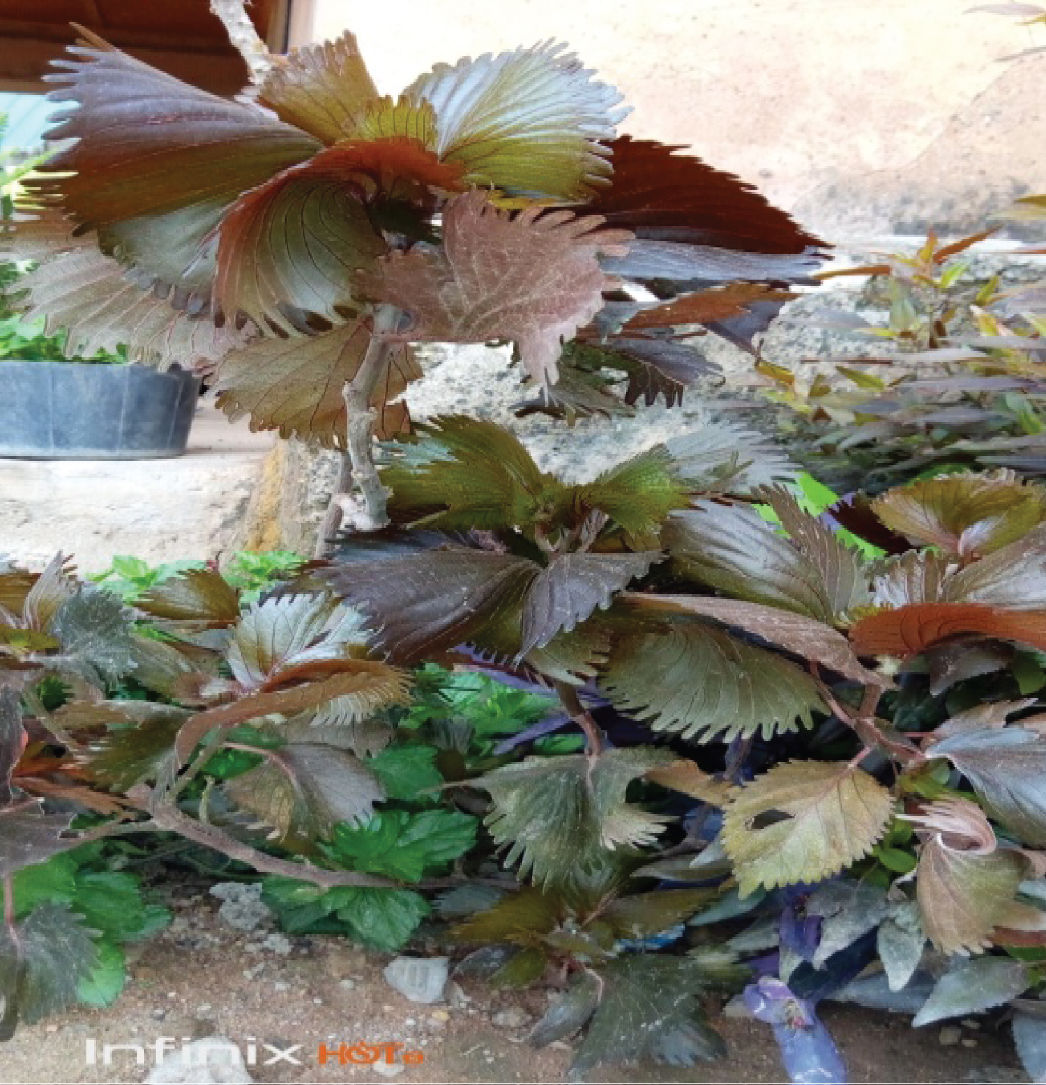
Acalypha wilkesiana is used in West Africa for the treatment of headache and cold and in Nigeria; the cold extract of the leaves is used to bath babies with skin infection [7] (Figure 1).
Acalypha wilkesiana is prominent in the traditional medicinal practice of most tribes in Africa and Asia [1,8,9]. Acalypha wilkesiana Muell Arg leaf has been reported to have medicinal properties for the treatment of malaria, dermatological and gastrointestinal disorders [10]. Some of the species are well known in traditional medicine and a few have actually appeared in the homeopathic pharmacopoeia of India [11]. A. wilkesiana was reported to be used in the treatment of hypertension, especially in managing the abnormal sodium and potassium metabolism that accompany hypertension [12]. The leaf poultice is deemed good for headaches, swellings and colds in Trinidad [13]. The use of Acalypha wilkesiana in the treatment of diabetes and cardiovascular related diseases, spurred investigation by Ikewuchi and Ikewuchi [14] who examined the effect of the plant extract administration on blood sugar and cholesterol levels using a rat model. They reported that the aqueous extract of Acalypha wilkesiana had a lowering effect on blood cholesterol level as well as blood sugar, thereby explaining its use in the treatment of cardiovascular related diseases.
Ogbuehi, et al. [15] investigated the protective effects of A. wilkesiana on biomarkers of oxidative stress in liver homogenates. 70% methanol was used for the extraction of A. wilkesiana leaves and the rats were intraperitoneally administered 50 mg/kg and 100 mg/kg of the extract for 14 days. The results showed significant decreases in malondialdehyde levels in the liver. There was a significant increase in the liver activity of superoxide dismutase and catalase in both the 50 mg/kg and 100 mg/kg administered groups compared to control. There was an insignificant increase in glutathione peroxidase activity in the A. wilkesiana administered groups compared to control and an increase in glutathione levels in liver homogenates of A. wilkesiana administered groups compared to control. The results suggest that A.wilkesiana enhanced the antioxidant capacity of the animals and decreased reactive oxygen species mediated oxidation of lipids.
However, few studies have mentioned the phytochemical constituents and elemental studies of A. wilkesiana. Akinde [11] reported the presence of sesquiterpense, monoterpenes, triterpenoids and polyphenols. Adesina, et al. [7] reported the presence of gallic acid, corilagin, geranin, quercentin, 3-0-rutinoside and Kaempferol in the leaves of A. wilkesiana. The preliminary phytochemical screening of the leaves of A. wilkesiana revealed the presence of alkaloids, carotenoids, flavonoids (catechins and flavones), saponins and tannins all of which have potential health promoting effects, at least under some circumstances [16]. The present study therefore investigated the Biochemical constituents and in-vitro antimicrobial effects of ethanolic and aqueous extracts of A. wilkesiana leaf on some gastrointestinal tract pathogens and bacteria causing skin infections in neonates.
Materials and Methods
Plant collection and identification
The leaf of Acalypha wilkesiana was collected from Federal College of Forestry Jos, Jos North Local Government Area of Plateau State. The specimen was identified in the Herbarium Department of College of Forestry by the Curator Mr. Joseph Azila. The voucher specimen was deposited in the herbarium with the voucher number FHJ 32820.
Preparation and extraction of the plant material
Fresh leaves of the plant material was washed with clean water and dried in a drying cabinet, the hot air oven at a temperature of 35-40 ℃ for 30 mins to remove the excess water on the surfaces of the leaves. The leaves were then dried in the shade for three weeks at room temperature. The dried leaf samples were ground to powder using pestle and mortar. Soxhlet apparatus was used for the extraction of the leaf powder of Acalypha wilkesiana. About 100 g of the sample was weighed on a Mettler weighing balance and dissolved in 70 ml of 70% ethanol. Soxhlet extractor was used for the extraction with duration of 72 hours. The extract was slowly evaporated to dryness using a water bath at 40 ℃. The dried crude residue was collected labeled and store in a refrigerator until required. The same procedure was also used for the aqueous extract.
Collection and preparation of test microorganisms
The test microorganisms were selected based on their availability from infected patients. Hence the bacteria pure cultures were collected from Our Lady of Fatima (OLA) Hospital, Jos and they included Staphylococcus aureus, Salmonella typhi and Escherichia coli. The test fungi included Candida albicans, Aspergillus niger. All the test bacteria were sub-cultured on Nutrient agar, selective and differential media for 24 hours while Candida albicans and Aspergillus niger were sub-cultured on Sabouraud Dextrose Agar for confirmation of their identities using biochemical tests [17].
Biochemical screening
The ethanolic and aqueous leaf extracts of A. wilkesiana were analyzed for Flavonoids, Pholabatannins, Glycosides, Saponins, Lipids, Tannins, Anthraquinones, Coumarins, Terpenoids, Reducing Sugars, Alkaloids, Acids, Phenols, Proteins and Amino acids. The methods of Harborne [18] and Soforowa [1] were employed for the determination of the biochemical composition of the plant extracts.
Determination of tannins
Ferric chloride (2 ml of 5%) was added to 1 ml of each of the plant extract and the formation of dark blue or greenish black indicating the presences of tannins.
Determination of saponins
A volume of 2 ml of distilled H2O was added to 2 ml of each plant extract and shaken in graduated cylinder of 15 minutes length wise. The formation of 1 cm layer of foam indicated the presence of saponins.
Determination of flavonoids
A volume of 1 ml Sodium hydroxide was added to 1 ml of each plant extracts. Yellow color indicates the presence of flavonoids.
Determination of alkaloids
A volume of 2 ml of concentrated hydrochloric acids was added to 2 ml of each plant extracts, then few drops of Mayer's reagent were added. Presence of green colour or white precipitate indicated the presence of alkaloids.
Determination of anthocyanin and betacyanin
One ml of Sodium hydroxide was added to 2 ml each of plant extract and heated for 5 minute at 100 ℃. Formation of bluish green color indicated the presence of anthocyanin and formation of yellow color indicates the presence of betacyanin.
Determination of glycosides
Chloroform (3 ml) and ammonium (10%) was added to 2 ml of plant extract. Formation of pink colour indicated the presence of glycosides.
Determination of phenols
Distilled H2O (2 ml) followed by few drops of 10% ferric chloride was added to 1 ml of the extracts. The formation of blue or green colour indicated the presence of phenols.
Determination of coumarins
A volume of 10% ash (1 ml) was added to 1 ml of the plant extracted formation of yellow colour indicated the presences of coumarins.
Determination of acid
One 1 ml of plant extract was treated with Sodium bicarbonate solution. Formation of effervescences indicated presence of acids.
Determination of protein and amino acids
Ninhydrin test: Few drops of 0.2% ninhydrin were added 2 ml of plant extract and heated for 5 minutes. Formation of blue colour indicated the presence of protein.
Determination of anthraquinones
A volume of 0.5 ml of the extract was boiled with 10 ml of sulphuric acids and filtered while hot. The filtrate was shaken with 5 ml of chloroform. Observe for colour change (gray).
Determination of phlobatannins
Plant powder sample was mixed with distilled H2O in a test tube, then it shaken it well, and filtered to take plant extract. To each plant extract, 1% aqueous hydrochloric (HCL) acid was added and each plant sample was boiled with the help of hot plate and stirred formation of red colour precipitate confirmed presence of Phlobatannins.
Determination of terpenoids
A weight of 0.8 g of selected plant sample was taken in a test tube and then 10 ml of methanol was added to it, shaken well and filtered. A volume of 5 ml of the extract was removed. Then 2 ml of chloroform was mixed with the extract and 3 ml of sulphuric acid was added to it. Formation of reddish brown colour indicated the presence of terpenoids.
Determination of reducing sugar
A weight of 0.5 g of powdered plant sample was added in 5 ml of distilled H2O. Then 1 ml of ethanol was added after which 1 ml of Fehling solution A & B was added in a test tube, heated to boil then was poured in aqueous ethanol. If a reaction was observed, it shows a positive result for reducing sugar.
Preparation and standardization of overnight culture of test bacteria and Candida albicans
The overnight cultures of the test bacteria and C. albicans were prepared by taking a loopful of each of the pure organisms into sterile nutrient and Sabouraud broth in MacCartney bottles. The loop was always flamed before and after each transfer of the microorganisms. The bottles were incubated at 370 ℃ for 24 hours. The serial dilution of this overnight culture was done to obtain a 10-5 dilution (Mac Farland standard) which was used for the sensitivity testing.
Preparation of stock solution and serial dilution of extract
Two gram of the extract was dissolved in 4 ml of sterile distilled water to give a stock solution of (500 mg/ml). A volume of the stock solution was dispensed into a test tube, 2 ml of sterile distilled water aseptically dispensed into the solution to obtain doubling serial dilution of 250 mg/ml. This was serially diluted to get the remaining 125 mg/ml and 62.5 mg/ml respectively. The bottles containing the concentration were labelled properly.
Antimicrobial sensitivity screening of both extracts using agar well diffusion method
The agar well diffusion method Esimone, et al. [19] and Ukwueze [20] were adopted for the antimicrobial sensitivity screening of both ethanolic and aqueous leaf extracts of Acalypha wilkesiana. The Nutrient and Sabouraud Agar media earlier prepared were poured into sterile Petri dishes and they were grouped into duplicate sets for each of the test organism and the control. The plates were allowed to set and were labeled appropriately. A sterile cork borer 3 mm in diameter was used to bore four wells of equidistance around the plates. Using sterile micropipette, 0.1 ml of 10-5 dilution (Macfarland standard) of each of the bacterial test organism and C. albicans was inoculated into each Nutrient and Sabouraud agar plates and was spread through the Petri dishes using sterile swab stick.
A volume of 0.1 ml of each extract concentrations (500, 250, 125, 62.5 mg/ml) was introduced into the holes while 0.1 ml of the standard drug (Gentamycin) 40 mg/ml was introduced into the holes of the plates that served as control. After the introduction of the concentrations, the plates were left on the bench for 1 hour for agar diffusion to take place. The plates were then incubated at 37 ℃ for 24 hours. The experiment was repeated for Aspergillus niger using the spore suspension of the fungus. The plates were incubated at 25 ℃ for 7 days. For C. albicans and A. niger, fluconazole 50 mg/ml was used as control. At the end of the incubation period, zones formed were measured in millimeters (mm) using a transparent ruler. All the tests were carried out in triplicates and their mean values were recorded.
Results
The percentage yield of the aqueous and ethanolic leaf extracts of Acalypha wilkesiana were 10.16% and 12.05% respectively (Table 1). The biochemical screening of both the ethanolic and aqueous leaf extracts of A. wilkesiana revealed the presence of some secondary metabolites which included tannins, flavonoids, anthocyanins/betacyanins, alkaloids, glycosides, Phenols coumarins, acids and reducing sugars. Saponins and phlobatannins were found to be present only in the ethanolic extract. Glycosides and terpenoids were present only in the aqueous extract. Proteins, amino acids and anthraquinones were completely absent in the two extracts. The details of the results are presented in Table 1.
The antimicrobial sensitivity screening of the ethanolic leaf extract of Acalypha wilkesiana revealed that it had significant effects on the test bacteria and the test fungus, Aspergillus niger. Candida albicans was not susceptible to the extract. The ethanolic extract inhibited the gram +ve and the gram –ve test bacteria at the concentration of 500, 250, 125 and 62.5 mg/ml used. Staphylococcus aureus had the highest inhibition zone of 23.33 ± 0.9 mm at 500 mg/ml. This was follows Salmonella typhi with inhibition zone of 26.33 ± 1.9 mm. E. coli recorded inhibition zone of 21.33 ± 1.5 mm. Aspergillus niger recorded the inhibition zone of 16.33 ± 2.2 mm at 500 mg/ml. All the inhibition zones recorded for the test organisms at 500 mg/ml significantly differ with that of the control except for A. niger that the control was higher recording 31.33 ± 0.9 mm for fluconazole. Candida albicans was not susceptible to the ethanolic extract. The details of the results are shown in Table 2.
The antimicrobial sensitivity screening of the aqueous leaf extract had effects only on E. coli at the different concentrations used. The other test organisms were not susceptible to the aqueous extract. The concentration of 500 mg/ml recorded the highest inhibition zone of 24.33 ± 0.7 mm while 62.5 mg/ml had the least, 14 ± 1.2 mm.
Discussion
The plant Acalypha wilkisiana var morea contains several secondary metabolites which account for its use in the treatment of various ailments. The Biochemical analysis revealed that tannins and phenols were present in large quantity in the aqueous extract than ethanolic extract while alkaloids and acids were present in ethanolic extract in higher quantities as compared to the aqueous extract. Saponins and phlobatannins were present only in ethanolic extract while glycosides and terpenoids were present only in aqueous extract. Proteins and amino acids, and anthraquinones were absent in both extracts. Ethanolic extract had more qualitative secondary metabolites when compared with aqueous extract (Table 1). The 70% ethanol used in the extraction of the leaf yielded more of the biochemical components than the aqueous extract used alone. These various secondary metabolites have been reported by various researchers to have medicinal properties. Flavonoids have demonstrated antimicrobial and strong antiviral activities and have been referred to as biological response modifiers because of their ability to modify the body reactions to allergies and other cancer causing substances. Tannins are known for their astringent properties which help to facilitate wounds healings as well as inflamed mucous membrane. They also help to reduce cell proliferation in bacteria. They do this by blocking the main enzymes of microbial metabolism and those that help the building blocks of bacterial cells. Glycosides have been reported to have anti hyperglycaemic activity while reducing sugars have anti hypoglycaemic effects [21-26].
The antimicrobial activity of the ethanolic extract of Acalypha wilkesiana displayed activity against S. aureus (23.33 mm), E. coli (21.33 mm), Salmonella typhi (26.33 mm) and Asperillus niger (16.33 mm) but did not show any inhibition on Candida albicans (0.00 mm) (Table 2). The sensitivity of the test organisms to the ethanolic extract could probably be as a result of the flavonoids, saponins and alkaloids components of the extract. Okerulu and Chinwe [27] reported similar results in their work where flavonoids component of the plant Tetracarpidium conophorum inhibited the growth of Staphylococcus epidermidis, Streptococcus viridans and Escherichia coli. Olaniyi [28] also reported that flavonoids, tannins and saponins also had inhibitory effects on the growth of Bacillus subtilis, Escherichia coli, S. aureus and Candida albicans. Saponins and alkaloids found more in the ethanolic extract have been reported to have antimicrobial properties. It was also observed that the extracts were dose dependent, as the concentration increased the sensitivity also increased and vice versa. This result is similar to previous reports. Iyekowa, et al. [29] carried out a similar research on some selected skin pathogens using Acalypha wilkesiana (Red acalypha) which also agreed with the results of the present study. Inference could be drawn on the plant having effects that were significantly higher than that of the control because the plant extract worked better than the control except for Aspergillus niger which the effect was not higher significantly since the sensitivity of the control was higher than those of the plant extract. Stephen and Sorbari [30] carried out a research using ethanolic leaf extract in creams for the treatment of microbial skin infection, which showed positive results similar to that of the present study. The presence of saponins in the ethanolic extract showed that it has potent effects on fungal skin infections in both adults and neonates. The saponins component could be responsible for the higher antimicrobial activity recorded for the ethanolic extract.
The aqueous extract (Table 3) was only significant against E. coli at all concentration but did not work against S. aureus, Salmonella typhi, Candida albicans and Aspergillus niger at each concentration. This shows that the active principles responsible for the antimicrobial activity of the aqueous extract are not in tannins which were found in large quantities there in. The extracts could be said to have broad spectrum of activity since their effects cut across gram positive and gram negative bacteria.
Previous reports on Acalypha wilkesiana revealed the potency of the plant in treatment of pathogenic infections. Oyelami, et al. [31] carried out a non-comparative study to evaluate the safety and efficiency of Acalypha wilkesiana ointment using 32 Nigerians with mycological infections as well as clinical evidence of mycosis. The ointment successfully controlled the mycosis in 73.3% of the affected patients. He concluded that Acalypha wilkesiana ointment can be used to treat superficial mycoses which are always presented by neonates. Akinyemi et al. [32] evaluated crude extracts from six important medicinal plants, namely Phylantus discoideus, Ageratum conyzoides, Terminalia avicennioides, Bridella ferruginea, Acalypha wilkesiana and Ocimum gratissimum, to find activity against methicillin resistant Staphylococcus aureus (MRSA). Water and ethanolic extracts of these plants were obtained locally and MRSA strains isolated from patients were used. Both ethanol and water extracts showed effects on MRSA with Minimum Inhibitory Concentrations (MIC) and Minimum Bactericidal Concentration (MBC) ranging from 18.2-24.0 μg/ml and 30.4-37.0 μg/ml respectively. This study provided scientific support for the use of Acalypha wilkesiana and other leaves against MRSA. According to Ogundaini [6], the expressed juice or boiled decoction A. wilkesiana is used for the treatment of gastrointestinal disorders and fungal skin infections such as Pityriasis versicolor, Impetigo contagiosa, Candida sp, Tinea versicolor, Tinea corporis and Tinea pedis.
In Southern Nigeria, the leaves of this plant are eaten as vegetables in the management of hypertension [12]. Agu [33] reported that the essential oils distilled from leaves of the plant possess microbiological activities against Staphylococcus aureus and Klebsiella aerogenes.
Seeds from Acalypha wilkesiana are essential components of a complex plant mixture used by traditional healers in Southwest Nigeria in the treatment of breast tumors and inflammation [11]. The large armamentarium of diseases reportedly treated using A. wilkesiana has necessitated scientific inquiry into the biochemical basis of its therapeutic value. Due to the reported use of the plant in the treatment of gastrointestinal disorders, Gotep, et al. [34] carried out in vitro antimicrobial screening using ethanol extracts of A. wilkesiana. They reported from their study that the ethanol extract of the plant had varying antimicrobial activity against Staphylococcus aureus, Yersinia enterocolitica, Escherichia coli, Salmonella typhi, Pseudomonas aeruginosa and Klebsiella aerogenes. Since some of these organisms have been implicated in gastrointestinal diseases and skin diseases, their results provide insight into the acclaimed therapeutic effect of this plant on skin and gastrointestinal related diseases.
The leaf and young shoot are used as vegetables, eaten with rice-dishes and popularly used for the treatment of gastrointestinal disorder and fungi infection particularly impetigo and Tinea versicolor which affects the back, chest and axilla (armpit) of many babies in Nigeria. This study was done using A. wilkesiana leaves growing in school compound in Ibadan South western Nigeria and some bacteria and fungi which had hitherto not been tested in those previous studies were also tested.
Conclusion
The ethanol and aqueous extracts of Acalypha wilkesiana morea has shown antimicrobial properties from the result of the test carried out on the plant. It has shown greatest effect in Staphylococcus aureus and Salmonella typhi and will be effective in treatment of Staphylococcus aureus, Salmonella typhi, E coli based infections. It can also be used in the treatment of aspergillosis caused by Aspergillus spp. The aqueous extract can be only used for the treatment of ailment related to only E coli. However, more work can be done on the effects of the aqueous extract using other bacterial and fungal species.
Acknowledgement
The authors are thankful to the Department of Plant Science and Biotechnology, University of Jos and Department of Environmental Biotechnology and Bio-conservation National Biotechnology Development Agency (NABDA) Abuja, Nigeria for providing the laboratory space and the reagents used for the research work. The authors also acknowledge the contributions and technical assistance of Mr. Thomas Yakubu of Pharmacognosy Laboratory, Faculty of Pharmaceutical Sciences and Mr. Jude Essessien of Chemistry Laboratory, College of Forestry, Jos.
References
- Sofowora A (1993) Medicinal Plants and Traditional Medicines in Africa. Spectrum Books Ltd Ibadan 289.
- Dahanukar SA, Kulkarni RA, Rege NN (2000) Pharmacology of Medicinal Plants and Natural Products. Indian Journal Pharmacol 32: 81-118.
- Personos GJ, Quimby MW (1967) Nigerian Plant III: Phytochemical Screning for Alkaloids, Saponons and Tanins. J Pharmachemical Sciences 56: 1512-1515.
- Lawinson E, Tadmer Y (2007) Biotechnology in medicinal crop improvement In: Handbook of medicinal plants India. Harworth Press 55.
- Riley HP (1963) Families of flowering plants of southern Africa. University of Kentucky Press 73.
- Ogundaini AO (2005) From Green into Medicine: Taking a lead from Nature. An inaugural lecture delivered at Oduduwa Hall,m Obafemi Awolowo 12-15.
- Adesina SK, Idowu O, Ogundaini AO, et al. (2000) Antimicrobial Constituent of the Leaves of A. Wilkesiana and A. hispida Phytotherapy research 14: 371-374.
- Mothana RAA, Abdo SA, Hasson S, et al. (2010) Antimicrobial, antioxidant and cytotoxic activities and phytochemical screening of some Yemeni medicinal plants. Evid Based Complement Altern Med 7: 323-330.
- Duraipandiyan V, Ayyanar M, Ignacimuthu V (2006) Antimicrobial activity of some Ethno medicinal plants used by Paliyar tribe from Tamil Nadu, India. BMC Complement Altern Med 6: 35.
- Akinde BE (1986) Phytochemical and Microbiological evaluation of the oils from the leaves of Acalypha Wilkesiana. In Safowora A. editor. The State Medicinal Plant Reseaarch in Niogeria. University of Ibadan Press 362-363.
- (1971) Homeopathic Pharmacopoiea of India, 1: 33.
- Ikewuchi JC, Anyadiegwu A, Ugono EY, et al. (2008) Effect of Acalypha wilkesiana Muell Arg on Plasma Sodium and Potassium Concentration of Normal Rabbits. Pakistan Journal of Nutrition 7: 130-132.
- Gills LS (1992) Ethno Medical uses of Plants in Nigeria. University of Benin Press 3-8.
- Ikewuchi C, Ikewuchi J (2010) Hypocholesterolcaemic effect of aqueous extract of Acalypha wilkesiana 'Godseffiana' Muell Arg on rats fed egg yolk supplemented diet: Implications for cardiovascular risk management. Research J Science Tech 2: 78-81.
- Ogbuechi I, Adikwu E, Oputiri D (2014) Effect of Acalypha wilkesiana Mulle Arg Leaf Extract on the axidative indices liver enzymes and liver integrity of rats infected with Plasmodium berghei. British Journal of Pharmacology and Toxicology 5: 76-82.
- Basu KS, Thomas JE, Acharya SN (2007) Prospects for Growth in Global Nutraceutical and Functional Food Markets: A Canadian Perspective. Aust J Basic Appl Sci 1: 637-649.
- Cheesbrough M (2003) District Laboratory practice in tropical countries. University of Cambridge Press 194-201.
- Harborne AJ (1993) Phytochemical Methods: A guide to Modern Techniques of Plant Analysis. (1st edn), Chapman and Hall, London, 279.
- Esimone CO, Adujwym MU, Okonta JM (1998) Preliminary antimicrobial screening of the ethanolic extract from the Lichen Usnea subfloridans (L). IJPRD 3: 99-102.
- Osadebe PO, Ukwueze SE (2004) A Comparative Study of the Phytochemical and Anti-microbial Properties of The Eastern Nigerian Species of African Mistletoe (Loranthus micranthus) Sourced from Different Host Trees. Bio-Research 2: 18-23.
- Awosika F (1991) Local Medicinal Plants and Health of Consumers. Clin. Pharm. Herbal Medicine 9: 28-29.
- Tyler VE, Braddy LR, Robets JE (1988) Pharmacognosy. Lea and Febriger 85-90.
- Robbinson T (1967) Organic Constituents of Higher Plants. Burgress Pub 20.
- Frantisck S (1991) The Natural guide to Medicinal Herbs and Plants Tiger Barks Inst. Twickenham UK 1-18.
- Watts JM, Brandwyk BMG (1962) The Medicinal and Poisonous Plants of South Africa. (2nd edn), E and S Livingstone Ltd. Edingburgh, UK 1457.
- Gotep J (2011) Glycosides fraction extracted from fruit pulp of Cucumis metuliferus E. Meyer Has antihyperglycemic effect in rats with alloxan-induced diabetes. J Nat Pharm 2: 48-51.
- Okerulu IO, Chinwe J (2001) The phytochemical analysis and antimicrobial screening of extracts of Tetracarpidium conophorum. J Chem Soc Nig 20: 53-55.
- Olaniyi AA (1998) Basic requirements and strategies for chemical standardization and evaluation of herbal medicines. Herbal Abstracts, Ibadan, Nigeria 11-12.
- Iyekowa O, Oviawe AP, Ndiribe JO (2016) Antimicrobial Activities of Acalypha wilkesiana (Red Acalypha) Extracts on some selected skin pathogens. Zimbabwe Journal of Science & Technology 11: 48-57.
- Stephen OM, Sorbari EN (2014) Formulation of Acalypha Wilkesiana Muell. Arg. Ethanol Leaf Extract into Creams for the Treatment of Microbial Skin Infections. International Journal of Pharmaceutical Science Invention 3: 45-53.
- Oyelami OA, Onayemi O, Oladimeji FA, et al. (2003) Clinical Evaluation of Acalypha Ointment in the Treatment of Superficial Fungal Skin Diseases. Phytotherapy Research 17: 555-557.
- Akinyemi KO, Oladapo O, Okawara CE, et al. (2005) Screening of crude extracts of six medicinal plants used in South-west Nigeria unorthodox medicine for anti-methicillin resistant Staphylococcus aureus activity. BMC Complementary and Alternative Medicine 5: 6.
- Agu SI (1980) Phytochemical Studies on the Volatile Products of some Nigerian Medicinal Plants in Treatment of Diseases. Master of Philosophy Thesis, University of Ife, Nigeria.
- Gotep JG, Agada GOA, Gbise D, et al. (2010) Antibacterial activity of ethanolic extract of Acalypha wilkesiana leaves growing in Jos, Plateau State, Nigeria. MJM 6: 69-74.
Corresponding Author
Ogbonna Abigail Ify, Applied Microbiology and Biotechnology Unit, Department of Plant Science and Biotechnology, University of Jos, Nigeria, Tel: 234-8033555188.
Copyright
© 2021 Ify OA, et al. This is an open-access article distributed under the terms of the Creative Commons Attribution License, which permits unrestricted use, distribution, and reproduction in any medium, provided the original author and source are credited.