Anesthesia Considerations for the Repair of a Left Ventricular to Right Atrial Shunt (Gerbode Defect) Post Double-Valve Repair
Abstract
The Gerbode defect is a rare type of ventricular septal defect (VSD) causing a left ventricle (LV) to right atrium (RA) shunt. These are usually congenital but acquired defects are increasingly being reported. Direct surgical repair remains one of the treatment options and presents considerable anesthetic challenges. We present a case report on anesthesia considerations for acquired Gerbode defect post mitral and tricuspid valve repair. Our case report highlights firstly, the crucial role of anesthetic management in preventing and managing perioperative complications by having a better understanding of physiology and technical aspects of the surgery. Secondly, the importance of high vigilance in a redo cardiac surgery. Thirdly, the utility of intraoperative monitoring using real time transesophageal echocardiography and interpretation of the findings. Fourthly, the pivotal role of close team work to improve patient outcomes, and lastly, post-operative analgesia and intensive care management.
Keywords
Anesthesia management, Gerbode defect, Left ventricular-right atrial communication, Redo cardiac surgery, Transesophageal echocardiography
Introduction
The Gerbode defect is a rare type of ventricular septal defect (VSD) causing a left ventricle (LV) to right atrium (RA) shunt [1]. This left ventricular to right atrial (LV-RA) shunt was first reported by Gerbode in 1958 and hence called as Gerbode defect [1]. These defects are typically congenital but in recent years the numbers of acquired defects have risen considerably due to increased numbers of cardiovascular procedures as well as improved diagnostic capability [2]. Infective endocarditis, previous cardiac surgery, trauma and myocardial infarction are some of the most common predisposing factors [3]. The defect has been classified by Sakakibira and colleagues into: Type I (supravalvular), Type II (infravalvular) and Type III (valvular, or an intermediate between Types I and II) [4]. The incidence of these defects is 76%, 16% and 8% respectively [5]. Type I (supravalvular) is a direct shunt between the left ventricle and right atrium across the atrioventricular portion of the membranous interventricular septum, while Type II (infravalvular) type is a VSD at the ventricular level associated with tricuspid regurgitation, with the high velocity shunt directed into the right atrium. A precise diagnosis and timely treatment of LV-RA shunt, predominantly the acquired type, may lead to better clinical results. We report a case of anesthesia for a patient with acquired Gerbode defect secondary to previous mitral valve and tricuspid valve repair.
Case Report
A 57-year-old gentleman was admitted for progressive dyspnea, orthopnea and peripheral edema. Significantly, he had a known Gerbode defect diagnosed after a mitral valve repair and tricuspid annuloplasty performed three years back for mitral and tricuspid regurgitation. Right and left heart studies post valve repair showed:
• An acquired Gerbode defect with shunting from the left ventricular outflow tract (LVOT) to RA with a Qp (Pulmonary flow):Qs (Systemic flow) ratio of 1.3.
• Step up of oxygen saturations from the mid-RA to right ventricle (RV) was 6-7%.
• Mean pulmonary arterial pressure (MPAP): 40-42 mmHg.
• Pulmonary vascular resistance (PVR): 3.7 Woods.
• Left ventricular end diastolic pressure (LVEDP): 17 mmHg.
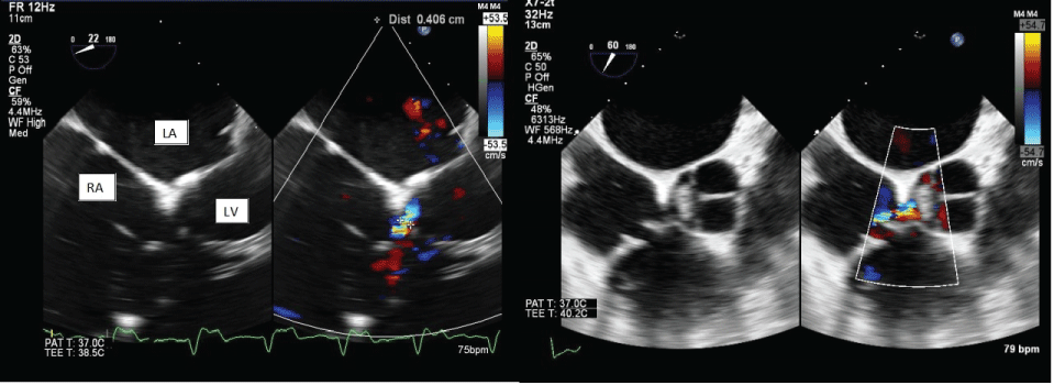
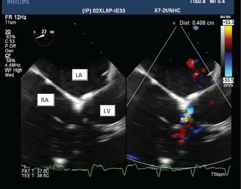
Transesophageal echocardiogram (TEE) showed a Gerbode defect measuring 0.63 cm at the mouth, 0.37 cm at the neck and color flow Doppler demonstrated bidirectional flow between the LV and RA. The RA and RV were both enlarged. Decision was made then to continue with medical management.
Additionally, the patient had a history of percutaneous coronary intervention, sick sinus syndrome with a permanent pacemaker in situ, recurrent strokes and atrial fibrillation on warfarin. He was followed up closely over a period of two years, with two transthoracic echocardiograms a year to monitor defect size, which remained stable.
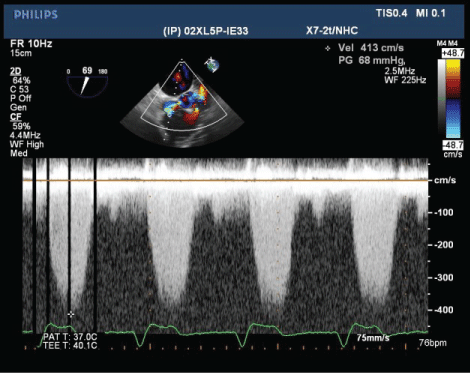
During this current admission intravenous diuresis was commenced and a chest drain was inserted which led to an improvement of symptoms. Repeat right and left heart studies were done, which showed a step up in oxygen saturation from the inferior vena cava (IVC) to the RA of 8% with Qp:Qs ratio of 1.6. TEE showed the defect of 4-6 mm in diameter with continuous flow seen. Systolic maximum velocity (Vmax) across the defect was at least 4.1 m/sec. Percutaneous closure was judged to be difficult as the defect rim was formed by tricuspid valve and hence surgical closure was offered.
An intra-arterial line and an internal jugular central venous catheter were inserted prior to anesthesia induction. Routine monitoring for cardiac surgery, a bispectral index monitor (BIS), bilateral cerebral oximetry and external pads were applied for pacing and defibrillation. The pacemaker was turned off and baseline arterial blood gas and activated clotting time were noted.
Anesthesia was induced with midazolam, etomidate, fentanyl and atracurium and airway secured by intubation. Milrinone and tranexamic acid were administered, and a TEE probe was inserted. Figure 1, Figure 2 and Figure 3 show 2D, color flow and velocity of the shunt.
In-spite of meticulous sternotomy, an inadvertent tear in the right atrium occurred due to dense adhesions from previous sternotomy coupled with a highly pressurized right atrium secondary to the Gerbode shunt. This led to profound hemorrhage and hypotension requiring fluid and blood resuscitation and inotropic support. The patient was heparinized straightaway and established on cardiopulmonary bypass.
Direct repair of the Gerbode defect was performed via the RA with interrupted pledgetted and reinforced mattress sutures. Pressurization of the left sided system did not show any leak of oxygenated blood through the repaired defect. Cardiopulmonary bypass lasted 147 min.
Transition from cardiopulmonary bypass was smooth and easy, terminated on first attempt with low doses of inotropic support. TEE showed disappearance of the previously seen LV-RA shunt.
Fresh frozen plasma, platelets, cryoprecipitate and pump blood were transfused. Further analgesia was with bolus morphine. The patient was then transferred sedated and ventilated to the cardiothoracic intensive care unit.
The patient was initially extubated on the first postoperative day. However, he developed respiratory failure from fluid overload secondary to acute kidney injury and required reintubation the next day. He was subsequently commenced on continuous renal replacement therapy, and successfully extubated on postoperative day 6. Recovery was otherwise uneventful and he was discharged home on postoperative day 20.
Discussion and Conclusion
The pathophysiology of the Gerbode defect is such that shunting occurs from the LV to the RA due to the large pressure gradient that exists between these cardiac chambers [6]. Shunting of blood into the RA leads to subsequent increased flow into the RV, leading to enlargement of the right heart chambers, pulmonary hypertension and eventually right heart failure. In a large shunt, the high pulmonary blood flow can also lead to volume overload of the left heart, resulting in dilatation, arrhythmias and impairment of function [6].
Yuan and colleagues performed a systematic review of 237 acquired LV to RA shunts in 234 patients and reported the underlying etiologies as iatrogenic in 51.1%, infective in 36.7%, traumatic in 9.3% and ischemic in 3% of patients [2]. The iatrogenic shunts were most commonly caused by valvular operations (56.6%) with sole mitral and aortic valve surgeries responsible for the majority of these shunts [2]. Of particular importance, however, as described below in our case report, is combined tricuspid annuloplasty ring insertion and mitral valve replacement as this may lead to damage to the membranous septum caused by excessive debridement of annular calcium [6]. Rarely, this complication can occur in a tricuspid annuloplasty in the absence of a left sided valve operation [7].
A definite diagnosis of acquired Gerbode defect prior to death or operation was obtained in 70% of patients by transesophageal, transthoracic echocardiography or cardiac catheterization [2]. Differential diagnoses include ruptured sinus of Valsalva aneurysm into the right atrium, paravalvular leak, aorta to right atrium shunt, tricuspid insufficiency or tricuspid insufficiency with pulmonary hypertension [2]. Dzwonczyk and colleagues suggest that color flow imaging in the parasternal short-axis, apical short-axis and subcostal projections are useful in eliminating the other possibilities and in confirming the diagnosis [8]. In addition, a disturbed color signal in the RA not originating from the tricuspid valve and with a velocity of 4.0 m/s or more should suggest a LV-RA communication [8].
Of the patients with an iatrogenic shunt, the majority experience symptoms such as dyspnea, or have a murmur [2]. These murmurs are typically pansystolic, are loud and harsh, and may be associated with a thrill along the left sternal border [6]. Percutaneous closure has been described with iatrogenic shunts, however, surgery is still the mainstay of treatment in 52% of patients with a minority of patients with small shunts treated conservatively [2].
As operative techniques evolve and survival after cardiac operations improves, the number of patients who have repeat sternotomy inevitably continues to increase [9]. Intraoperative adverse events (injury to bypass grafts, heart chambers, great vessels, life threatening arrhythmias and severe lung injury) occur in 7% of cardiac re-operations [9]. Kamdar and colleagues recommend that patients for cardiac re-operations should be assessed preoperatively using computed tomography (CT) with volume rendered and multiplanar reconstruction 3-dimensional techniques [10]. Planned alternative routes of mechanical circulatory support, standardized opening and dissection techniques together with a pre-induction operative team 'huddle' optimizes the efficiency of the team in event of an intraoperative adverse occurrence [9]. In our patient, close communication between the surgeons and anesthetists and prompt action at the time of right atrial injury minimized an adverse outcome.
Goals of anesthesia for a patient with Gerbode defect involve preventing a change in the shunt flow and maintaining cardiovascular and respiratory stability for adequate tissue perfusion and oxygenation. Increases in pulmonary vascular resistance should be prevented with close attention to avoiding airway obstruction, hypoxia, hypoventilation, acidosis and hypothermia. On the other hand, significant decreases in pulmonary vascular resistance will increase shunt flow and should be avoided as well. Inotropes such as milrinone and inhaled nitric oxide upon weaning from bypass may be considered in those with significant pulmonary hypertension, in conjunction with a carefully balanced anesthetic technique to maintain Qp:Qs ratio. In patients with complications such as pneumonia from prolonged hospitalization as a result of congestive cardiac failure from the defect, treatment and optimization should be commenced before elective surgical repair.
The acquired Gerbode defect is a rare and complex defect which may present as a diagnostic challenge in a patient presenting with progressive dyspnea after surgery, trauma or infective endocarditis. In patients with this defect post cardiac surgery, dense adhesions from a previous sternotomy pose an additional challenge for the surgical and anesthetic teams. These patients should preferably be managed in experienced centers with the expertise and capability in dealing with the potential diagnostic challenges and complications.
In conclusion, our case reports successful anesthetic management of surgical repair for an iatrogenic Gerbode defect. Prudence during the perioperative period and intraoperative assessment and confirmation of successful repair by TEE is necessary.
Competing Interests
No external funding and no competing interests declared by the authors.
References
- Gerbode F, Hultgren H, Melrose D, et al. (1958) Syndrome of left ventricular-right atrial shunt; successful surgical repair of defect in five cases with observation of bradycardia on closure. Ann Surg 148: 433-446.
- Yuan SM (2015) A systematic review of acquired left ventricle to right atrium shunts (Gerbode Defects). Hellenic J Cardiol 56: 357-372.
- Demirkol S, Gurkan YF, Bozlar U, et al. (2013) Multimodality imaging of a congenital Gerbode defect. Kardio Pol 71: 104.
- Sakakibara S, Konno S (1963) Left ventricular-right atrial communication. Ann Surg 158: 93-99.
- Sinisalo JP, Sreeram N, Jokinen E, et al. (2011) Acquired left ventricular-right atrium shunts. Eur J Cardiothorac Surg 39: 500-506.
- Saker E, Bahri GN, Montalbano MJ, et al. (2017) Gerbode defect: A comprehensive review of its history, anatomy, embryology, pathophysiology, diagnosis, and treatment. J Saudi Heart Assoc 29: 283-292.
- Dadkhah R, Friart A, Leclerc JL, et al. (2009) Uncommon acquired Gerbode defect (left ventricular to right atrial communication) following a tricuspid annuloplasty without concomitant mitral surgery. Eur J Echocardiogr 10: 579-581.
- Dzwonczyk T, Davidson WR (2009) The spectrum of left ventricular-right atrial communications in the adult: Essentials of echocardiographic assessment. Journal of the American Society of Echocardiography 8: 263-269.
- Roselli EE (2011) Reoperative cardiac surgery: Challenges and outcomes. Tex Heart Inst J 38: 669-671.
- Kamdar AR, Meadows TA, Roselli EE, et al. (2008) Multidetector computed tomographic angiography in planning of reoperative cardiothoracic surgery. Ann Thorac Surg 85: 1239-1245.
Corresponding Author
Dr. Shah Shitalkumar S, MBBS, DNB, FCARSCI, MRCA, FAMS, Consultant, Anesthesiology, Singapore General Hospital, Singapore.
Copyright
© 2018 Koh LY, et al. This is an open-access article distributed under the terms of the Creative Commons Attribution License, which permits unrestricted use, distribution, and reproduction in any medium, provided the original author and source are credited.