Prolonged Cardiac Arrest and Localized Thoracic Spinal Cord Ischemia
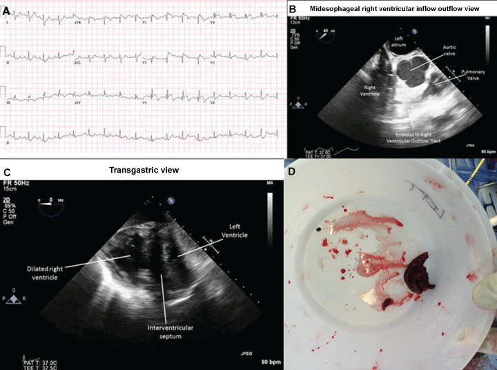
A 29-year-old man presented to the emergency with a sudden onset of shortness of breath and syncope. Electrocardiogram showed a right bundle branch block, ST elevation in leads V1-V4 (Figure 1A). A transthoracic echocardiography showed an embolus in transit at the inferior vena cava and right atrial junction. He had two episodes of Pulseless Electrical Activity (PEA), lasting 2 and 62 minutes. ACLS protocol [1] was followed and LUCAS device was used to provide chest compressions. Multiple epinephrine [2] and atropine boluses were administered in addition to tissue plasminogen activator. With no signs of circulation and resuscitation chart review, a single dose of vasopressin (40 IU) intravenously was administered by anesthesia team [3,4]. A Return of Spontaneous Circulation (ROSC) was observed. Later patient underwent a sternotomy and pulmonary embolectomy on cardiopulmonary bypass in the operating room. Transesophageal echocardiography also showed an echogenic material in the RVOT (Figure 1C) in addition to dilated right ventricle (Figure 1D) and flattened interventricular septum. Embolic material was retrieved from the Right Ventricular Outflow Tract (RVOT) and bilateral distal pulmonary arteries (Figure1B).
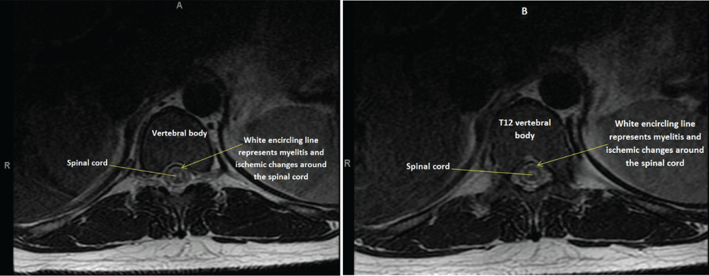
Patient was transferred to the ICU and hypothermia protocol was used. He was extubated on day three and a neurological exam revealed bilateral lower extremity motor deficit. An MRI of the spine showed an abnormal increased T2, STIR signal in the central gray matter of the spinal cord from T9 to T12 vertebral level (Figure 2A and Figure 2B). Short TI Inversion Recovery (STIR) is a global and homogenous method to null the signal from fat, to distinguish the two tissue components. Increased STIR signal reflects a prolonged ischemic episode. After three months of rehabilitation, patient was seen walking with a cane and currently undergoing physiotherapy until today. This is a rare case of survival after an hour of resuscitation. Powerful chest compressions were helpful and administering vasopressin was instrumental in achieving ROSC. However, motor paralysis and localized spinal cord changes were unexpected which resolved over period of few months.
References
- American Heart Association (2005) Part 7.2: Management of Cardiac Arrest. Circulation 112: 58-66.
- Wenzel V, Krismer AC, Arntz HR, et al. (2004) A comparison of vasopressin and epinephrine for out-of -hospital cardiopulmonary resuscitation. N Engl J Med 350: 105-113.
- Gueugniaud PY, David JS, Chanzy E, et al. (2008) Vasopressin and epinephrine vs. Epinephrine alone in cardiopulmonary resuscitation. N Engl J Med 359: 21-30.
- Brandon O (2011) Is Vasopressin indicated in the Management of Cardiac Arrest? Clinical Correlation.
Corresponding Author
Sridhar Musuku, Department of Anesthesiology, Albany Medical Center, 43 New Scotland avenue, Albany 12208, USA, Tel: 518-262-4300, Fax: 518-262-4736.
Copyright
© 2017 Musuku S. This is an open-access article distributed under the terms of the Creative Commons Attribution License, which permits unrestricted use, distribution, and reproduction in any medium, provided the original author and source are credited.


