Parathyroid Carcinoma Management in an Underdeveloped Country: A Case Series
Abstract
Parathyroid carcinoma (PC) is a rare type of cancer that presents in individuals during their 4th or 5th decade of life; most cases are sporadic and of unknown etiology [1]. Approximately 1-2% of cases of hyperparathyroidism are caused by PC; with the average serum calcium levels higher in patients with PC than parathyroid adenomas (15.9 vs 12 mg/dL). Clinical diagnosis is often difficult and almost always invariably obtained after postoperative histopathological confirmation; making this disease rare and likely under diagnosed. Para fibromin immunostaining is currently the gold standard for definitive diagnosis; albeit its availability may be limited in resource-poor settings. We report a case series of five PCs who were diagnosed with primary hyperparathyroidism (pHPT) and underwent parathyroidectomy in a 10-year period. All of the patients in this case series presented post-surgery hungry bone syndrome (HBS). Efforts should be taken to establish more reliable and precise diagnostic tools to establish concrete guidelines for the management of PCs.
Keywords
Parathyroid carcinoma, Parathyroidectomy, Hyperparathyroidism, Hypercalcemia
Introduction
Parathyroid Carcinoma (PC) is an exceedingly rare malignancy and its prognosis is highly variable [2]. It has an incidence of approximately 0.5-5% in patients with primary hyperparathyroidism (PHPT), as parathyroid adenoma is the most common cause of hyperthyroidism in 87% -91% of all cases [3]. The 5-year survival rate ranges from 20% -85% [4]. Despite low mortality, PC is accompanied by serious clinical manifestations, in contrast with its benign counterpart parathyroid adenoma [4-5]. Most of these tumors are sporadic, although patients with Hyperparathyroidism-jaw tumor syndrome (HPT-JT), and multiple endocrine neoplasia (MEN) types 1 and 2A, have a higher risk of developing PC, ranging from 15% to 37.5% in different case series [2].
Parathyroid Carcinoma is a rare type of cancer that could present in individuals during their 4th or 5th decade of life. The clinical diagnosis of PC is often difficult and is almost always obtained only after a postoperative histopathological examination [1]. The real incidence of PC is difficult to establish, due to variable diagnostic criteria. Its histological features are not specific, as they have also been found in adenomas or even local recurrences of benign tumors [6] For this reason, the tumor's malignancy must be unequivocally confirmed only after prolonged follow-up, bearing in mind both histological (capsular and vascular invasion) and clinical features (invasive appearance, recurrences, distal metastasis) [1,6]. Parafibromin immunostaining is currently the gold standard for definitive diagnosis. We report a case series of five PC, which presented with malignancies such as co-synchronous Papillary Thyroid Carcinoma in one of the cases and double adenomas in another, who were surgically treated by the same surgeon. The American Association of Endocrine Surgeons guidelines recommend patients with functional PC should undergo regular surveillance by testing serum calcium and PTH levels; however timeframe for follow-up is not specified due to rarity of this under diagnosed malignancy and drive by expert opinion [7]. Furthermore, there are no available guidelines in Dominican
Republic for follow up of Parathyroid Carcinoma - especially at 5-10-years periods, the primary surgeon in conjunction with the Endocrine team had at least 1-10 years follow-up period, with serum calcium, PTH control and ultrasound imaging.
Methods
A consecutive case series study was done in a single, tertiary-level, university-based, referral center with a retrospective data collection. The criteria of selection of these cases included adult (age >18-years) patients who underwent surgery having a diagnosis of pHPT. The data was extracted from the patients in the Department of Endocrine Surgery of CEDIMAT during study period January 2012 until December 2020. The exclusion criteria omitted patients diagnosed with primary hyperparathyroidism at the expense of benign parathyroid disease or that did not meet the pathological criteria for PC. During this period of time a total of 69 parathyroidectomies were performed in this center. A total of 5 of these cases out of the 69 were found to have Parathyroid Carcinoma (Table 1).
Consequently, this study was carried out in compliance with international ethical regulations, including the relevant aspects of the Declaration of Helsinki, and the guidelines of the Council for International Organizations of Medical Sciences (CIOMS). The study protocol and the instruments designed were approved by CEDIMAT's Institutional Review Board (IRB); each patient, additionally, provided informed written consent.
Preoperative laboratory assessments included a PTH test, measurement of calcium serum and phosphorus, alkaline phosphatase, 24h urine calcium, glomerular filtration rate (GFR), and bone densitometry. Neck ultrasound and gammagraphy studies were done to verify the location and existence of an abnormal parathyroid gland. All patients underwent thyroid pathology screening before undergoing surgery.
Preoperative PTH measurements were taken before resection, and repeated 10 minutes intra-operatively after resection of the tumor in all patients, confirming in all cases a descent of more than 50%. Calcium levels were measured 24 hours after the surgery as part of standardized protocol follow-up for downtrend of levels back to normal. The extracted gland was then sent for frozen histopathology and immunohistochemistry studies.
In these case series, all diagnosis confirmations were histopathological and validated by immunohistochemistry studies. Diagnostic pathology was confirmed by two pathologists concurrence. One of them is from our center (CEDIMAT), the other one from a Molecular Oncopathology Laboratory in the U.S., validating the diagnosis via immunochemistry studies. The markers used to confirm the diagnosis in this study included Pan Cytokeratin cocktail (PCK), Antibody against PTH (PT Hab), K1-67, Proliferation marker, Thyroglobulin, and Calcitonin. These are the normal ranges in our institution taken in consideration for laboratory values:
Case series
Case -1
Clinical Presentation: A 54-year-old Hispanic female, with a past medical history (PMH) of arterial hypertension (HTA), chronic kidney disease with nephrolithiasis, type -2 diabetes mellitus (T2DM), Peripheral Vascular Disease (PAD), and osteoporosis, presented to surgical consultation for primary hyperparathyroidism initially diagnosed in the setting of osteoarthralgia.
Biochemical workup: Total serum calcium levels were elevated with 13.4 mg/dL, BUN of 75.2 mg/dL, creatinine of 3.81 mg/dL, high phosphorus levels (5.0 mg/dL), as well as an elevated intact PTH (2,182 pg/mL) and a 24h calcium in urine 91.3mg/day (Table A and Table B and Table C).
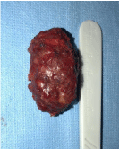
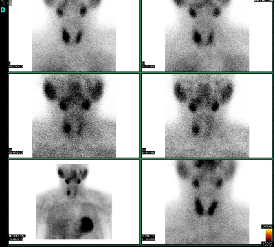
Radiological workup: Thyroid/Parathyroid ultrasound and gammagraphy with technetium 99m sestamibi revealed increased parathyroid gland size and uptake, respectively, in the inferior right pole (Figure 1).
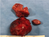
Surgical details: During the surgery, the affected lower right parathyroid gland was identified, displaying malignant macroscopic features. The affected gland was adhered to the carotid sheath, as well as its surrounding tissues. The 5 cm tumor infiltrated the retro-clavicular and retrosternal structures which protruded into the thorax and was attached to the right lower lobe of the thymus. A decision was made on-site to perform en-bloc parathyroidectomy along with ipsilateral hemithyroidectomy and dissection and emptying of zone lV and Vl, due to suspected malignancy.
Histopathology/Immunochemistry findings: Cytologic atypia, dystrophic calcification was found including capsular and vascular invasion. Immunohistochemical studies results showed positive PTH and PCK antibodies.
Immediate post-surgical issues: Temporary dysphonia as well as severe hypocalcaemia (6 mg/dL) due to HBS, 6 days post-op.
Long-term follow-up: A year after the procedure, the patient presented at follow-up with no laboratory results abnormalities was detected.
Additional data: Figure 1
Case -2
Clinical Presentation: A 62-year-old Hispanic male, with a known PMH of acute kidney injury and depressed mood.
Biochemical workup: Elevated serum calcium levels were found with 13.9 mg/dL, and elevated intact PTH levels (160.9 pg/mL) (Table A and Table B and Table C).
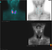
Radiological workup: Gammagraphy with technetium 99m sestamibi demonstrated an increased uptake activity adjacent to the left lower pole (Figure 2).
Surgical details: En-bloc resection of the affected parathyroid gland was conducted, along with ipsilateral hemithyroidectomy with lymph node dissection due to malignant suspicion during surgery.
Histopathology/Immunochemistry findings: Cytologic atypia as well as capsular and vascular invasion were found. Immunohistochemical studies results showed positive PTH and PCK antibodies, positive thyroglobulin, and high proliferation index of KI-67, consistent with a diagnosis of PC.
Immediate post surgical issues: Patient presented to outpatient clinic on day 7 post op for numbness. His labs were consistent with HBS.
Long term follow-up: By 11-months, follow-up laboratory work-up showed normal serum calcium levels at 9 mg/dL. At 8-year follow-up, no laboratory results abnormalities were detected.
Additional data: Figure 2
Case- 3
Clinical Presentation: A 72-year old Hispanic female, with PMH of hypertension, arthritis and osteoporosis presented to surgical consultation due to a previous diagnosis of secondary hyperparathyroidism.
Biochemical workup: Preoperative laboratory test results revealed high calcium levels (12.1 mg/dL), elevated intact PTH levels (4,038 pg/mL), BUN of 75.2 mg/dL, creatinine of 3.81 mg/dL, and high phosphorus levels (3.5 mg/dL) (Table A and Table B and Table C).
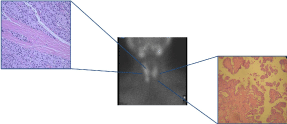
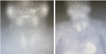
Radiological workup: An ultrasound and gammagraphy revealed an abnormal lower right parathyroid gland and suspicious thyroid nodules (Figure 3A).
Surgical details: Fine needle aspiration (FNA) of a thyroid nodule revealed synchronous papillary carcinoma of the thyroid. Patient underwent en-bloc resection of the affected parathyroid gland, along with a total thyroidectomy and dissection and emptying of zone lV and Vl was performed. Neither loco-regional nor distant metastasis was found during the surgery.
Histopathology/Immunochemistry findings: Cytologic atypia and ischemia. FNA revealed synchronous papillary carcinoma of the thyroid of category V Bethesda (Figure 3B).
Immediate post surgical issues: HBS.
Long term follow-up: A 6 month follow-up post-operative, repeat serum calcium levels were 9.2 mg/dL. No laboratory results abnormalities were detected.
Additional data: Figure 3A and Figure 3B.
Case -4
Clinical Presentation: A 58 year-old hispanic female with PMH of osteoporosis, depression and renal lithiasis, presented to consultation with lethargy. An initial diagnosis of primary hyperparathyroidism was made prior.
Biochemical workup: Preoperative laboratory test results revealed high calcium levels (12.3 mg/dL), elevated intact PTH levels (179 pg/mL), BUN of 75.2 mg/dL, creatinine of 3.81 mg/dL, and high phosphorus levels (3.5 mg/dL) (Table A and Table B and Table C).
Radiological workup: Gammagraphy assessment demonstrated increased uptake activity adjacent to both inferior poles (Figure 4).
Surgical details: An inferior bilateral en-bloc resection of the parathyroid gland was conducted. After biopsy findings revealed PC, reintervention with hemithyroidectomy and dissection and emptying of zone lV and Vl was performed.
Histopathology/Immunochemistry findings: Cytologic atypia as well as capsular and vascular invasion was found in one of the affected parathyroid glands. Immunohistochemical studies results showed positive PTH and pan-cytokeratin antibodies and high proliferation index of KI-67, consistent with a diagnosis of PC.
Immediate post surgical issues: HBS
Long term follow-up: A 6 year follow-up after the procedure, the patient presented with serum calcium levels of 9.0 mg/dL. No laboratory results abnormalities were detected.
Additional data: Figure 4
Case -5
Clinical Presentation: A 51-year-old male hispanic patient with PMH hypertension and stroke, presented to the emergency department with lethargy. He was found to have a relevant recent history of fatigue, tremors, new-onset seizures, bone pain, abdominal pain, asthenia, and severe hypercalcemia.
Biochemical workup: Elevated calcium levels at 19 mg/dL, creatinine 3.11 mg/dL, phosphorus 2.3 mg/dL, and intact PTH 2,946 pg/mL, and alkaline phosphatase 136.3ng/mL (Table A and Table B and Table C).
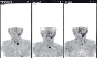
Radiological workup: An ultrasound and gammagraphy showed an abnormal right inferior parathyroid gland (Figure 5).
Surgical details: A decision was made on-site to also conduct an en-bloc parathyroidectomy with an ipsilateral hemithyroidectomy, along with dissection and emptying of zone lV and Vl, due to suspected malignancy.
Histopathology/Immunochemistry findings: Cytologic atypia with extensive areas of ischemia and hemorrhage, solid trabecular tumor proliferation, and mitotic figures consistent with a diagnosis of carcinoma. Immunohistochemistry studies were positive only for PTH antibodies, which are typically seen in parathyroid adenomas. However, parafibromin, which is the most specific marker for PC, was not measured due to unavailability of the test.
Immediate post surgical issues: HBS
Long term follow-up: At 1 year follow-up, the patient demonstrated persistent increased PTH levels (290 pg/ml).
Additional data: Figure 5A and Figure 5B
Discussion
The most common symptoms among patients in our case series with this condition were as follows; bone pain, osteopenia, depressed mood, lethargy, kidney lesions, nephrolithiasis, and/or osteoporosis, consistent with severe hypercalcemia signs and symptoms presented in other studies regarding PC presentation [1]. According to recent studies, the average serum calcium level in patients with PC is higher (15.9 mg/dL) than that reported in patients with parathyroid adenomas (12 mg/dL) [6]. However, 10 % of patients with PC have serum calcium of 13 mg/dL or less, which was observed in this case series, as mean laboratory values showed pre-operatory serum calcium of 13.5 pg/dL, with an average pre-operatory PTH of 1,529 pg/mL.
Calcium was not corrected in our case series as the formula is only routinely used to transform the calcium results in those patients who had hypoalbuminemia, so that the distribution of results would match the distribution of calcium results in the patients with normal serum albumin concentrations [8]. Although the elevation of PTH and calcium levels is not sufficient to make a diagnosis of PC, it could be suggestive of a malignant lesion since, in very rare cases, a benign lesion manifests with such extreme elevations of PTH levels. Due to its ambiguous clinical-histopathological presentation, the diagnosis is seldom made pre-operative, as patients may only present elevated calcium and PTH levels [3-4].
In the context of surgical exploration, adds a valuable asset to the determination of the diagnosis and the localization of the tumor, this is accompanied by neck ultrasound, 4D computed tomography, sestamibi scintigraphy (MIBI), and venous selective sampling [3]. In our case series, a neck ultrasound, as well as gammagraphy, was conducted with Technetium-99 m methoxy-isobutyl-isonitrile to confirm the existence and location of the tumors. These imaging tests are conducted to confirm pHPT when suspected, however, they do not lead to a definitive diagnosis of PC, as increased Tc-99m MIBI uptake by the affected gland can be due to an adenoma or thyroid pathology [9]. Fine needle aspiration is usually not recommended due to disruption of the neoplastic capsule with subsequent neoplastic spread. Key findings of PC, such as capsular and vascular invasion, are not easily identifiable with an FNA sample, and FNA cytology of parathyroid tissue can mimic a thyroid lesion [10].
Referral to Endocrine Surgery should be made after pHPT is diagnosed. The case is then studied and screened for abnormal parathyroid behavior with serum calcium levels, PTH, vitamin D, and further imaging studies. Parathyroidectomy of the abnormal gland is performed and sent for biopsy. In this case series, after PC was diagnosed by histopathology, the patients were re-intervened by the surgeon for an ipsilateral thyroidectomy and excision of any adjacent structures involved, to provide the best opportunity for better local disease control and significantly improved long term survival. Efforts should be made not to rupture the capsule of the tumor and spill tumor cells in the field. In this case series, all diagnoses were made by histopathological findings and validated by immunohistochemistry studies.
One of the patients underwent reintervention to complete the ipsilateral hemithyroidectomy and dissection of neck lymph nodes. In three other cases, there was suspicion of malignancy during the surgery due to macroscopic infiltration of the capsule and the ipsilateral lobule, therefore a decision was made on-site to perform en-bloc parathyroidectomy with ipsilateral hemithyroidectomy. In the one remaining case a total thyroidectomy was performed due to a suspicious thyroid nodule.
All of the patients in this case series presented post-surgery HBS. HBS usually reflects rapid mineralization after correction of hyperparathyroidism, causing severe and prolonged hypocalcaemia. It's a rare complication, however, it's often seen after a parathyroidectomy due to hyperparathyroidism, and it's relatively common after removal of a PC [11]. According to recent literature, the more severe the bone disease before surgery, the more prone the patient is to HBS after surgery [12]. Despite being an unfavorable outcome, the HBS state suggests that surgical removal of hypersecretory parathyroid tissue was accomplished [12]. In this study, HBS was observed in all of the patients, 4 of whom presented severe bone disease before surgery. Notably, all patients presented with relevant history of Chronic Kidney Disease. To the authors current knowledge, there are no have been no studies looking at association between Chronic Kidney Disease and development of Parathyroid Carcinoma (or vice-versa). PTH and Calcium values remained normal at 1-year follow-up in all cases.
Conclusion
Due to its low incidence, PC is considered an extremely rare tumor that continues to present many challenges in diagnosis, resulting in a more difficult presentation to treat. Despite this pathology being extremely rare, 5 cases were reported in this center in the past 10-years, out of 69 patients that underwent parathyroidectomy, for a rough estimate of 7.2% of all cases.
Diagnosis may be suspected when there is an extreme elevation of PTH and serum calcium levels, but can only be confirmed after histopathological and immunohistochemistry study after the tumor is resected. Due to the later years seemingly increased frequency of PC and in the absence of a gold standard test, a multidisciplinary approach, considering all clinical, biochemical, and structural aspects of the disease, offers the best chance for accurate diagnosis.
HBS should be expected in patients with severe hyperparathyroidism due to PC after correction with surgical resection of the affected gland. Efforts should be taken to establish more reliable and precise diagnostic tools to establish concrete guidelines for the management of PC.
References
- Takenobu M, Moritani S, Kawamoto K, et al. (2020) Parathyroid carcinoma coexisting with multiple parathyroid adenomas: A case report. Endocr J 67: 963-967.
- Sandelin K (2001) Parathyroid carcinoma. In: Holzheimer RG, Mannick JA, Surgical treatment: evidence-based and problem-oriented. Munich: Zuckschwerdt.
- Smith JC (2021) Parathyroidectomy .overview, technique, periprocedural care. Medscapes.
- Rotolo N, Cattoni M, Imperatori A (2017) Complications from tracheal resection for thyroid carcinoma. Gland Surg 6: 574-578.
- Sturniolo G, Gagliano E, Tonante A, et al. (2013) Parathyroid carcinoma: Case report. G Chir 34: 170-172.
- Fernandes J, Paiva C, Correia R, et al. (2018) Parathyroid carcinoma: From a case report to a review of the literature. IJSCR 42: 214-217.
- Ohe MN, Santos RO, Hojaij F, et al. (2013) Parathyroid carcinoma and hungry bone syndrome. Arq Bras Endocrinol Metabol 57: 79-86.
- Rathi MS, Ajjan R, Orme SM (2008) A case of parathyroid carcinoma with severe hungry bone syndrome and review of literature. Exp Clin Endocrinol Diabetes. 116: 487-490.
- Kitapçi MT, Tastekin G, Turgut M, et al. (1993) Preoperative localization of parathyroid carcinoma using Tc-99m MIBI. Clin Nucl Med 18: 217-219.
- Simons J (2019) Correcting the myth of calcium correction, This changed my practice. The University of British Columbia.
- Wilhelm SM, Wang TS, Ruan DT, et al. (2016) The American association of endocrine surgeons guidelines for definitive management of primary hyperparathyroidism. JAMA Surg 151: 959-968.
- Sriphrapradang C, Sornmayura P, Chanplakorn N, et al. (2014) Fine-needle aspiration cytology of parathyroid carcinoma mimic Hürthle cell thyroid neoplasm. Case Rep Endocrinol 2014: 680876.
Corresponding Author
Sylvia Batista, Centros de Diagnóstico y Medicina Avanzada y de Conferencias Médicas y Telemedicina (CEDIMAT), Dominican Republic.
Copyright
© 2022 Cabrera-Luis J. This is an open-access article distributed under the terms of the Creative Commons Attribution License, which permits unrestricted use, distribution, and reproduction in any medium, provided the original author and source are credited.