Renal Pelvis Leiomyoma: A Novel Percutaneous Approach and Literature Revision
Abstract
Renal pelvis leiomyomas are very rare benign neoplasms that derive from smooth muscle cells. These tumors occur in vessels and in the urothelial mucosa of urinary tract [1]. This report describes a 50-year-old woman with a contrast enhanced lesion in the right renal pelvis, measuring 20 × 23 × 25 mm, which was found incidentally during an abdominal-pelvic CT scan. Histopathologic analysis of a cold biopsy revealed a chronic inflammatory process without dysplasia. The mass was resected percutaneously; it was diagnosed as a leiomyoma. Only 13 cases of ureteral leiomyoma have been reported to date; to our knowledge, this is the first such tumor resected using a percutaneous approach [2].
Keywords
Kidney-sparing surgery, Ureteral leiomyoma, Percutaneous approach
Introduction
Ureteral leiomyomas are very rare benign neoplasms that usually present with symptoms of ureteral obstruction [3]. Since first described in 1955 [4], only 13 cases of leiomyoma of the ureter have been reported to date. Advances in ureteroscopy have enabled more conservative treatments of these tumors, allowing preservation of the kidney [3]. This report describes a patient with a renal pelvis leiomyoma who underwent kidney-sparing surgery using a percutaneous approach.
Case Report
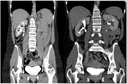
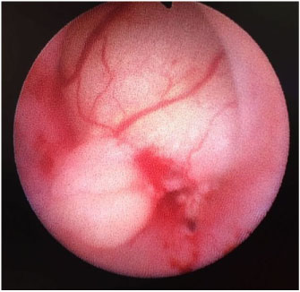
A 50-year-old woman was referred to the Urology Department for an incidentally discovered mass in the renal pelvis during an abdominal-pelvic CT scan for diverticulitis. The patient originally presented with a non-specific auto inflammatory syndrome, which had been treated with immunosuppressants. She did not experience low back pain or episodes of hematuria. An abdominal-pelvic CT scan revealed a contrast-enhanced lesion in right renal pelvis, measuring 20 × 23 × 25 mm and compatible with a pelvic tumor (Figure 1). A selective urinary cytology from right kidney showed inflammatory cells mixed with reactive urothelial cells with atypia. A semi-rigid diagnostic ureteroscopy showed a rounded yellowish solid lesion, 20 mm in diameter, with well-defined borders and a wide base of implantation (Figure 2). Histopathologic examination of a cold biopsy sample showed evidence of a chronic inflammatory process with Von Brunn's nests cells without urothelial dysplasia. The mass was resected percutaneously and diagnosed histologically as a renal pelvis leiomyoma. At last follow-up, nine months after surgery, there was no evidence of tumor recurrence (Figure 1).
Discussion
Renal pelvis leiomyomas are very rare benign neoplasms that derive from smooth muscle cells.
These tumors occur in vessels and in the urothelial mucosa of urinary tract. They are usually found incidentally during imaging for other indications, although they may also present with pain in the flank and, less frequently, macroscopic hematuria. Thirteen such tumors have been reported to date, six cases were female and eight cases were male. No difference in renal side was observed, since seven cases were on the left side and other seven cases on the right side.
The highest age incidence is at fourth decade, varying from 4 to 60-years-old, like our case. In the previous cases published, six of the thirteen cases were in the upper ureter, like our case, and five of these cases were treated with nephroureterectomy. Four cases were at middle ureter, three partial ureterectomies and one nephroureterectomy. Finally, four cases were located at lower ureter, all treated with distal ureterectomy [2]. Imaging does not allow differentiating from malignancy. Recent developments in ureteroscopy enable a proper diagnosis in benign tumors [5]. Exact mechanisms of development of ureteral leiomyoma are unclear, chronic inflammation or trauma are some of the hypothesis [2]. The patient originally presented with a non-specific auto inflammatory syndrome, which had been treated with immunosuppressants, we don't know if this condition play some role in the pathogenesis of this case.
Surgical treatment consists normally of nephroureterectomy or distal ureterectomy (Table 1). We decided to perform a kidney-sparing surgery after long explanation of risks and benefits with the patient, and influenced by the fact that we had a biopsy showing no malignant cells. We think that it is a difficult case for ureteroscopy management due to its large size and its wide base tumor. For this reason we decided to perform a percutaneous approach [6-15].
Conclusion
Renal pelvis leiomyomas is very rare disease often managed with radical surgical treatment. Due to its benign condition, kidney-sparing surgery could be offered. Here in, we present the first case reported in the literature treated by percutaneous approach with good results.
Acknowledgements
I would like to express my very great appreciation to all contributors to this communication, especially to Dr. Pieras.
References
- Brunocilla E, Pultrone CV, Schiavina R, et al. (2012) Renal leiomyoma: Case report and literature review. Can Urol Assoc J 6: E87-E90.
- Zehri AA, Ali A, Iqbal F, et al. (2013) Primary leiomyoma - a rare tumour of ureter. J Pak Med Assoc 63: 268-270.
- Englund M, McCredie S, Robson S (2017) Leiomyoma of the ureter. ANZ J Surg 87: E92-E93.
- Leighton KM (1955) Leiomyoma of the ureter. Br J Urol 27: 256-257.
- Yashi M, Hashimoto S, Muraishi O, et al. (2000) Leyomioma of the ureter. Urol Int 64: 40-42.
- Kao VC, Graff PW, Rappaport H (1969) Leiomyoma of the ureter. A histologically problematic rare tumour confirmed by immunohistochemical studies. Cancer 24: 535-542.
- Mondschein LJ, Sutton AP, Rothfeld SH (1976) Leiomyoma of the ureter in a child: The first reported case. J Urol 116: 516-518.
- Sekar N, Nagrani B, Yadav RV (1980) Ureterocele with leiomyoma of ureter. Br J Urol 52: 400.
- Zaitoon MM (1986) Leiomyoma of ureter. Urology 28: 50-51.
- Cussenot O, Teillac P, Billebaud T, et al. (1989) Leiomyoma of the urinary excretory tract. Ann Urol (Paris) 23: 305-308.
- Igarashi OSH, Nakada J, Nishida A, et al. (1994) A case of Leiomyoma of the ureter. Jpn J Clin Urol 48: 328-330.
- Ikota H, Tanimoto A, Komatsu H, et al. (2004) Ureteral leiomyoma causing hydronephrosis in Type 1 multiple endocrine neoplasia. Pathol Int 54: 457-459.
- Naruse K, Yamada Y, Aoki S, et al. (2007) A case of primary leiomyoma of the ureter. Int J Urol 14: 248-250.
- Shailesh AS, Ranka P, Dodiya S, et al. (2004) Leiomyoma of ureter - a rare cause of intractable hematuria and clot retention. Indian J of Urol 20: 181-182.
- Nouralizadeh A, Tabibi A, Mahmoudnejad N, et al. (2010) Partial ureterectomy for a huge primary leiomyoma of the ureter. J Pak Med Assoc 60: 62-64.
Corresponding Author
Luis Ladaria Sureda, Urology Department, C/Valldemossa 79, Hospital Universitari Son Espases, Palma de Mallorca, Spain, Tel: +34-871205008.
Copyright
© 2019 Ladaria SL, et al. This is an open-access article distributed under the terms of the Creative Commons Attribution License, which permits unrestricted use, distribution, and reproduction in any medium, provided the original author and source are credited.