

Screening of Abnormalities of External Genitalia in Adolescents: About 240 Cases Observed in Public Secondary School of Brazzaville
Abstract
Introduction : Abnomalities of the external genitalia can impact sexual and testicular function; therefore, a delay in their management can, in some cases, lead to fertility problems. Therefore, we conducted this study, the aim of which was to screen for external genitalia abnormalities in adolescents at public secondary school of Brazzaville.
Patients and methods: This was a prospective, descriptive, and cross-sectional study conducted over a 10-month period. Data collection took place in four public general education middle schools, using random cluster sampling, with parental consent. We studied epidemiological and clinical parameters. Data processing was performed using EPI INFO software version 7.2.4.0 and Excel. The Pearson chi-square test was performed with a significance level of 0.05.
Results: among 768 adolescents examined, 240 presented at least one abnormality of external genitalia (31.2%). Scrotal pain was found in 42.3% of cases, followed by scrotal discomfort (37.5%) and inguinal and/or scrotal swelling (19.4%). The 240 middle school students presented 291 EG abnormalities, and the most common were varicocele (35.7%), inguinal hernia (23.4%), and retractile testicle (14.1%). The main reasons for lack of treatment were ignorance and fear of surgery (40.3% and 21%, respectively).
Conclusion: abnormalities of external genitalia are common among adolescents in school settings, dominated by testicular pathologies, particularly varicocele, which carries risks of impaired reproductive function.
Keywords
Penis, Testicles, Hernia, Varicocele, Adolescent, Brazzaville
Introduction
Abnomalities of the external genitalia (AEG) in boys represent a group of congenital or acquired conditions affecting the penis, scrotum, testicle, and its adnexa [1]. The incidence of external genitalia malformations (hypospadias, ectopic testis) with perfectly differentiated sex is 1/200 to 1/300 births [2]. They can have long-term consequences on sexuality and/or reproduction, and consequently on fertility, as well as psychological repercussions, hence the need for early management [3]. However, the observation of late hospital consultations and the absence of school medicine in our setting led us to conduct this study, the aim of which was to screen for abnormalities of the external genitalia in adolescents in school.
Patients and Methods
This was a cross-sectional, observational study with prospective data collection. It took place from January 1st to November 1st, 2021, a period of ten (10) months in four public secondary school of Brazzaville. It involved male adolescents, aged between 10 and 19 years, who agreed to be examined and whose parents had given their consent. This was a two-stage cluster sampling with simple random selection without replacement. The sample size was calculated using the Schwartz formula and took into account the cluster effect. The sample consisted of 768 individuals. The clinical examination of the respondents was performed in the presence of a parent and under the supervision of a senior pediatric surgeon or urology specialist. The variables studied were:
- epidemiological: Age, parental education level, socioeconomic status
- diagnostic: Functional and physical signs, pathologies.
We also investigated the causes of the lack of care.
Data analysis was performed using EPI INFO software version 7.2.4.0 and Excel. The Pearson chi-square test was applied with a significance threshold of 0.05.
Results
Epidemiological Aspects
Of the 768 adolescents examined, 240 presented with at least one abnormality of the external genitalia, representing a prevalence of 31.2%. The mean age was 14.2 years ± 1.9 (range: 11 to 19 years).
The distribution of adolescents with AEG by school establishment is in the (Table 1).
The majority of parents had a secondary education (Table 2).
The adolescents came from families with a low socioeconomic status in 83 cases (34.6%), a medium one in 142 cases (59.2%), and a high one in 15 cases (6.2%).
Diagnostic Aspects
Pubertal development
Adolescents with AGE were pubertal in 113 cases (47.1%), prepubertal in 77 cases (32.1%), and 50 cases (20.8%) had adult morphology.
Signs
Sixty-two adolescents (25.8%) were previously aware of their condition, while 178% were unaware.
We noted intermittent inguinal and/or scrotal pain in 42.3% of cases (Table 3).
Pathologies
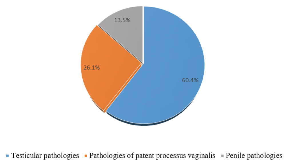
The 240 middle school students presented with 291 congenital and acquired external genitalia anomalies. We observed 11 types of AEG, and the three main pathologies were the varicocele (104 cases (35.7%), inguinal hernia (68 cases (23.4%), and retractile testis (41 cases (14.1%)). We had divided the AEG into 3 groups as illustrated in (Figure 1).
One hundred and ninety-three (80.4%) middle school students presented with a single anomaly, we found while pathological associations in 47 cases (19.6%), including 43 cases (17.9%) with two external genitalia anomalies and 4 cases (1.7%) with three external genitalia anomalies.
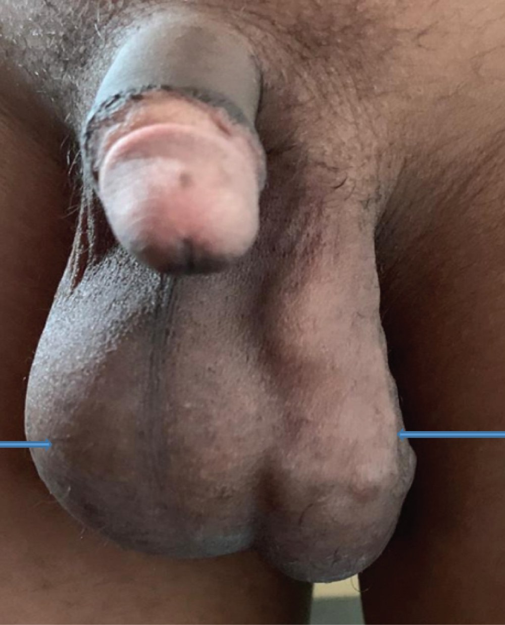
Figure 2 shows the association of a right hydrocele and a left varicocele found in a 12-year-old adolescent.
Testicular and annexes pathologies
These ranked first with 175 cases (60.4%), including 104 cases of varicocele (59.4%), 51 cases (29.2%) of testicular migration abnormalities, 13 cases (7.4%) of epididymal cysts, and 7 cases (4%) of testicular hypotrophy.
- Varicoceles were grade I in 27 cases (26%), grade II in 62 cases (59.6%), and grade III in 15 cases (14.4%).
- Testicular migration abnormalities were represented by retractile testicles in 41 cases (80.4%) and cryptorchidism in 10 cases (19.6%).
Pathologies of the patent processus vaginalis
Patients of the peritoneal-vaginal canal ranked second with 76 cases (26.1%); these included inguinal or inguinoscrotal hernias with 68 cases (90%), hydroceles with 4 cases (5%), and spermatic cord cysts with 4 cases (5%).
Penile Pathologies
These ranked third with 40 cases (13.7%) and were represented by a micropenis in 20 cases (50%), followed by penile curvature with 16 cases (40%), and hypospadias with 4 cases (10%). Penile curvatures were left lateral in 10 cases, right lateral in 4 cases, and ventral in 2 cases.
All cases of hypospadias were glandular.
Causes of lack of treatment
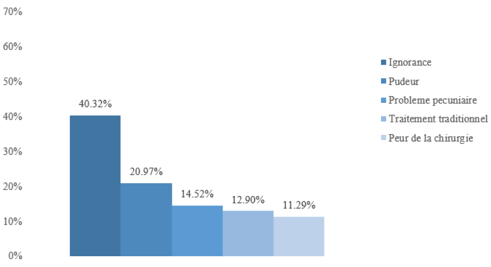
The lack of treatment for adolescents was often due to lack of awareness of the condition (Figure 3).
Discussion
Epidemiological aspects
In our study, 31.2% of the adolescents examined presented with at least one abnormality of the external genitalia. Our prevalence is close to that of Adekanye, et al. [4], who found 36.2% in primary school children, while some authors report a lower prevalence, such as Fiogbé, et al. [5] and Ghazzel, et al. [6], with 9.29% and 8.2%, respectively. This difference could be explained by methodological differences related to the age range studied and/or sample size. In our series, adolescents with AGE had a mean age of 14.1 ± 1.97 years (range, 11 to 19 years), which is close to the result of Fiogbé, et al. [5], with a mean age of 15.00 ± 2.30 years, ranging from 11 to 19 years.
Parental socioeconomic and educational levels
In our study, more than half of the adolescents (59.6%) with AEG had an average socioeconomic level and 34.2% had a low level. Fiogbé, et al. [5] reported a low socioeconomic level in 76.68% of cases in their series. Most parents had an average secondary education level.
Diagnostic aspects
Pubertal development
Among the adolescents with AEG, nearly half (47.1%) were pubertal and 32.1% were prepubertal. Our results are similar to those of Fiogbé, et al. [5] who found more clinically visible genital abnormalities in pubertal adolescents (87.4%) than prepubertal adolescents (12.6%).
Clinical signs
Scrotal pain in 42.3% of cases, followed by scrotal discomfort (37.5%) and finally inguinal and/or scrotal swelling with 19.4% dominated them. Fiogbé, et al. [5] had found scrotal pain, discomfort and swelling in 13.44%, 16.60% and 15.02% of cases respectively.
Pathologies
In our study, 19.6% of middle school students had more than one AGE, Adekanye, et al. [4] and Fiogbé, et al. [5] reported 5.28 and 8.30% respectively. Overall, eleven types of AGE were found, the most common being varicocele with a frequency of 35.7%, followed by inguinal and/or scrotal hernia with 23.4%. These results are similar to those of Fiogbé, et al. [5], while Ghazzel, et al. [6] and Gooran, et al. [7] reported more cases of inguinal hernia than varicocele. Our study focused on adolescents (10-19 years old) while that of Gooran, et al. [7] concerned a younger population between 7 and 12 years old; which explains the low frequency of varicocele in their study because it is rarely observed before puberty [8].
Pathologies of the testicle and its annexes occupy the first place in AOGE in middle school students.
Varicocele is a dilation of the veins of the pampiniform plexus secondary to valvular dysfunction, it is rare in children, its frequency increases with age to reach 15% in adolescence [8]. It is most often discovered incidentally during a systematic clinical examination. It is the most common cause of male infertility [8,9]. We found a predominance of grade 2 varicocele with (59.6%), as reported by Fiogbé, et al. [5] with 44% However in the literature, grade 1 is more frequent (10%), followed by grades 2 and 3 with respectively 1.7% and 1% of adolescents [9]. Varicoceles can lead to testicular atrophy and it is estimated that 15-20% of men with varicoceles suffer from sterility [8,9].
The frequency of cryptorchidism in our series, 3.4% of middle school students, is higher than that reported by Sano, et al. [10] and Gueye, et al. [11] with 1.2% and 1.41% of children examined, respectively. This difference can be explained by the methodology; the series of Sano, et al. [10] was larger with 7334 children aged 2 to 18 years examined. The delay in diagnosis and management of testicular positioning anomalies is criticized in several african series [10-12], while management is earlier in the West [13].
Pathologies of the patent processus vaginalis (PPV) represent the 2 nd group of AGE pathologies found in middle school students, with a frequency of 26.2%.
Inguinal or inguinoscrotal hernia is the most frequent pathology with 68 cases (90%), followed by hydrocele (5%) and spermatic cord cyst (5%). These are the most frequent surgical pathologies in children [14-17]; because only 18% of full-term children would have their peritoneal-vaginal canal closed at birth [14]. Indeed, according to Rahman [18], the prevalence of PPV would be nearly 80% in full-term newborns and the peritoneal-vaginal canal can still close spontaneously after birth. Of these 3 nosological entities, inguinal hernia is the most serious because it can be complicated by strangulation with the risk of intestinal and testicular necrosis [14].
Penile pathologies represent the third cause of AOGE in our study. In the United States, the incidence of penile anomalies is around 45-85/10,000.
Micropenis is the first penile pathology that we report. It is a penis of normal structure with a length < 2.5 SD below the average for age [18]. It is linked to defects in the secretion or action of testosterone. Micropenis must be treated to ensure normal urination, normal sexual intercourse and a good self-image [19]. Its incidence in the United States is 1.5 per 10.000 boys [20].
Congenital penile curvature represents the 2nd cause of penile pathologies in our study. Of 17 cases of objectified penile curvature, we found 15 lateral curvatures and 2 ventral curvatures were lateral; Adekanye, et al. [4] reported 37 cases of penile curvatures including 31 lateral, 2 dorsal, 1 ventral. Congenital penile curvature, uncommon (37/100.000), results from an asymmetry of development of the corpora cavernosa and sometimes associated with the urethra. It is not bothersome up to 20° angulation, but can become an obstacle to sexual life beyond this angulation [21].
For hypospadias, all was balanic. Ghazzel, et al. [6] reported 81.4% of glandular hypospadias; Yegane, et al. [22] found 64% of balanopreputial hypospadias and 36% of balanic hypospadias. Fiogbé, et al. [5] found 6 cases of hypospadias including 3 balanic, two anterior penile and one middle penile. Hypospadias is the most common malformation of the urethra in boys with 1 case/300 [23]. The predominance of distal forms reported by the different authors is explained by by the facts that balanic and balanopreputial forms are more frequent and represent 50% of cases [24] and that being minor they can go unnoticed or not be considered pathological. Odzébé, et al. [25] found an average age of 5.2 years at the time of diagnosis while in developed countries it is 16-24 months [23].
Causes of lack of care
In our study, 40.3% of parents were unaware of their children's condition. Yegane et al. [20] reported that 60.1% were. We also noted a lack of parental information related to adolescent modesty (20.9%) and financial problems (14.5%). Fear of surgery also appears to be a barrier to care. With all the consequences that these AEG can cause, such as potentially fatal complications such as hernia strangulation or cancer, or functional ones such as infertility or sexual dysfunction with a significant risk of psychosocial repercussions, the lack of care can be considered a form of neglect or even abuse [26]. It is also important to highlight the role of the system in the lack of medical checkups in public schools on the one hand and social security coverage for disadvantaged populations on the other.
Conclusion
Anomalies of the external genitalia are common among adolescents in school settings in Africa, particularly in Brazzaville. Varicoceles and inguinal hernias are the most common in our setting. Their presence in this age group is explained by parental negligence, ignorance, and lack of financial means, on the one hand, and by the lack of social and health coverage and school medicine, on the other.
Conflict of Interest
The authors declare that they have no conflict of interest.
References
- Rouvière H (1970) Urinary system, Male genital system. In: Descriptive and topographical human anatomy, Trunk. Masson, Paris 2: 415-492.
- Grapin Dagorno C (2023) Surgical treatment of genital malformations in children-legal context. Bull Acad Nat Med 207: 97-105.
- Nordenvall AS, Frisen L, Nordenstrom A, et al. (2014) Population based nationwide study of hypospadias in Sweden, 1973 to 2009: Incidence and risk factors. J Urol 191: 783-789.
- Adekanye AO, Adefemi SA, Onawola KA, et al. (2017) Abnormalities of the external genitalia and groins among primary school boys in Bida, Nigeria. Afr Health Sci 17: 1120-1125.
- Fiogbé MA, Gbénou AS, Metchihoungbé S, et al. (2013) Epidemiological and clinical aspects of clinically visible urogenital malformations in adolescent students in Cotonou. Prog Urol 23: 1428-1434.
- Ghazzel AM (2006) Inguinal hernias and genital abnormalities in young Jordanian males. East Mediter Health J 12: 483-488.
- Gooran S, Fazeli F, Asghari-Sheikhi M, et al. (2014) Prevalence of inguinal hernias and genital abnormalities among elementary-school boys. Zahedan J Res Med Sci 16: 28-31.
- Vasavada S, Ross J, Nasrallah P, et al. (1997) Prepubertal varicoceles. Urology 50: 774-777.
- Fontaine E, Benoit G, Jardin A, et al. (2000) La varicocèle de l’adolescent. Prog Urol 10: 1099-1107.
- Sano D, Wandaogo A, Tapsoba TL, et al. (1999) Testicular position anomalies: Epidemiological survey in schools in Ouagadougou. Med Afr Noire 46: 75-77.
- Gueye SM, Ba M, Ndoye A, et al. (1996) Cryptorchidism in Senegalese school-age children. Androlog 6: 287-292.
- Bouya PA, Odzébé AWS, Avala PP, et al. (2012) Management of cryptorchidism at the University Hospital of Brazzaville. Androlog 22: 108-111.
- Penna FJ, Nguyen HT, Passerotti CC, et al (2011) The concordance of testicular anatomic location in bilateral cryptorchidism. J Pediatr Urol 7: 52-56.
- Galinier P, Kern D, Bouali O, et al. (2005) Urgent pathologies of the peritoneo-vaginal process in children. EMC-Médecine 2: 215-223.
- Lau ST, Lee YH, Caty MG (2007) Current management of hernias and hydroceles. Semin Pediatr Surg 16: 50-57.
- Mboutol-Mandavo C, Ondima IPL, Miéret JC, et al. (2019) Pathologies of the processus vaginalis: A report on 285 cases operated on at the Brazzaville University Hospital. Rev Afr Chir Spéc 13: 10-13.
- Sewa EV, Tengue KK, Kpatcha MTK, et al. (2016) Clinical and therapeutic aspects of peritoneo-vaginal canal pathologies at the Dapaong regional hospital center (TOGO). J Conf Ouest Afr Uro Andro 6: 1-15.
- Rahman N, Lakhoo K (2009) Patent processus vaginalis: A window to the abdomen. Afr J Paediatr Surg 6: 116-117.
- Aaronson IA (1994) Micropenis: Medical and surgical implications. J Urol 152: 4-14.
- Hatipoglu N, Kurtoglu S (2013) Micropenis: Etiology, diagnosis and treatment approaches. J Clin Res Pediatr Endocrinol 5: 217-223.
- Faix A, Ferretti L, Terrier JE, et al. (2014) Congenital curvature of the penis. Prog Urol 24: 26-33.
- Yegane RA, Kheirollahi AR, Bashashati M, et al. (2005) The prevalence of penoscrotal abnormalities and inguinal hernia in elementary-school boys in the west of Iran. Int J Urol 12: 479-483.
- Nelson CP, Park JM, Wan J, et al. (2005) The increasing incidence of congenital penile anomalies in the United States. J Urol 174: 1573-1576.
- Mohannad Ashou, Haleseh SA, Ashour M (2016) Its Embryology, etiology and surgical techniques. Springer A, Van den Heijkant M, Baumann S: Worldwide prevalence of hypospadias. J Pediatr Urol 12: 152.e1-152.e7.
- Odzébé AWS, Mboutol-Mandavo C, Ondima IPL, et al. (2019) Congenital urinary tract anomalies: About cases 80 cases at the university hospital of Brazzaville. Open J Urol 10: 8-15.
- (2025) World Health Organization. Child Maltreatment.
Corresponding Author
Docteur Caryne Mboutol Mandavo, Associated professor, Faculty of Health Sciences, University Marien Ngouabi, Brazzaville, Congo, Tel: +242055456601.
Copyright
© 2026 Mboutol MC, et al. This is an open-access article distributed under the terms of the Creative Commons Attribution License, which permits unrestricted use, distribution, and reproduction in any medium, provided the original author and source are credited.