

Ureteropelvic Junction Obstruction in Children Caused by Crossing Vessels a Case Report
Abstract
The uretero-pelvic junction obstruction (UPJO) also known as essential hydronephrosis is the most common obstructive congenital uropathy in children. It’s diagnosis may be pre-natal or post- natal and it has multiple etiologies. Among these etiologies is the compression by a lower polar vessel (crossing vessel). standard treatment is open or laparoscopic pyeloplasty using the Kuss-Anderson-Hynes technique.
Keywords
Pyelo-ureteral junction obstruction, Crossing vessel, Pyeloplasty
Introduction
Ureteropelvic junction syndrome is a congenital or acquired obstruction between the renal pelvis and ureter, causing a dilation of the pyelocaliceal cavities and a normal-caliber downstream ureter, impairing urine transport. In children, the obstruction may be primary and due to intrinsic factors such as abnormal amounts of muscle and collagen deposition or secondary, extrinsic factors due to the presence of a lower polar vessel compressing the ureter. In 1842, Von Rokitansky was first described the association of a lower polar vessel (LPV) with UPJO.
Diagnosis of a lower polar vessel is challenging with conventional ultrasound but has improved with Doppler ultrasound. The standard treatment is open or laparoscopic pyeloplasty via the Kuss-Anderson-Hynes technique (desmembraned pyeloplasty). UPJO can be revealed mostly by intermittent lumbar pain or be completely asymptomatic and discovered incidentally during an imaging test. Whatever the cause, the effect of the obstruction is the same and can lead, in some cases, to complications (pyelonephritis, lithiasis and in rare cases urinoma) that may affect the fate of the kidney.
The surgical procedure of choice is dismembered pyeloplasty which can be successfully adapted to almost all clinical situations. We present a case of UPJO caused by an inferior polar vessel discovered intra-operatively.
Case description
A 3-year-old boy with no medical history presented to the emergency department with left lumbar pain.
At the exam, there were no signs of dehydration or malnutrition, and no break in the weight curve, normal blood pressure referred to his age, no bladder globe, no mass, and no other malformations.
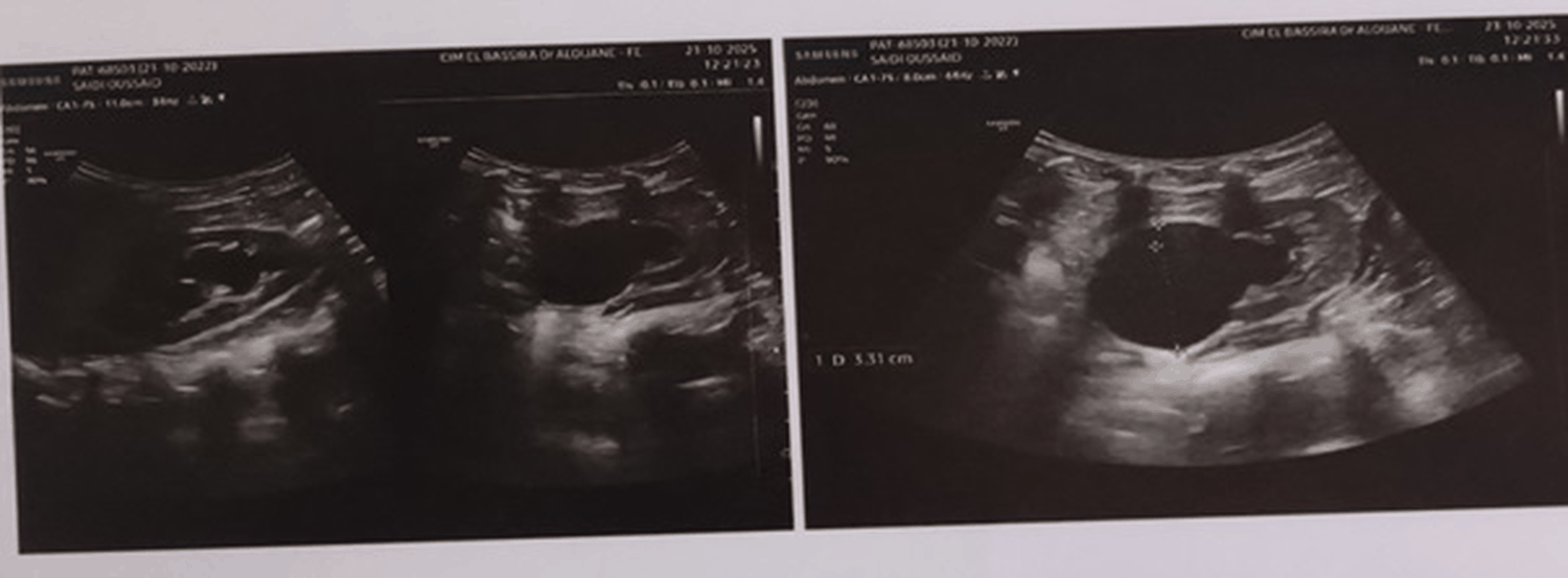
An abdominal ultrasound exam was performed and was suggestive of a pyeloureteral junction syndrome in the left kidney with an antero-posterior diameter of the renal pelvis measured at 33 mm (Figure 1), as well as significant dilatation of the calyceal cavities laminating the renal parenchyma.
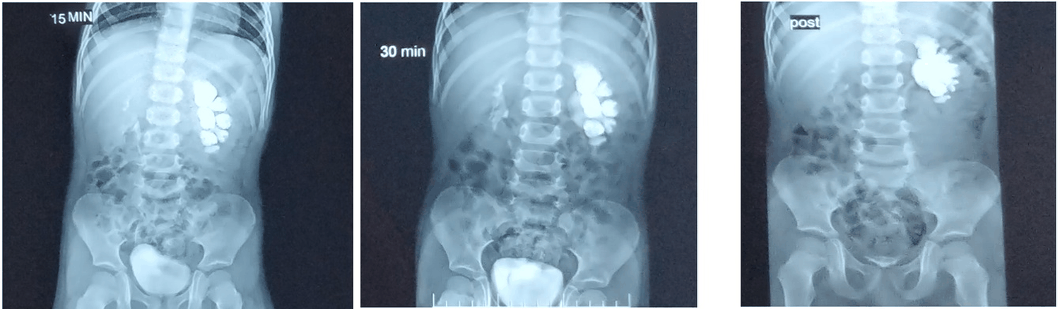
Intravenous Urography (Figure 2).
Dilated Collecting System: The renal pelvis and ureter appear enlarged and swollen (grade 3)
Delayed Washout: Contrast stays longer in the dilated area.
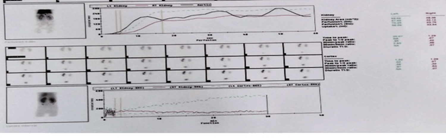
DTPA Scintigraphy (Figure 3) showed a retentionnel plateau curve of the left kidney.
DMSA scintigraphy: didn’t show renal scares.
Based on these data of investigation we made a decision of a surgical treatment.
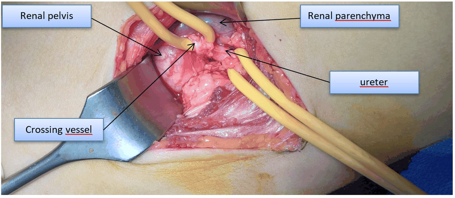
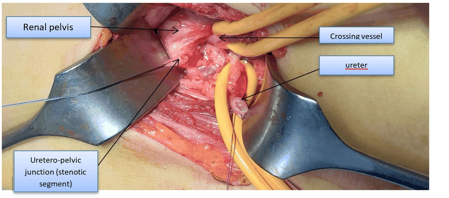
The patient was admitted to the operating room under general anesthesia, intubated and ventilated, and placed in the right lateral decubitus position. An incision was made midway between the twelfth rib and the iliac crest, and after approaching the retroperitoneal region, the proximal ureter was identified retroperitoneally, looped, and dissected to the uretero-pelvic junction where we found a dilated renal. A lower polar vessel was found crossing and compressing the ureter, with significant renal pelvis dilation (Figure 4).
The stenotic segment (Figure 5) was excised. The vessel was decrossed and transposed posteriorly, and the ureter was sectioned 1cm below the stenotic area, spatulated, and the renal pelvis was trimmed and reshaped and Kuss-Anderson-Hynes Pyeloplasty (dismembraned pyeloplasty) was performed anterior to the polar vessel.
A transureteral catheter, Redon drain, and Foley urinary catheter were inserted.
The surgical site was closed.
Blood loss was minimal.
Postoperative recovery was uneventful except the accidental drainage extraction by the patient himself in the 3 th day after the surgery with no consequences.
Discussion
Hydronephrosis is a frequent uropathology in pediatric population often linked to stenosis of the uretero-pelvic junction.
It’s diagnosis can be made antenatally or postnataly and based on clinical exam and radiological explorations.
The low rate of antenatal diagnosis could be explained by the insufficiency of prenatal consultation and by the realization of antenatal ultrasounds by practitioner’s non-specialists in most cases. In our case the diagnosis was made postnatally.
Male predominance has been reported by several authors like in our case.
Frequent involvement of the left kidney is reported in the literature, which is the case in our patient.
Low-grade hydronephrosis often evolve towards regression.
High-grade hydronephrosis often requires surgery.
In our case, it is a high-grade hydronephrosis and we opted for surgical treatment.
Open or laparoscopic Pyeloplasty using the Kuss- Anderson-Hynes technique is the gold standard for the treatment of uretero-pelvic junction syndrome, with a 90% success rate.
In our patient we performed an open pyeloplasty due to the absence of laparoscopy in our structure.
The presence of crossing vessels is described in literature and should be searched before surgery if possible, by ultra sound and Doppler and uro-scanner.
The treatment should decrosse the vessels and not ligature them to prevent ischemia and hemorrhage.
In our case we decrossed the polar vessel and did our pyeloplasty in front of the vessel.
The postoperative complications include infection, hemorrhage and urinary fistules
We did not record postoperative complications except the accidental drainage extraction with no consequences [1-9].
Conclusion
Uretero-pelvic junction syndrome is a frequent uropathology in pediatric population it is not very symptomatic, mainly affecting male infants.
Ultrasound and Uro scanner have a very important role in the exploration of this pathology.
The presence of crossing vessels should be present in the mind of surgeons at the diagnosis and should be preserved during surgery by doing a pyeloplasty in front of the vessels after decrossing them by open or laparoscopic surgery.
Conflict of interest statement
None of the authors have any financial or personal relationships with other people or organizations that could inappropriately influence this work. We have nothing to disclose.
Patient consent
Consent to publish the case report was not obtained. This report does not contain personal information that could lead to the identification of the patient.
Funding
No funding or grant support.
Authorship
All authors attest that they meet the current ICMJE criteria for Authorship.
References
- Harriet J Corbett, Liam McCarthy (2013) Hydronephrosis in children: Pelviureteric junction dysfunction. Surgery (Oxford) 31: 135-139.
- Horatio N Dorman (1931) Hydronephrosis with Aberrant Polar Vessels: Report of a case. Journal of Urology 26: 121-130.
- Dongyan Zhao, Long Sun, Chang Tao, Daxing Tang, Guangjie Chen (2024) Ureteropelvic Junction Obstruction Caused by Crossing Vessels in Infants and Young Children. J Pediatr Surg 59: 1835-1840.
- Bumbu GA, Berechet MC, Nacer K, Gheorghe Bumbu, Octavian Adrian Maghiar, et al. (2018) Clinical, surgical and morphological assessment of the pyeloureteral syndrome. Rom J Morphol Embryol 59: 1173-1177.
- Deming CL (1951) The effects of intrarenal hydronephrosis on the components of the renal cortex. J Urol 65: 748-753.
- Autorino R, Eden C, El-Ghoneimi A, Giorgio Guazzoni, Nicolòmaria Buffi, et al. (2014) Robot-assisted and laparoscopic repair of ureteropelvic junction obstruction: A systematic review and meta-analysis. Eur Urol 65: 430-452.
- Värelä S, Omling E, Börjesson A, Salö M (2021) Resolution of hydronephrosis after pyeloplasty in children. J Pediatr Urol 17: 102e1-102e7.
- Frech-Dörfler M, Durand S, Prüfer F, Holland-Cunz S, Rudin C (2022) Giant bilateral hydronephrosis in a newborn-a case report. Children (Basel) 9: 1890.
- Carpenter CP, Tolley E, Tourville E, Sharadin C, Giel DW, et al. (2018) Hydronephrosis after pyeloplasty: "will it go away?" Urology 121: 158-163.
Corresponding Author
Dr. Aries Aissam, Surgery Department, EPH Ferdjioua Hospital, Mila, Algeria.
Copyright
© 2026 Aissam A. This is an open-access article distributed under the terms of the Creative Commons Attribution License, which permits unrestricted use, distribution, and reproduction in any medium, provided the original author and source are credited.