Triple Testis in Pediatric: The Story and Management
Abstract
The presence of more than two testicles is rare congenital anomaly and has different names in the literature like Polyorchidism, triochidism & supernumerary testis. The majority of cases were triorchidism with occasional bilateral duplication. The extra testis is classified into type A and type B according to its reproductive potential and connection to vas deferens. The condition has a wide range of presentation and hence different management. Here we report the case of a 2-months-old infant presented with palpable extra testis in right upper scrotum and underwent elective orchiectomy after confirmation of the condition by ultrasound examination. An elaborated discussion of the condition and its management with reviewing the related literature is provided here.
Keywords
Polyorchidism, Triple testis, Supernumerary testis, Orchiectomy, Testicular tumor
Introduction
Supernumerary testis is a very rare congenital anomaly. The first case was reported by Lane in 1895 [1]. It may have scrotal, inguinal, or abdominal location but most commonly found in the scrotum, on the left side [2] with preserved function of sperm production [3]. The condition is usually asymptomatic & found incidentally during surgical exploration for another condition in the majority of cases or the patients may present with complications like pain or inguinoscrotal swelling due to inguinal hernia, cryptorchidism, torsion, hydrocele, varicocele, epididymitis and malignancy [4].
The Polyorchidism is classified into type-A: Connected to a vas (with preserved reproductive function) and type-B: Not connected to a vas (no reproductive function). Type-A further divided into A I: Separated epididymis and vas. Type A II: Separated epididymis but shared vas. Type A III: Shared epididymis and vas. While type-B is further divided into type B I: With its own epididymis. Type B II: Only separated testicular tissue [4].
There is no consensus about standard management of supernumerary testis due to its rare occurrence and wide spectrum of presentation. More than half of the cases have been diagnosed between ages 15-45 years, and rarely seen in infancy or above 45-years-old [5].
Although regular follow-up examination is suggested for stable cases; orchiectomy is the preferable option in other situations. Here we present the story of triple testis in an infant and highlights the rationale for its removal with literature review.
Statement of Human Rights
This work has been approved by the local scientific - ethical committee of university of Mustansiriyah before being considered for publication.
Committee Name: Scientific Ethical Committee
Head of Committee: Professor Ehab Taha Yassen
Approval Letter Number: 239
Date of Approval by the Ethics Committee: 15-June-2020
Patient Consent
The parents of this patient have given their informed consent for participation in this work and given their permission for publishing the related photographs.
Case Presentation
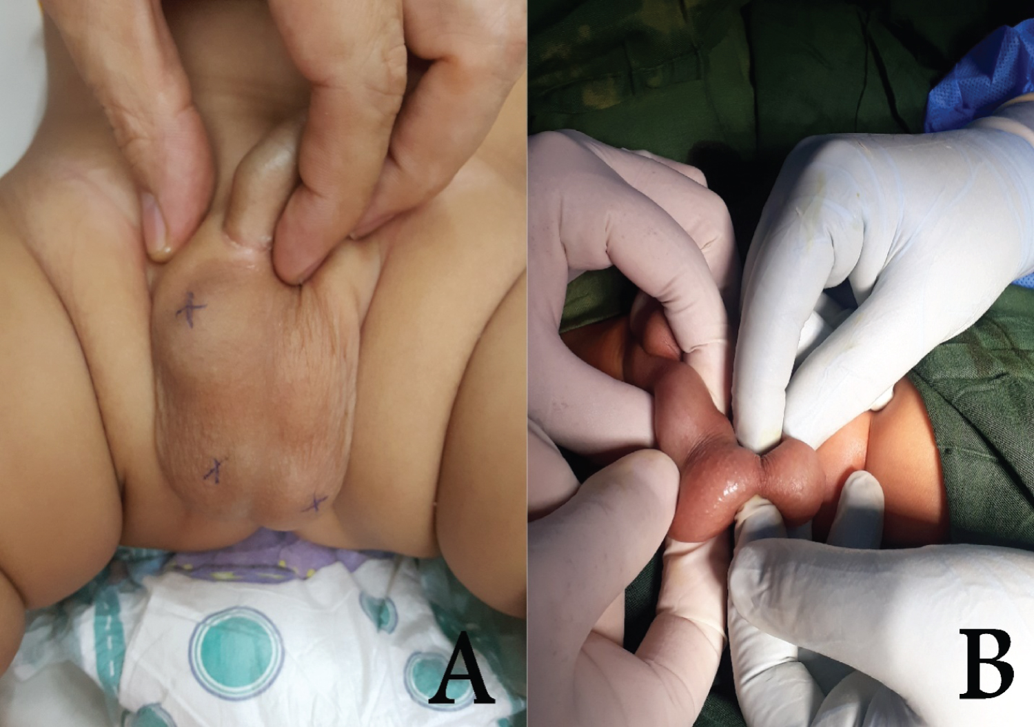
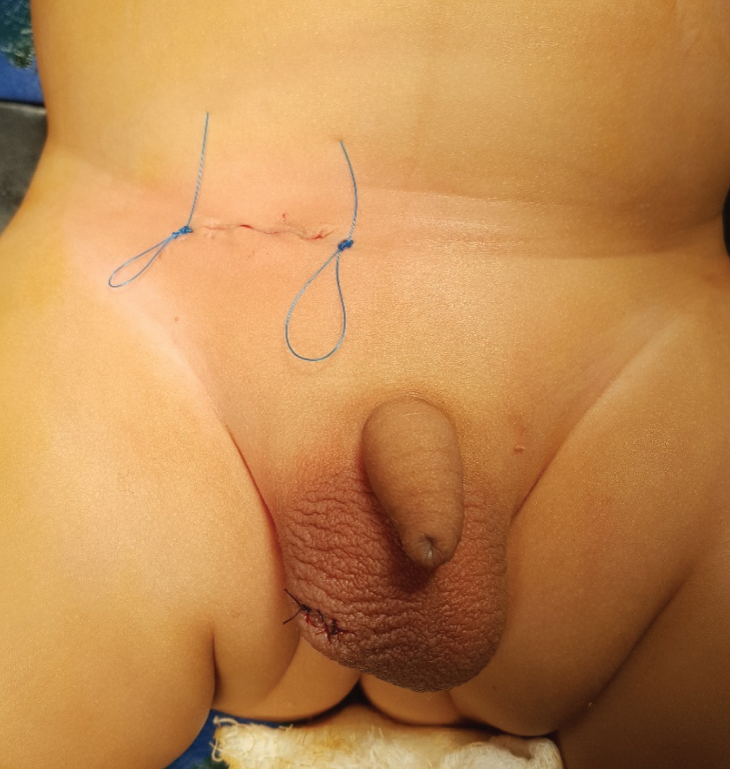
A 2-month-old infant presented by his parents to my private clinic of pediatric surgery after they noticed an abnormal lump in upper scrotum few days ago. The infant was completely normal without family history of similar condition. Physical examination revealed normal phallus, normal palpable both testes in scrotum but with lump in upper right hemi-scrotum, it was solid, mobile & non-tender with a little pit larger than the size of ipsilateral testis. It also has an abnormal texture compared with the native testis, negative trans-illumination test and no feature of inguinal hernia or hydrocele (Figure 1). The two testes were of normal texture and size for his age & palpation of both inguinal regions reveals no palpable inguinal lymph nodes.
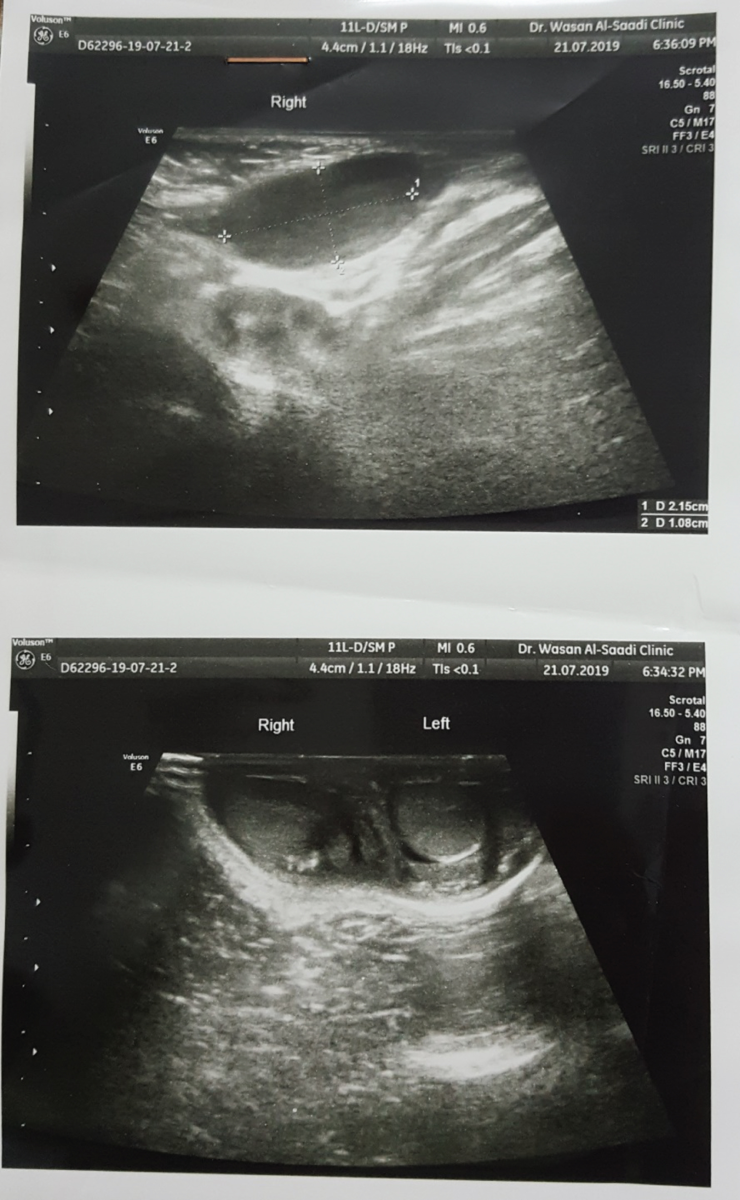
Two different Ultrasound examinations demonstrated well defined oval shape mass lesion isoechoic with the native testis located at the exit of right inguinal canal or upper scrotum measured 21 × 11 × 9 mm with internal & peripheral vascularity by Doppler study, with some areas of hyper echogenicity otherwise normal surrounding tissue. The picture was highly suggestive of a third testis (Figure 2).
We decided to go for surgical exploration on the basis of a combination of clinical suspicious size and texture and ultrasonography finding rather than continuous regular follow-up examination.
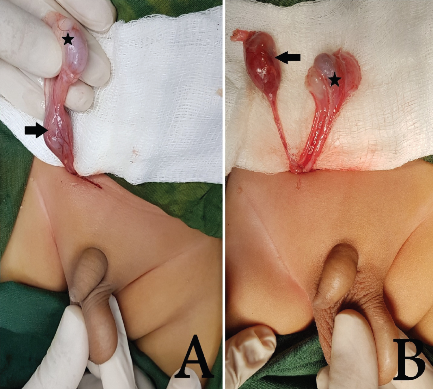
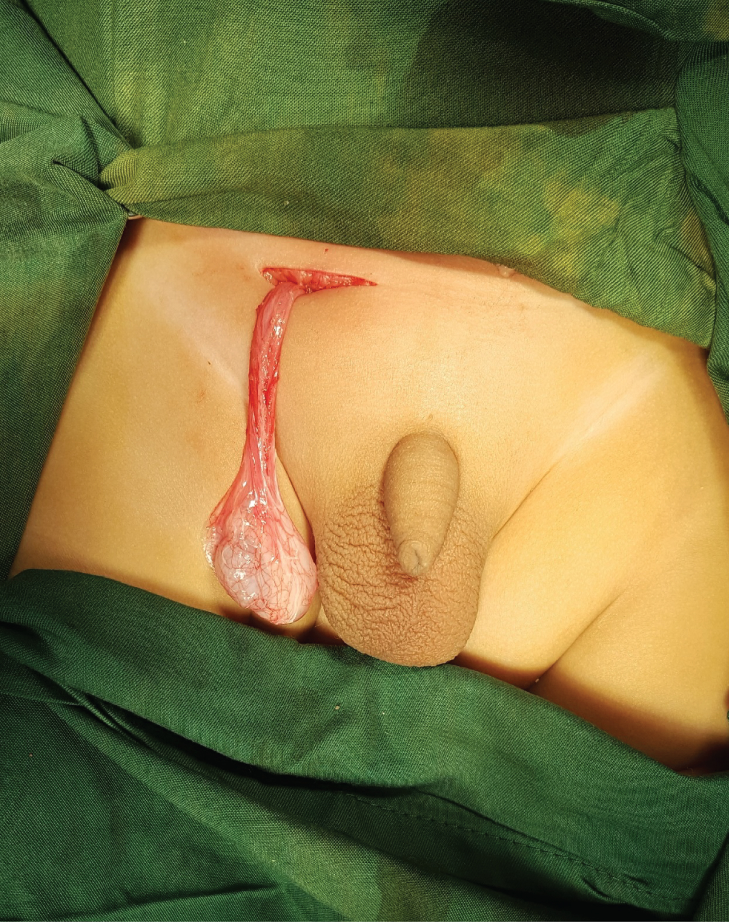
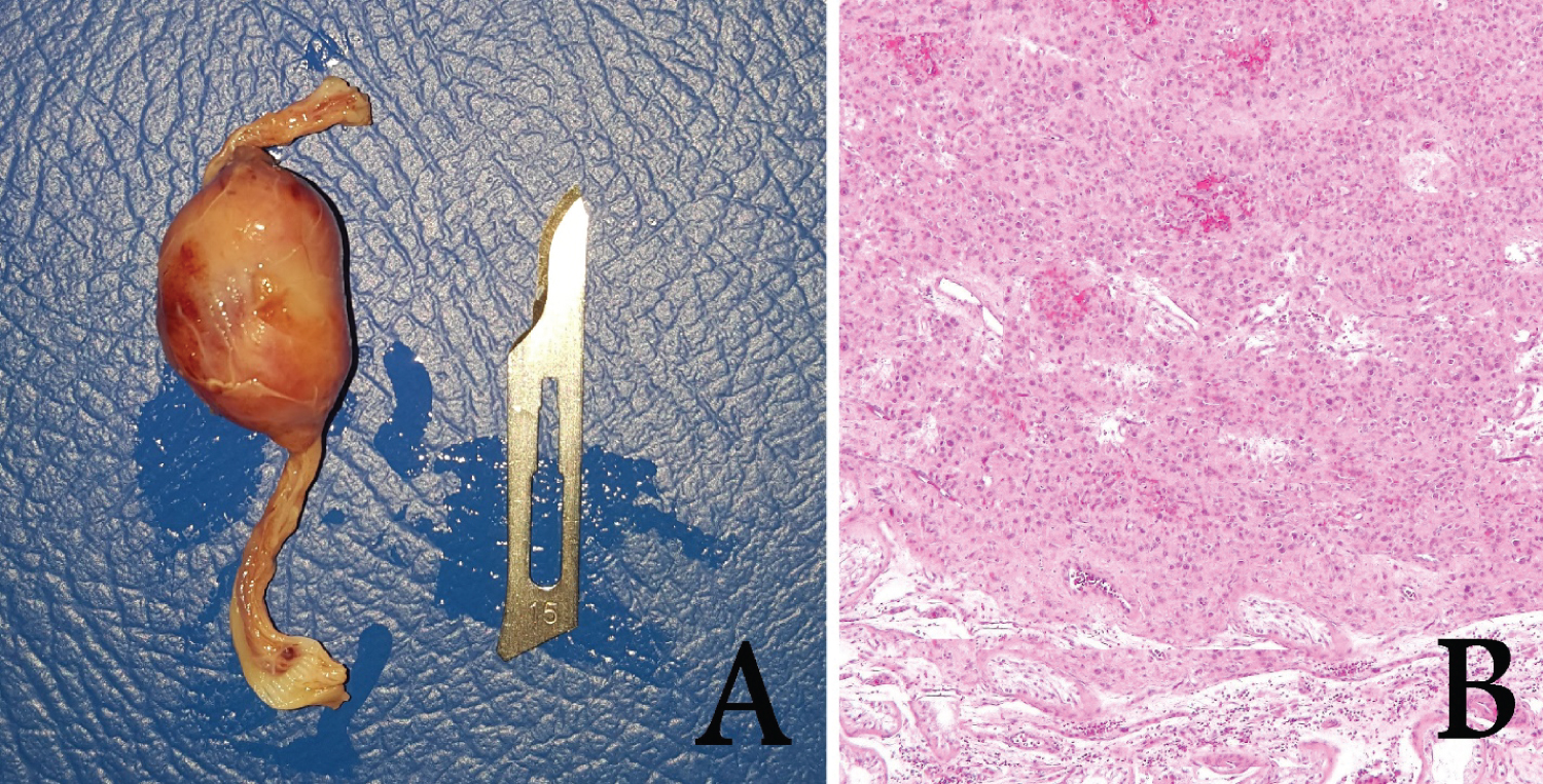
An inguinal exploration was performed, delivery of whole spermatic cord structures to the wound which reveals a third testis above the native right testis with its own separated epididymis and vas, it was larger in size with different color and texture compared to native testis (Figure 3). Complete orchiectomy with careful preservation of the cord structures of original testis is ensured (Figure 4). Orcheopexy of native right testis done with closure of inguinal incision (Figure 5). The orchiectomy specimen was send for pathologic examination which revealed leydig cells hyperplasia with some micro dysplastic changes (Figure 6). The patient recovered well from anesthesia and now has completely normal follow up period.
Discussion
The rare condition of supernumerary testis is usually discovered incidentally during groin exploration and it is believed to result from an abnormal division of the genital ridge during early embryonic development [4]. Although it is mostly seen in adult & on the left side, our case was on the right side of an infant's scrotum. There is argument about the left side predilection of triple testis as the left testicle has relatively larger size & different vascular anatomy compared to right testicle making it more prone to embryonic subdivision [6]. We found a third testis in upper scrotum which is goes with the finding of Bostwick, et al. that 75% of triorchidism were scrotal & 20% of the testes were inguinal while just 5% were discovered in abdomen [7].
The patients most often present with a painless scrotal mass or detected incidentally during evaluation for other symptoms such as inguinal hernia and cryptorchidism. The minority of cases were presented because of complications mostly torsion due to absence of the gubernaculums, absence of the normal attachment of the epididymis to scrotal wall, and the extra mobility of supernumerary testis that is why have increased risk for twist. Sometimes it may lead to development of hydrocele, varicocele, epididymitis and malignancy [4]. It is true that Polyorchidism has no specific clinical presentation but one should put this condition in mind during evaluation of any solid mass in the groin or scrotum. The clinical examination alone is not sufficient to make a diagnosis for this condition because it may be mistaken for other conditions like the encysted hydrocele, spermatocele, varicocele, Morgagnian cyst or testicular neoplasm [8]. Testicular duplication should be differentiated from some variants of ectopic testis like transverse testicular ectopia especially in child with an empty hemi-scrotum [9].
The ultrasound examination can confirm the diagnosis of supernumerary testis as in our case because it usually shows similar echo texture or echogenicity to normal testes with similar vascular flow pattern on color Doppler [10], but these findings may be variable or give equivocal results and in these cases a magnetic resonance imaging will be of great help in confirming the diagnosis with again the same characteristics of signal intensity on T1- and T2 weighted images [11]. Some old studies recommended testicular biopsy to verify scrotal or inguinal mass as supernumerary testis [12-14], but now the recent advances in the ultrasound technology with high resolution images can give the accurate diagnosis without need for surgical exploration and biopsy. Many advantages can be obtained by the use of MRI in the evaluation of scrotal pathology like differentiation between malignant and non-tumorous testicular lesions, not operator dependent imaging technique, and give multiplanar imaging with higher soft tissue resolution and contrast [15]. Although MRI is superior to ultrasound & provide additional information in complicated cases of supernumerary testes, several articles supported the use of high resolution ultrasound examination alone as an effective non-invasive method for the diagnosis of Polyorchidism without the need of anesthesia for doing MRI in children or surgical exploration [16,17]. Another rare congenital anomaly should be considered during evaluation of unusual scrotal mass which is spleno-gonadal fusion which also give similar appearance of Polyorchidism but should be confirmed by technetium sulfur colloid scan to detect the presence of ectopic splenic tissue [18]. Laparoscopy sometimes detect intra-abdominal supernumerary testis during management of cryptorchidism.
The histopathology of supernumerary testis is variable, it may be normal or shows abnormal tubular architecture and absent spermatogenesis [19], the testicular malignancy is usually preceded by dysplastic features or cellular hyperplasia as our case when we detect leydig cell hyperplasia in orchiectomy specimen.
The reported risk of malignancy in Polyorchidism is 6-7% with increased incidence in the last decades according Huyghe E, et al. [20], the most important risk factor for development of malignancy in supernumerary testes is cryptorchidism this is evident by the early presentation of testicular cancer (median age 19 years) when both conditions of cryptorchidism and Polyorchidism coexist together compared to median age of 34 years for development of testicular cancer in normal testes [21]. The most common types of malignancies reported in supernumerary testis are embryonal carcinomas, germ cell tumors and seminomas. Extra testicular rhabdomyosarcoma and adenoma of rete testis also have been reported [3].
The argument about standard management of supernumerary testis is evident in many literatures without consensus till now, previously the surgical removal was encouraged regardless location of supernumerary testis due to increased risk of torsion and malignancy but with the development of radiological imaging techniques, more conservative approach is suggested. The dilemma is when the surgeon incidentally detect extra testis during surgical exploration for other conditions, should he leave the testis in site, doing orcheopexy if needed, take a biopsy and then manage accordingly, or remove it surgically? The answer to this question is by determining the functional potential of testis from its connection to vas deferens and the risk of development malignancy. Although many factors determine whether a conservative approach or surgical exploration is undertaken like age, fertility potential, risk of malignancy, location of the testis, associated cryptorchidism; our opinion is removal of all supernumerary testes because even in type-A where there is vas deferens the patency of the duct is not guarantee by just gross intra-operative appearance and might not add to fertility potential as well as the majority of supernumerary testes have histologically reduced or absent spermatogenesis [22], meanwhile it carry risks of torsion and malignancy especially when it is undescended or when watchful observation is unlikely keeping in mind that frequent ultrasound or MRI screening will not be cost-effective. Furthermore, high resolution imaging study might not detect tumor changes of supernumerary testis thus we must perform serological test for tumor markers if surgical exploration is not the choice [4].
Conclusion
Triple testis is a rare congenital anomaly that is usually encountered accidentally in asymptomatic individual or incidentally during surgical exploration for another condition. It can be diagnosed with Doppler ultrasound in majority of cases without need for MRI. Pediatric surgeon, urologists and radiologists should put this condition in the differential diagnosis of any inguinal or scrotal mass. Surgical removal of the triple testis is advised because of risk of complications like torsion of malignancy and it usually does not contribute to fertility especially when lifelong follow up by early ultrasound of MRI is unlikely in infants or children.
Authors' Contribution
The author Ali Egab Joda is the only contributor of this paper, he read and approved the final version of the manuscript.
Conflicts of Interest
The author certifies that there is no conflict of interest with any financial organization regarding the material discussed in the manuscript.
Funding
There are no funding sources or sponsors have been involved with this work.
References
- Spranger R, Gunst M, Kuhn M , et al. (2002) Polyorchidism: A strange anomaly with unsuspected properties. J Urol 168: 198.
- Wolf B, Youngson GG (1998) Polyorchidism. Pediatr Surg Int 13: 65-66.
- Kharrazi SM, Rahmani MR, Sakipour M, et al. (2006) Polyorchidism: A case report and review of literature. Urology 3: 180-183.
- Bergholz R, Wenke K (2009) Polyorchidism: A Meta-Analysis. The Journal of Urology 182: 2422-2427.
- Nistal M, Paniagua R, Martin-Lopez R, et al. (1990) Polyorchidism in a newborn: Case report and review of the literature. Pediatr Pathol 10: 601-607.
- Wilson WA, Littler J (1953) Polyorchidism A report of two cases with torsion. Br J Surg 41: 302-307.
- Bostwick DG (1997) Spermatic cord and testicular adnexa. In: Bostwick DG, Eble JN, Urologic Surgical Pathology. Mosby, St Louis, MO, 647-674.
- Mor Y, Leibovitch I, Duvdevani M, et al. (2004) Scrotal epidermal inclusion cyst clinically mimicking polyorchidism in a child: Ultrasonic characteristics. Pediatr Radiol 34: 175-176.
- Joda AE (2019) Five different cases of ectopic testes in children: A self-experience with literature review. World Jnl Ped Surgery.
- Arslanoglu A, Tuncel SA, Hamarat M, et al. (2013) Polyorchidism: Color Doppler ultrasonography and magnetic resonance imaging findings. Clinical Imaging 37: 189-191.
- Arslanoglu A, Tuncel SA, Hamarat M, et al. (2013) Polyorchidism: Color Doppler ultrasonography and magnetic resonance imaging findings. Clin Imaging 37: 189-191.
- Leung AK (1988) Polyorchidism. Am Fam Physician 38: 153-156.
- German K, Mills S, Neal DE, et al. (1993) Polyorchidism. Br J Urol 72: 515.
- Ozok G, Taneli C, Yazici M, et al. (1992) Polyorchidism: A case report and review of the literature. Eur J Pediatr Surg 2: 306-307.
- Cramer BM, Schlegel EA, Thueroff JW, et al. (1991) MR imaging in the differential diagnosis of scrotal and testicular disease. Radiographics 11: 9-21.
- Chung TJ, Yao WJ (2002) Sonographic features of polyorchidism. J Clin Ultrasound 30: 106-108.
- Yalçınkaya S, Şahin C, Şahin AF, et al. (2011) Polyorchidism: Sonographic and magnetic resonance imaging findings. Can Urol Assoc J 5: 84-86.
- Joda A-E, Aziz A (2017) Splenogonadal fusion as a cause of left undescended testis: Case report & review of literature. Journal of Pediatric Case Report 21: 22-25.
- Thum G (1991) Polyorchidism: A case report and review of literature. J Urol 145: 370-372.
- Huyghe E, Matsuda T, Thonneau P (2003) Increasing incidence of testicular cancer worldwide: A review. J Urol 170: 5-11.
- Bahrami A, Ro JY, Ayala AG (2007) An overview of testicular germ cell tumors. Arch Pathol Lab Med 131: 1267-1280.
- Abbasoglu L, Salman FT, Gun F, et al. (2004) Polyorchidism presenting with undescended testes. Eur J Pediatr Surg 14: 355-357.
Corresponding Author
Ali Egab Joda, Department of Surgery, College of Medicine, University of Mustansiriyah; Department of Pediatric Surgery, Central Child Teaching Hospital, Baghdad, Iraq, Tel: 00964-7725-465-090
Copyright
© 2021 Joda AE. This is an open-access article distributed under the terms of the Creative Commons Attribution License, which permits unrestricted use, distribution, and reproduction in any medium, provided the original author and source are credited.

