Experiences in The Care and Treatment of Children and Adolescents with Phimosis
Abstract
Phimosis is the inability or difficulty to expose the glans by retracting the prepuce due to changes in the prepuce. It is the most commonly observed disorder in boys and accounts for a large number of visits in all levels of pediatric care. The objective of the present study is to report an experience of treating boys diagnosed with Phimosis in a pediatric surgery referral center. This was a retrospective observational study of pediatric patients referred with an initial diagnosis of Phimosis. Phimosis was classified as grade I, grade II, moderate preputial ring, and loose preputial ring. The following data were collected: Age at admission; history of balanoposthitis, paraphimosis, and previous treatments: Rate of diagnostic confirmation of phimosis; indicated treatment; and therapeutic outcome. Of the 944 boys who received care, diagnosis of phimosis was confirmed in 475 (50.32%), of which 417 (87.79%) were successfully treated through topical treatment. Surgical treatment was more frequent among patients with grade I phimosis, and all cases of moderate or loose preputial ring were resolved with topical treatment. History of balanoposthitis or paraphimosis did not interfere with the treatment, and previous treatment with corticosteroids was associated with low risk of surgery. It was difficult for physicians in non-specialized healthcare settings to establish a diagnosis of phimosis. Topical treatment was effective in most cases of phimosis, and surgery was reserved for boys with inflammation of the foreskin that was refractory to local treatment and who were usually older.
Keywords
Phimosis, Children, Teenagers, Epidemiology, Treatment
Introduction
Phimosis is the most common disorder of the penis in children and adolescents. In the first year of life, it occurs in 95-96% of children, although by age 3-4 years, it spontaneously resolves in up to 90% of cases [1-4]. Therefore, approximately 10% of children aged more than 3-4 years have some degree of phimosis and will probably receive treatment for it [5-6]. Phimosis is the inability or difficulty to expose the glans by retracting the foreskin due to changes in the dermis of the prepuce, including the loss of collagen and tissue elasticity and foreskin scar tissue resulting from repeated inflammation or local trauma [1-8].
The distinction between normal genitalia and phimosis remains difficult. For this reason, and because phimosis affects 1-10% of all boys aged less than 18 years, there is a high demand for management and treatment of this disorder in pediatric health settings [1,3,5]. When Phimosis is diagnosed, it is vital to classify it and define its degree [1,3,4,9].
The treatment of patients with Phimosis after diagnosis remains controversial [2,6,8,10,11]. In some countries, especially in the US and Canada, circumcision is advocated for all boys, even during the neonatal period, regardless of cultural or religious backgrounds [8,12-15]. In other countries, it is believed that surgical treatment should only be considered for children with preputial scarring, chronic inflammatory changes such as balanitis xerotica obliterans, or repeated urinary tract infections, with or without associated urinary malformations [3-6,16]. In the last two decades, the emergence of topical treatment for Phimosis with corticosteroids has promoted discussions on this topic [4, 6,10-11,17].
In this context, the present report describes the experience of treating children and adolescents with suspected Phimosis who were referred to a pediatric surgery referral center. Another aim is to recommend standardization of the nomenclature used in the classification of Phimosis and to assess the treatment outcomes, including the success rate of non-surgical treatment and rate of surgical indication in this series of patients.
Materials and Methods
Ethical aspects
The present study was approved by the Research Ethics Committee of the FEPECS/SES-DF through Plataforma Brasil, under CAAE no. 10433119.3.0000.5553, and was in accordance with all ethical aspects described in Resolution CNS/MS 466/2012.
Study design and sample
This was a retrospective observational study and data was collected from physical and electronic records of pediatric patients referred with suspected phimosis (ICD N47). It was conducted over a period of 30 months, between April 1, 2017 and September 30, 2019, in a pediatric surgery referral center.
Diagnostic evaluation
All children and adolescents were evaluated by the same pediatric surgeon. A diagnosis of normal-for-age genitalia was made when the glans could be exposed without difficulty. A patient whose prepuce was adherent to the glans without signs of changes in the foreskin dermis that could prevent its opening was also considered as not having Phimosis [1,5,9,11]. These boys' parents or guardians were given guidance on general care and hygiene, and the patients were scheduled for outpatient follow-up.
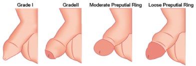
Phimosis was defined as changes in the prepuce that hindered or prevented the exposure of the glans. In this series of patients, Phimosis was classified as grade I, grade II, moderate preputial ring, and loose preputial ring, based on classifications proposed by other authors who recommended grades ranging from the absence of Phimosis to tight Phimosis [3, 9,12,18-20].
In grade I Phimosis, also designated as tight Phimosis, the pathological prepuce prevented the exposure of the glans. In grade II Phimosis, children exhibited a pathological prepuce that allowed partial exposure of the glans. Moderate preputial ring classification was used for cases with complete or almost complete exposure of the glans but with moderate retraction difficulty and high risk of developing paraphimosis after erection. Finally, loose preputial ring classification was used in cases with complete exposure of the glans, albeit with the presence of a preputial ring along with a mild risk of paraphimosis. The described grading is shown in [Figure 1].
Treatment
Based on clinical criteria, patients with grade I phimosis could be treated with primary surgery or topical treatment. Non-surgical topical treatment was initially indicated for patients with grade II phimosis or moderate or loose preputial ring. This treatment involved the application of 0.5% clobetasol propionate for five minutes, twice a day, for at least four weeks but no longer than nine weeks [4-5,10,18-19]. Cases that did not resolve with topical treatment were referred for surgery.
Data Collection and Statistical Analysis
The following parameters were investigated: Age at admission; history of balanoposthitis, paraphimosis, or previous treatments; correlation between referral of patients with suspected Phimosis and diagnostic confirmation of the disorder: Grade of Phimosis: And rate of Phimosis resolution with non-surgical (topical) treatment or with surgery.
Data were collected using Excel® spreadsheets. The study data were subjected to descriptive analysis and correlation analysis. Data analysis was performed using the IBM SPSS (Statistical Package for the Social Sciences) software, version 23. Values of p < 0.05 were considered statistically significant.
Qualitative variables were presented using frequency and percentage. The normality null hypothesis of the age distribution data was rejected, therefore, the Kruskal-Wallis test (nonparametric) was chosen to verify the association between age and Phimosis classifications. For the comparison in pairs, the Dunn-Bonferroni post-hoc test was performed.
Pearson's chi-square test was used to analyze the association between diagnostic confirmation with previous history (previous treatment with glucocorticoid, balanoposthitis and paraphimosis), with continuity correction when necessary (at least one cell expected a value less than 5). To assess the association between final resolution and age at admission, the Mann-Whitney U nonparametric test was used. And the analysis of the association of the clinical or surgical outcome with the classification of Phimosis and previous history of the patients was performed using Pearson's chi-square test.
Results
During the study period, 944 patients referred with suspected Phimosis received care at our department. Their mean age was 8.08 ± 3.16 years (range 1 to 18 years, median 8.00). Of these, 544 (56.57%) patients had not received treatment previously, and only 2.86% (27/944) and 0.11% (1/944) had a history of balanoposthitis and paraphimosis, respectively [Table 1].
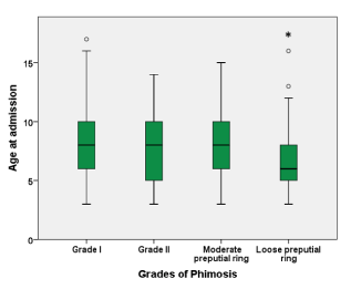
Phimosis was confirmed in 475 children, who accounted for about half of the patients referred for pediatric surgery evaluation. Approximately half of the boys (46.53%) had grade I phimosis [Table 2]. There was a positive correlation between a diagnosis of Phimosis and history of previous treatment with topical agents (p < 0.001-RR 2.224, 95% confidence interval [CI] 1.170-2.893). Patients with a loose preputial ring were significantly younger than the patients with other grades of Phimosis [Figure 2].
Nine children (1.89%) were lost to follow-up, and Phimosis resolution was achieved with non-surgical treatment in 87.79% of patients [Table 2]. Surgery was significantly more frequent among patients with grade I Phimosis than among those with other grades of the disorder (p < 0.001 - Pearson's chi-square test). All boys with moderate and loose preputial rings had their Phimosis resolved without surgical intervention.
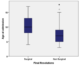
There was a significant association between age at admission and final resolution. Patients who required a surgical approach to the treatment of Phimosis were significantly older than those whose Phimosis resolved with topical treatment, as shown in [Figure 3].
The analysis of the correlation between previous treatment and final resolution showed that patients who had previously undergone topical treatment were 1.905 times more likely than those who did not receive such treatment to achieve Phimosis resolution with non-surgical treatment (Pearson's chi-square test, p = 0.035, 95% CI 1.038-3.4). The only patient who had a history of paraphimosis had his phimosis resolved with topical treatment. A previous history of balanoposthitis did not significantly influence the final resolution, i.e., the same proportion of patients with balanoposthitis was observed in the surgical and non-surgical treatment outcomes (Pearson's chi-square test, p = 1.00, RR 1.001, 95% CI 0.224-4.469).
Discussion
Approximately one in every two boys referred for pediatric surgery had their diagnosis of Phimosis confirmed. This finding provides further evidence of the difficulty that physicians not specialized in pediatric surgery or pediatric urology have in differentiating between normal and altered male genitalia (Phimosis in this case) [3,5,9]. In the present study, the boys whose diagnoses of phimosis were not confirmed had the prepuce adherent to the glans or some excessive foreskin, but they had no difficulty in exposing the glans due to changes in the prepuce. In addition, some children had normal-for-age genitalia [3,5,9,11,18,20]. The development of strategies for training these physicians in common pediatric disorders, such as Phimosis, could be useful in improving the care of pediatric patients.
Different classifications have been proposed by some authors with the aim of helping the diagnostic evaluation of Phimosis: these grades range from total inability to expose the glans to mild preputial rings with less difficultly in glans exposure [3,9,12,18-20]. Given that there is no established, uniform, and widely used classification in the publications on this topic, we herein proposed the standardization of the nomenclature used in the classification of Phimosis: We recommend using four grades and exclude the absence of phimosis as a grade. We aimed to test this simpler version of the previously used classifications in order to facilitate the assessment and therapeutic management of these patients.
The analysis of the correlation between age and grade of Phimosis showed that children with a loose preputial ring (those at a low risk of complications) tended to be younger and that there was no significant difference in the age at admission of children with other grades of the disorder. Some studies have reported that younger boys with a loose preputial ring should not be treated and only be monitored over the course of development of the genitalia as their disorder usually resolves without needing any additional treatment [3,5, 9,20].
However, other studies indicate that treatment is required for all patients, regardless of the grade of Phimosis, to avoid any further complications; the most serious complication being increased risk of cancer of the penis [8,10,14-19]. Thus, given the socio-economic characteristics of the population that received care in our pediatric surgery department, and with the aim of avoiding potential local complications and difficulties accessing specialized medical care, all boys with Phimosis (regardless of the grade of Phimosis and their age) were treated and monitored until the disorder was resolved. According to our experience, topical treatment should be administered initially. When this approach is not successful, or in complicated cases, surgical intervention is indicated.
After the diagnosis of Phimosis was confirmed by the surgical team, the children's parents or guardians received guidance on the use of topical corticosteroids (0.5% clobetasol propionate), which were to be applied for a minimum of five minutes, twice a day, for at least four weeks, as recommended in established protocols [5,10,19,21-22]. This treatment routine led to resolution of the disorder in 87.89% of all cases in this series, and resolution was achieved in all children with moderate or loose preputial rings. These findings confirm the results obtained in similar studies on Phimosis that reported resolution with topical corticosteroid therapy in 65% to 90% of patients [6,16, 21-23].
Surgical intervention, either by circumcision or posthectomy, for Phimosis should be consider only in specific cases because more complications are associated with this treatment than with topical treatment [4,12,17,24]. The need for surgical intervention was more frequent among older children with grade I Phimosis whose prepuce is probably less responsive to treatment with corticosteroids [5-6,9, 25-26].
Regardless of their age, patients who received topical treatment with corticosteroids on the foreskin were less likely to need surgery. This can be explained by the protective effect of this medication against the development of local inflammation, balanitis xerotica obliterans, and local fibrosis, which are conditions associated with Phimosis that require surgical correction [2,7,21,27].
This was a retrospective observational study that focused on the experience of a pediatric surgery referral center. The study did not involve random assignment of groups: Therefore, some factors might constitute limitations to the analysis of the obtained results. Moreover, it was not possible to control all the potential confounding factors during the study.
Despite the study's limitations, this was an important study given the size of the sample included and the limited loss to follow-up (1.89%). In accordance with the protocols used in the treatment of Phimosis in various pediatric surgical centers, all children and adolescents in the present study were evaluated by a single pediatric surgeon. This might have favored the standardization of the diagnosis, grading, and management of phimosis.
Considering the prevalence of Phimosis in the pediatric population, there are relatively few studies on the topic conducted in Latin countries. Studies like ours are important to help improve the knowledge and management strategies of this common disorder that is a great burden on healthcare.
Acknowledgements
Special thanks to Isabel Cristina, supervisor of the outpatient care section of our hospital, for her invaluable support in the organization of the process of outpatient care, which was essential in carrying out the study.
References
- Morris BJ, Matthews JG, Krieger JN, et al. (2020) Prevalence of Phimosis in Males of All Ages: Systematic Review. Urology 135: 124-132.
- Zampieri N, Frigo I, Caliò A, et al. (2021) Histology and immunohistochemical evaluation of phimotic prepuce: the role of steroid therapy. Andrologia 53: 13967.
- Lourenção PLTA, Queiroz DS, Oliveira Junior WE, et al. (2017) Ortolan EVP: Observation time and spontaneous resolution of primary phimosis in children. Rev Col Bras Cir 44: 505-510.
- Esposito C, Centonze A, Alicchio, et al. (2008) Topical steroid application versus circumcision in pediatric patients with phimosis: A prospective randomized placebo controlled clinical trial. World J Urol 26: 187-190.
- McGregor T, Pike JG, Leonard M, et al. (2017) Pathologic and physiologic phimosis: Approach to the phimotic foreskin. Canadian Family Physician 53: 445-448.
- Liu J, Yang J, Chen Y, et al. (2018) Is steroids therapy effective in treating phimosis? A meta-analysis. Int Urol Nephrol 48: 335-342.
- West DS, Papalas JA, Selim MA, et al. (2013) Dermatopathology of the foreskin: an institutional experience of over 400 cases. J Cutan Pathol 40: 11-18.
- Dave S, Afshar K, Braga LH, et al. (2018) CUA guideline on the care of the normal foreskin and neonatal circumcision in Canadian infants. Can Urol Assoc J 1212: 76-99.
- Yang C, Liu X, Wei GH, et al. (2009) Foreskin development in 10 421 Chinese boys aged 0-18 years. World J Pediatr 5: 312-315.
- Reddy S, Jain V, Dubey M, et al. (2012) Local steroid therapy as the first- line treatment for boys with symptomatic phimosis - a long-term prospective study. Acta Paediatr Int J Paediatric 101: 130-133.
- Chan IH Y, Wong K Y (2016) Common urological problems in children: prepuce, phimosis, and buried pênis. Hong Kong Med J 22: 263-269.
- Prabhakaran S, Ljuhar D, Coleman R, et al. (2018) Circumcision in the paediatric patient: A review of indications, technique and complications. J Paed Child Health 54: 1299-1307.
- Morris BJ, Kennedy SE, Wodak AD, et al (2017) Early infant male circumcision: Systematic review, risk- benefit analysis, and progress in policy. World J Clin Pediatr 6: 89-102.
- Many BT, Rizeq Y K, Vacek J, et al. (2020) A contemporary snapshot of circumcision in US children's hospitals. J Pediat Surg. 55: 1134-1138.
- Wang H, Huang Z, Zhou J, et al. (2019) Clinical Outcomes And Risk Factors In Patients Circumcised By Chinese Shang Ring: A Prospective Study Based On Age And Types Of Penile Disease. Ther Clin Risk Manag. 15: 1233-1241.
- Chen CJ, Satyanarayan A, Schlomer BJ, et al. (2019) The use of steroid cream for physiologic phimosis in male infants with a history of UTI and normal renal ultrasound is associated with decreased risk of recurrent UTI. J Ped Urology 15: 472-472.
- Quaba O, MacKinlay GA (2004) Changing trends in a decade of circumcision in Scotland. J Pediatr Surg 39: 1037-1039.
- Kikiros C, Beasley S, Woodward A, et al. (1993) The response of phimosis to local steroid application. Pediat Surg Int 8: 329-332.
- Kuehhas FE, Miernik A, Sevcenco S, et al. (2012) Predictive power of objectivation of phimosis grade on outcomes of topical 0.1% betamethasone treatment of phimosis. Urology 80: 412- 416.
- Hsieh TF, Chang CH, Chang SS, et al. (2006) Foreskin development before adolescence in 2149 schoolboys. Int J Urology 13: 968-970.
- Favorito LA, Balassiano C, Rosado J, et al. (2012) Structural analysis of the phimotic prepuce in patients with failed topical treatment compared with untreated phimosis. Int Brazj Urol 38: 802-808.
- Lund L, Wai KH, Mui LM, et al. (2000) Effect of Topical Steroid on Non-retractile Prepubertal Foreskin by a Prospective, Randomized, Double-blind Study. Scand J Urol Nephrol 34: 267-269.
- Favorito LA, Gallo C, Costa W, et al. (2016) Ultrastructural analysis of the foreskin in patients with true phimosis treated or not treated with topical betamethasone and hyaluronidase ointment. Urology 98: 138-143.
- Earp BD (2015) Do the benefits of male circumcision outweight the risks? A critique of the proposed CDC guidelines. Frontiers in Pediatrics 3: 1-6.
- Korkes F, Carlos A, Pompeo L, et al. (2012) Circumcisions for medical reasons in the Brazilian public health system: Epidemiology and trends. Einstein 10: 342-346.
- Sneppen I, Thorup J (2016) Foreskin Morbidity in Uncircumcised Males. Pediatric 137: 20154340.
- Folaranmi SE, Corbett HJ, Losty PD, et al. (2018) Does application of topical steroids for lichen sclerosus (balanitis xerotica obliterans) affect the rate of circumcision? A systematic review. J Ped Surg 53: 2225-2227.
Corresponding Author
Rodrigo Pinheiro de Abreu Miranda, Endereço: SHIN QI 08 conjunto 12, casa 18. Lago Norte, Brasília, DF, CEP 71520-320, Brazil; Tel: 61-999832251.
Copyright
© 2021 Miranda RPA, et al. This is an open-access article distributed under the terms of the Creative Commons Attribution License, which permits unrestricted use, distribution, and reproduction in any medium, provided the original author and source are credited.


