Rare Anatomic Finding of a Common Origin of the Coeliac and Superior Mesenteric Arteries in a Term Neonate
Case Report
A male infant was born term at 39 + 4 weeks gestation and birth weight of 3.98 kg. His mother was a 33-year-old primigravid with no relevant past medical history. The conception was natural and gestational diabetes mellitus identified in pregnancy was diet controlled. The baby was delivered via spontaneous delivery, did not require resuscitation and Apgars of 9 and 10 were documented.
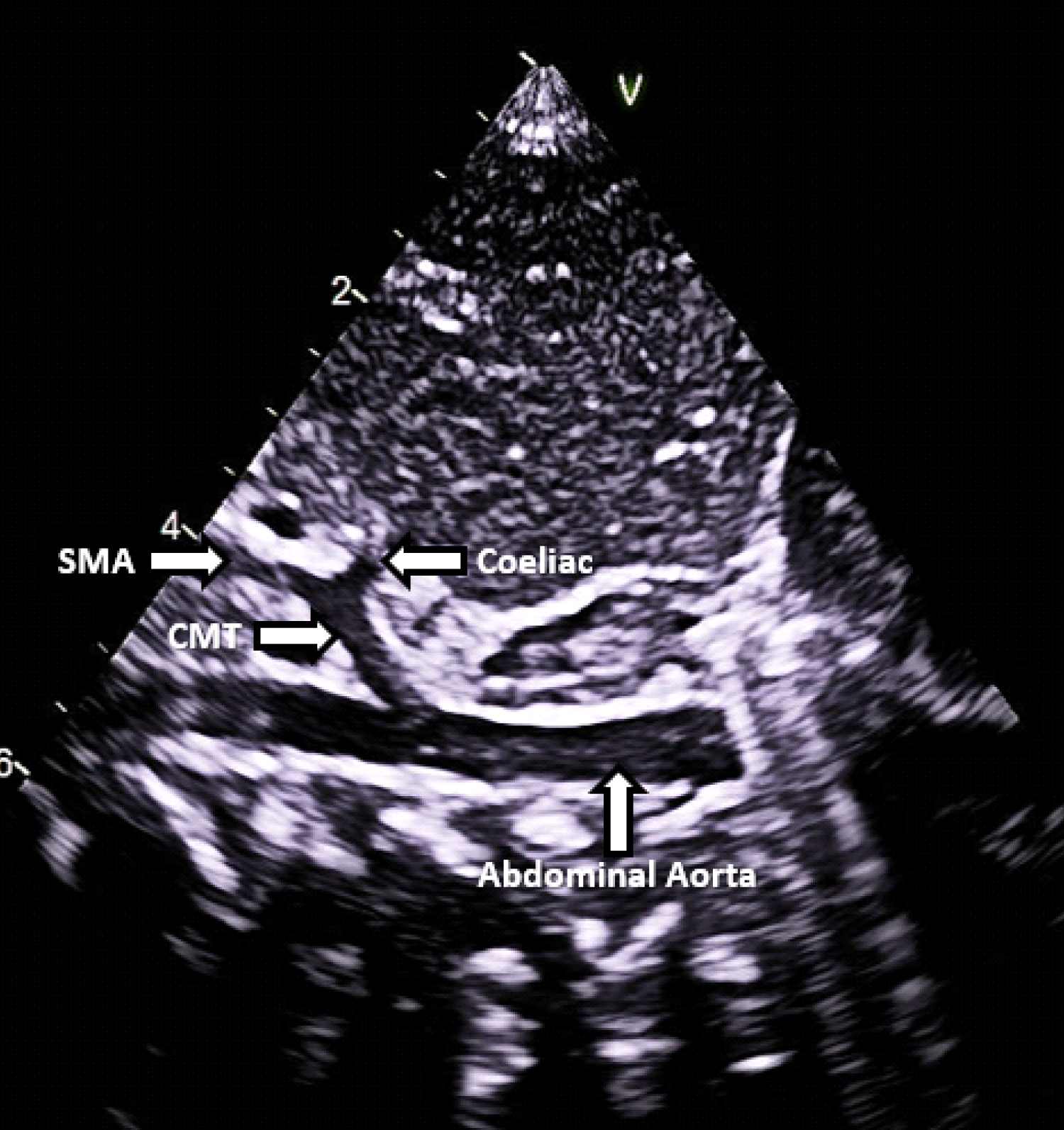
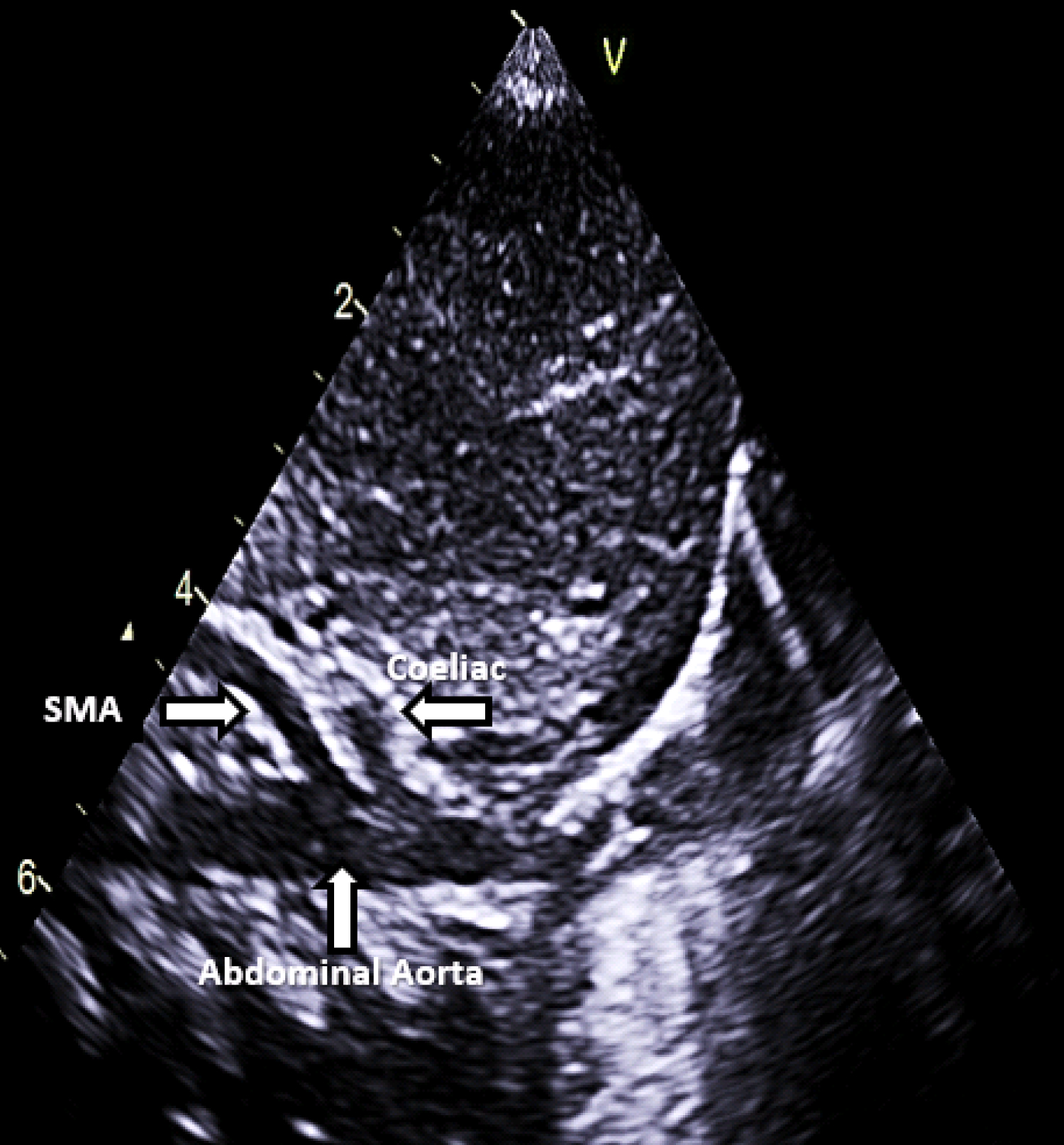
The infant was enrolled into an echocardiography research study assessing myocardial function in infants of gestational diabetic mothers. The echocardiogram confirmed a structurally normal heart, however an incidental finding of a common origin of the coeliac and superior mesenteric arteries was identified (Figure 1a). At the time the baby was clinically well, breathing spontaneously on room air and breastfeeding successfully. On physical examination there was no evidence of jaundice, hepatomegaly or poor perfusion. An abdominal ultrasound performed by a consultant radiologist (SR) confirmed that the superior mesenteric artery (SMA) and coeliac artery had a common trunk. The liver appearance was normal as was the remainder of the abdominal ultrasound scan. The baby's images were reviewed at a paediatric gastroenterology radiology conference and the vascular findings of a common SMA/coeliac trunk were deemed consistent with a variant of normal anatomy. No further investigations or follow up were advised.
The coeliac trunk is the first branch of the abdominal aorta. Three arteries usually originate from the coeliac trunk; namely the common hepatic artery, the left gastric artery and the splenic artery [1]. The superior mesenteric artery (SMA) is the second branch of the abdominal aorta and typically arises separately from the aorta below the coeliac trunk (Figure 1b). The coeliac trunk and SMA share a close anatomical and functional connection and various configurations in their branching patterns have been reported in the literature [2,3]. The celiac axis, SMA and hepatic artery are the most important branches of abdominal aorta due to their large vascularization fields [4].
Coeliomesenteric trunk (CMT) is a rare congenital variation in which the coeliac artery and the SMA originate from the abdominal aorta via a common trunk [5]. The estimated incidence of CMT ranges from 0.4% to 2.7% [6]. Recently a new classification system has been proposed to further classify CMT variants identified on multidetector computed tomography (MDCT) angiography [6]. This classification system describes five types of CMT variants according to the length of the single common trunk, origin of the left gastric artery and specific branching variation as assessed by MDCT angiography imaging. Gender is not thought to influence CMT variations however some publications suggest that they may be more prevalent in Japanese and Korean people than the Caucasian population [7]. Reported clinical consequences of CMT can include arterial aneurysm, left renal vein compression (also known as 'Nutcracker phenomenon') and a potentially increased risk of mesenteric ischaemia [5,6,8]. With growing trends in the use of laparoscopic surgery, invasive radiological procedures, pancreatic surgery and organ transplantation accurate identification of vascular anomalies and anatomical variations prior to such interventions is essential [9]. A study reviewing CT scans of 36 adults with CMT reported that in 88% of cases the CMT was not documented in the official radiology report, illustrating that such anatomic anomalies are frequently unnoticed [10]. Further knowledge and awareness of such vascular anomalies among radiologists, physicians, and surgeons may enhance pre-operative planning and prevent vascular injury during such procedures.
Funding
The authors have not declared a specific grant for this research from any funding agency in the public, commercial or not-for-profit sectors.
Competing Interests
None declared.
Patient Consent
Parental consent obtained.
References
- Satheesha Nayak B, Shetty SD, Sirasanagandla SR, et al. (2018) Concurrent variations of celiac and superior mesenteric arteries. Kathmandu Univ Med J (KUMJ) 16: 345-347.
- Yan J, Nagasawa Y, Nakano M, et al. (2014) Origin of the celiac and superior mesenteric arteries in a common trunk: Description of a rare vessel variation of the celiacomesenteric trunk with a literature review. Okajimas Folia Anat Jpn 91: 45-48.
- Wang Y, Cheng C, Wang L, et al. (2014) Anatomical variations in the origins of the celiac axis and the superior mesenteric artery: MDCT angiographic findings and their probable embryological mechanisms. Eur Radiol 24: 1777-1784.
- Farghadani M, M Momeni, A Hekmatnia, et al. (2016) Anatomical variation of celiac axis, superior mesenteric artery, and hepatic artery: Evaluation with multidetector computed tomography angiography. J Res Med Sci 21: 129.
- Peterson J, Hage AN, Diljak S, et al. (2017) Incidental anatomic finding of celiacomesenteric trunk associated with 'Nutcracker Phenomenon,' or compression of the left renal vein. Am J Case Rep 18: 1334-1342.
- Tang W, J Shi, LQ Kuang, et al. (2019) Celiomesenteric trunk: New classification based on multidetector computed tomography angiographic findings and probable embryological mechanisms. World J Clin Cases 7: 3980-3989.
- Panagouli E, Venieratos D, Lolis E, et al. (2013) Variations in the anatomy of the celiac trunk: A systematic review and clinical implications. Ann Anat 195: 501-511.
- Wang Y, P Chen, N Shen, et al. (2010) Celiomesenteric trunk with concurrent aneurysm: Report of a case. Surg Today 40: 477-481.
- Ozgokce M, Ayyıldız VA, Ogul H, et al. (2018) Common coeliacomesenteric trunk: A computed tomography radiological study. Folia Morphol (Warsz) 77: 683-686.
- Maldjian PD, MA Chorney (2015) Celiomesenteric and hepatosplenomesenteric trunks: Characterization of two rare vascular anomalies with CT. Abdom Imaging 40: 1800-1807.
Corresponding Author
Aisling Smith, Department of Neonatology, The Rotunda Hospital, Dublin, D01P5W9, Ireland.
Copyright
© 2020 Smith A, et al. This is an open-access article distributed under the terms of the Creative Commons Attribution License, which permits unrestricted use, distribution, and reproduction in any medium, provided the original author and source are credited.