Primary Endobronchial Tumors in Children: Four Cases
Abstract
Primary lung tumors are very infrequent in childhood. According their degree of malignancy, they can be classified into two groups: Benign endobronchial tumors and malignant tumors, which are the most frequent.
Herein, we show our experience in the management of four endobronchial tumors: One bronchial carcinoid tumor, one myofibroblastic tumor, one Hodgkin lymphoma metastases and one endobronchial polyp. We describe the clinical presentation, diagnostic method, histology, treatment and outcome.
Although primary endobronchial tumors are uncommon, it is a diagnosis to consider with persistent bronchial obstruction, infection or recurrent lung collapse that does not improve despite standard treatment. Bronchoscopy and pathology studies will allow us to confirm the exact diagnosis. Treatment is usually surgical or adjuvant chemotherapy associating with the type of tumor diagnosed. The evolution is generally good after suitable treatment.
Keywords
Endobronchial, Tumors, Children
Introduction
Primary lung tumors are extremely rare in childhood; the exact incidence remains unknown [1-4]. They can be classified into two major groups according to their degree of malignancy [2,5]; within benign endobronchial tumors the most frequent are inflammatory myofibroblastic tumor (IMT) and hamartoma; in the group of malignant tumors, which constitute approximately 70-75%, we have metastases, usually extracranial tumors and primary tumors [5]. We include in primary tumors bronchial carcinoid tumors (the most common primary lung tumor), followed in frequency by mucoepidermoid tumor, bronchogenic tumor, pleuropulmonary blastoma and lymphoma among others [1,2,5].
The low incidence of this pathology often leads to a delay in diagnosis [2,5] which is based on imaging tests and pathological studies [2]. The overall treatment is surgical with a favorable outcome in pediatric ages [2].
Material and Methods
We analyzed four cases of endobronchial tumors diagnosed at our center between 2000 and 2017, in order to describe the clinical presentation, diagnostic method, histology, treatment and outcome.
Results
Four patients, three girls, one boy, aged from 3 months to 12 years diagnosed with endobronchial tumors were found. The four patients were symptomatic and their pathological diagnoses were bronchial carcinoid tumor [1], myofibroblastic tumor [1], Hodgkin lymphoma metastases [1] and endobronchial polyp [1]. Each case is presented hereafter with more detail.
Case 1
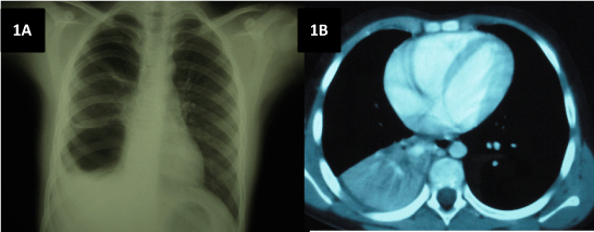
A 9-year-old girl was transferred from another hospital to complete imaging study condensation/persistent right lower lobe atelectasis (LID). She did not have personal or family history of interest, except episode asthma. She complained of intermittent fever syndrome for 2 months associated with cough and weight loss. Initially, she was diagnosed with pneumonia, having carried out antibiotic treatment without clinical or radiological response. The personal history included frequent episodic asthma mites and pollen sensitization and 3 previous episodes of pneumonia, 2 of which have required hospitalization. She had no family history (AF) of special interest. Physical examination revealed the presence of hypoventilation in the lower third of the right hemithorax. The complementary tests performed were: CBC, biochemistry, liver function and kidney: Normal; ESR 113 mm/h; mantoux 0 × 0 mm, negative sweat test, α-1 antitrypsin normally. Chest x-ray (Figure 1A): Condensation in right lung base; lung CT (Figure 1B) showed condensation LID, atelectasis that level associated with small pleural effusion and posterior right paratracheal lymphadenopathy, ipsilateral hilar and subcarinal; urinary catecholamines were normal; bronchoscopy: Tumor at entry of right basal bronchus pyramid that almost completely obstructs the bronchial lumen; pathological anatomy was consistent with bronchial carcinoid tumor with low proliferative index. Based in this diagnosis, middle and lower lobectomy is performed with favorable evolution. The patient did not require additional treatment with chemotherapy or radiotherapy.
Case 2
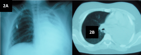
A 12-year-old boy is derived from their hospital of origin in order to study massive atelectasis of the left lung. He complained of dry cough with 2-3 months of evolution, associated with intermittent vomiting and fever. He has received antibiotic treatment in 2 occasions with transient improvement of the fever. Given the persistence of symptoms, a chest radiography was performed, appreciating massive atelectasis of the left lung. The boy did not have personal or family history of interest. Physical examination showed normal findings except hypoventilation in left chest. Investigations performed were: Blood count, biochemistry, liver function and kidney normal, ESR 94 mm/h, LDH 1207 IU/L, enolase, catecholamines: Normal; aspirate and biopsy of bone marrow (BM): Normal; cytochemical CSF: Normal; Chest x-ray (Figure 2A) and lung CT (Figure 2B) showed massive atelectasis of the left lung; abdominal CT revealed right intrarenal mass which caused obvious nephromegaly; bronchoscopy: Left intrabronchial mass causing atelectasis; biopsy intrabronchial mass: Lymphocyte lineage cells positive for CD45 and CD20, compatible with Non-Hodgkin lymphoma B cells; FNA right renal tumor: Round cell neoplasia positive for CD45, compatible with Non-Hodgkin lymphoma (NHL) B-cell lymphoma Ki negative. In view these tests the patient is diagnosed with NHL immunophenotype B stage III, with primary location doubtful. Treatment and evolution: The patient began chemotherapy. At the end of the induction phase of imaging and bio-hematologic shows complete clinical remission. At this time, after 5 years of follow-up in outpatient, he is discharged free of disease.
Case 3
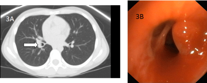
10-year-old girl who was referred for consultation by hemoptysis and contacting with a cousin diagnosed of tuberculosis. Additional tests: Mantoux 0 × 0 mm; lung CT: Occupation air space right lower lobe basal and lateral segments. These images of occupancy simulate a tree in bud, findings suggestive of infectious bronchiolitis and endobronchial spread of infectious process. Peripherally patchy areas seen in "ground glass". Probable right hilar nodal formations. In this situation, she began treatment with isoniazid, rifampicin, pyrazinamide and ethambutol for 2 months, after she continued with isoniazid and rifampicin. The second mantoux was also negative, so a new CT (Figure 3A) was performed and it showed a hyperdense image of polipodideo aspect that partially occupies the lumen of the right intermediate bronchus. Fibrobronchoscopy (Figure 3B) two reddish bumps, pedunculated and well defined that partially occupy the right intermediate bronchus which are removed along with service of Pediatric Surgery. The pathology was consistent with an inflammatory polyp with no evidence of granulomas, culture and PCR of bronchoalveolar lavage and sputum were negative. Treatment and evolution: After the removal of the endobronchial polyps, the patient presented a favorable evolution.
Case 4
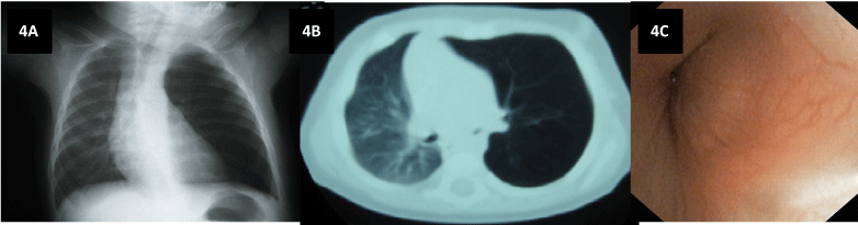
Infant 3-month-old was admitted for study rejection of shots in the last 10 days, remaining afebrile without signs of fatigue or breathlessness. Personal or family history did not provide remarkable information. Physical examination: hypoventilation in left chest, with the rest of the scan being normal. Chest x-ray (Figure 4A): Air trapping in the left lung without significant displacement of the mediastinum and contralateral lung. Lung CT (Figure 4B): Stenosis of the left main bronchus and left lung hyperinflation; bronchoscopy (Figure 4C): Tumor non-pulsatile output level of the left main bronchus stenosing 70-80% of the bronchial lumen. Post excision pathology was consistent with myofibroblastic tumor. After tumor resection by thoracotomy, she presented favorable evolution, remaining disease-free at present time.
Discussion and Conclusions
In our series, we dealt with an inflammatory myofibroblastic tumor (IMT) or inflammatory pseudotumor, cataloged within benign endobronchial tumors, a bronchial carcinoid tumor, an endobronchial polyp and endobronchial metastases from Non-Hodgkin type B. Most of patients have symptoms at the time of diagnosis [2-4], the most frequent are persistent cough, recurrent respiratory infections and persistent bronchospasm [2-4]. In our serie, all patients were symptomatic at diagnosis, appearing in 2 of the cases dry cough and intermittent fever, hemoptysis in one case and rejected shots in the three-month infant. Three patients presented pathological cardiopulmonary auscultation, being hypoventilation the most common finding.
Myofibroblastic tumor behaves as a benign tumor and it is considered as an inflammatory response to a previous stimulus [5-7] It is one of the most frequent benign endobronchial tumors and may also appear in intraparenchymal level 4. Its growth is slow and progressive and tends to local invasion and recurrence without treatment [5,6]. The treatment of choice is total excision if possible [4]. In our case nº .4 after the surgical removal of the endobronchial tumor, evolution was favorable.
Bronchial carcinoid tumor, regarded as the most common primary malignant endobronchial tumor, is a tumor of neuroendocrine origin, usually affecting the larger bronchi [5,7]. Often patient presented hemoptysis or repeated infections [5-9]. Carcinoid syndrome is rare in bronchial carcinoid tumors. Typical bronchial carcinoids are central, distinct and show favorable prognosis after resection [1,2,5,9].
Endobronchial polyps found in our case nº .3, may appear as a rare manifestation of a endobronchial tuberculosis [3,9,10] which in our case could not be verified as the mantoux was repeatedly negative, cultures and PCR Mycobacterium tuberculosis were negative and no granulomas were evident in the resected piece.
Pulmonary lymphoma is defined as lung/endobronchial involvement appears with no extrapulmonary disease previously diagnosed [2,11]. In the three-month time following the diagnosis of primary pulmonary lymphoma, no extrathoracic disease [2,11] should appear. Non-Hodgkin lymphoma primary endobronchial type B, although it may appear, is extremely rare and cannot rule out hematogenous or lymphatic spread from a primary tumor undiagnosed [11], as confirmed in our case nº .2 when performing abdomen CT.
In conclusion, the primary endobronchial tumors are uncommon in pediatric patients but it is a diagnosis to consider with persistent bronchial obstruction, infection or recurrent lung collapse that does not improve despite antibiotic treatment. The suspected diagnosis is given by imaging tests, bronchoscopy and pathology studies that allow us to confirm the exact diagnosis. They have a wide variety and different histological grades of malignancy. Treatment is usually surgical or adjuvant chemotherapy associating with the type of tumor diagnosed. In metastases will be necessary to treat the primary tumor, in some cases avoiding surgery of endobronchial tumor. The evolution is generally good after suitable treatment, as shown in our series of cases, where 100% of patients remain disease-free after five years after diagnosis. However so long-term follow-up of these children is recommended [12].
References
- Tardío Torío E, Sánchez Sánchez E, Vila Torres J, et al. (1998) Carcinoide bronquial: A propósito de un caso. An Esp Pediatr 49: 171-173.
- Otero E, Rizzi A, Maurizi M, et al. (2006) Tumores primitivos de pulmón en niños: Imágenes y hallazgos. Medicina Infantile 13: 132-138.
- Al-Qahtani AR, Di Lorenzo M, Yazbeck S (2003) Endobronquial tumors in children: Institutional experience and literature review. J Pediatr Surg 38: 733-736.
- Andrés Martín A, Alfageme Michavilla I, Escalada Berta J, et al. (2007) Tumor carcinoide bronquial en una niña de 10 años. Neumosur 19: 109-111.
- López Día M, Antón-Pacheco Sánchez JL, Tejedor Sánchez R, et al. (2006) Tumores broncopulmonares primarios. Cir Pediatr 9: 223-227.
- Karnak I, Haliloglu M, Orhan D, et al. (2014) Pure endobronchial inflammatory myofibroblastic tumor in children. J Pediatr Hematol Oncol 36: 108-110.
- Camela F, Gallucci M, Di Palmo E, et al. (2018) Pulmonary inflammatory myofibroblastic tumor in children: A case report and brief review of literature. Fronto Pediatr 6: 35.
- Cano García J, Baamonde Laborda C, Algar Algar FJ, et al. (2007) Cirugía broncoplástica en el tumor carcinoide bronquial típico. Descripción de tres casos en la infacia. Revista Neumosur 19: 218-221.
- Hullo E, Cotta L, Rabeyrin M, et al. (2011) Tumeurs carcinoïdes bronchiques de l'enfant. Bulletin du Cancer 98: 709-715.
- Andrés Martín A, Pérez Pérez G, Borja Urbano G, et al. (2012) Síndrome del lóbulo medio y tuberculosis bronquial. Aportación de la fibrobroncoscopia. Acta Pediatr Esp 70: e51-e55.
- Taisuke Kobayash, Masamitsu Hyodo, Nobumitsu Honda (2013) Primary endobronchial Burkitt's lymphoma in a child: A case report. Int J Pediatr Otorhinolaryngol 77: 875-878.
- Eyssartier E, Ang P, Bonnemaison E, et al. (2014) Characteristics of endobronchial primitive tumors in children. Pediatr Pulmonol 49: 121-125.
Corresponding Author
Marta González Fernández-Palaciosa, Unidad de Neonatología, Hospital Universitario Virgen del Rocío, Sevilla, España.
Copyright
© 2018 Fernández-Palaciosa MG, et al. This is an open-access article distributed under the terms of the Creative Commons Attribution License, which permits unrestricted use, distribution, and reproduction in any medium, provided the original author and source are credited.