Bronchial Mucoepidermoid Carcinoma in a 14-year-old Patient
Abstract
A bronchial mucoepidermoid carcinoma is a rare malignant tumour. It usually appears in young subjects and most often involves the salivary glands. We report the case of a 14-year-old patient who presented with a pulmonary mucoepidermoid carcinoma that was discovered during the evaluation of a case presenting with recurrent pneumopathies. Recurring pneumopathies must lead to suspicion of various aetiologies and therefore require thorough evaluation so that rare tumours are not ignored, not missed, particularly in children.
Keywords
Mucoepidermoid carcinoma, Recurrent pneumopathies, Segmentectomy
Introduction
Bronchopulmonary tumours are rare among children. Bronchial mucoepidermoid carcinoma is a malignant tumour that accounts for 0.1% to 0.2% of pulmonary tumours [1]. They are usually observed in young subjects who are less than 30-years-old. These lesions are often found in salivary glands. They are composed of mucus-secreting cells, squamous cells and intermediate cells. To date, surgical resection remains as the first treatment. Its prognosis is good for low-grade lesions; high-grade lesions can result in local recurrences and metastases. We present the case of a 14-year-old girl patient who was treated for a pulmonary mucoepidermoid carcinoma that was discovered in a patient presenting with recurrent pulmonary infections.
Observation
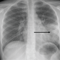
A 14-year-old girl child presented with a history of recurrent pulmonary infections that was always involving the upper lobe of left side lung (Figure 1). In November 2010, she presented with the first acute pulmonary infection. The resolution was good after receiving empirical antibiotic therapy. During February to June 2011, she presented with 3 more attacks in the same pulmonary territory that resolved with antibiotic treatment. Chest X-rays taken after recovery were considered normal every time.
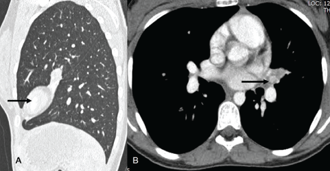
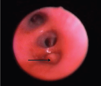
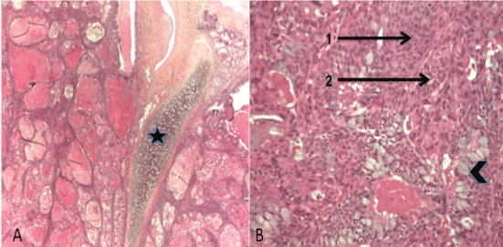
In April 2012, a chest CT scan was first performed to evaluate the acute pain in the left side chest in the patient (Figure 2). It demonstrated a 7 millimetre diameter tumour that was responsible for atelectasis of the anteroinferior segment of upper left lobe. Bronchial endoscopy confirmed the obstruction of the proximal lingual (Figure 3). A carcinoid tumour was first suspected from the endoscopic aspect. Several endoscopic biopsies of the obstructive lesion did not allow a pathological diagnosis. Surgical treatment, performed through transaxillary thoracotomy, consisted of a lingulectomy with lymph node dissection in areas 4, 5, 7, 10 and 11 L. Four lymph nodes were dissected. Pathological examination of the surgical resection demonstrated a 2 centimetre diameter low-grade malignant mucoepidermoid carcinoma. The diagnosis was confirmed by routine histology stains (Figure 4). Immunochemistry was not performed because it was not considered necessary in this case. The analysis confirmed the complete removal of tumor with a healthy 1.5 centimetre margin of the bronchial section. Three peribronchial lymph nodes were analysed and none showed metastasis. The lesion was classified as pT1aN0M0.
The postoperative course was uneventful. The patient was discharged one week after the operation. The patient is still followed up with regular clinical examination and chest X-rays. Five years after the surgery she is well and has not presented with any more pulmonary infection.
Discussion
Smetana, et al. first described the mucoepidermoid carcinoma in 1952 [2]. It occurs in all ages ranging from 4 to 80-years-old with prevalence in young adults between 20 and 30 years of age. It accounts for only 0.1% to 0.2% of primary bronchopulmonary cancers [3]. No supporting factor has found to explain the occurrence of this tumor. This tumour is classified as low-grade or high-grade according to histological criteria such as cellular atypias, mitotic activity, tumoural necroses, or local extension of the disease. This tumour mainly occurs in the lobar or segmental bronchi but rarely occurs in the most distal bronchi. In the same ages, the most common tumour types were carcinoid, inflammatory myofibroblastic tumour and pleura pulmonary blastoma. It is possible to find all types of bronchogenic carcinoma that are described in adults in the pediatric age group also. Carcinoid tumours accounted for 11% to 13% of all paediatric pulmonary tumours [4].
Among the two histological subtypes of mucoepidermoid carcinomas, low-grade lesions account for approximately 95% as compared to only 5% of high-grade lesions. Histologically, low-grade lesions primarily consist of a glandular structure, clear limits and a small few mitoses. There is no necrosis in low-grade lesions in contrast to high-grade lesions, the later present with a majority of squamous component [4]. Mitoses are numerous in high-grade lesions. For high-grade lesions, diagnosis is often difficult because they have very similar histological characteristics of adenosquamous carcinomas (Figure 4).
Low-grade lesions have a good prognosis with rare local recurrence, which is opposite in case of high-grade lesions that have a prognosis that seems poorer according to the small number of reported cases.
The clinical presentation of these tumours is not specific and initially consists of an irritation of one of the air routes, gradually progressing to complete obstruction. Thus, cough, haemoptysis, dyspnoea or recurrent pulmonary infections can be observed [3,5]. In this context, several diagnoses were possible in children like the ingestion of any object. Chest X-ray is usually not conclusive, but in the absence of a clear explanation repeated pneumopathy in the same territory of the lung should allow suspicion of the existence of a tumour. Bronchial endoscopy can then confirm the existence of a grey rosy nodular Polyp Endobronchial lesion; pathological examination of biopsies should allow the diagnosis.
In parallel, an injected CT scan typically shows oval or round well-defined lesions. Hyper vascularisation of the tumour can lead to misdiagnosis with carcinoid tumours [6,7]. Calcifications can also be noted.
The treatment of these tumours consists of complete surgical resection. Association with radiotherapy for lesions with a high rank has been proposed [8]. In our case, we performed a minimally invasive surgery, a transaxillary muscle-sparing thoracotomy. The aim was to preserve muscle and aesthetics for the young child.
Conclusion
Mucoepidermoid tumours with pulmonary locations are rare tumours that are difficult to diagnose because of variable clinical presentations. It is necessary to repeat routine examinations and bronchial endoscopy in order to understand the etiology of repeated respiratory episodes, particularly in young patients.
Regardless of the minimal frequency of these lesions, it is necessary to remember that mucoepidermoid tumours occur in young patients without particular risk factors and that the risk of metastases exists in the absence of treatment. Continuation of clinical and radiological monitoring seems necessary in order to detect local recurrences.
References
- Heitmiller RF, Mathisen DJ, Ferry JA, et al. (1989) Mucoepidermoid lung tumors. Ann Thorac Surg 47: 394-399.
- Smetana HF, Iverson L, Swan LL (1952) Bronchogenic carcinoma; An analysis of 100 autopsy cases. Mil Surg 111: 335-351.
- Terrier JP, Chetaille B, Reynaud-Gaubert M, et al. (2000) Bronchial mucoepidermoid carcinoma: A series of three cases. Ann Pathol 20: 623-625.
- Yu DC, Grabowski MJ, Kozakewich HP, et al. (2010) Primary lung tumors in children and adolescents: a 90-year experience. J Pediatr Surg 45: 1090-1095.
- Blawat P, Kowalewski J, Dancewicz M, et al. (2010) Muco-epidermoid carcinoma of the lung in a 20-year-old woman. Rev Mal Respir 27: 1092-1095.
- Ishizumi T, Tateishi U, Watanabe S, et al. (2008) Mucoepidermoid carcinoma of the lung: high-resolution CT and histopathologic findings in five cases. Lung Cancer 60: 125-131.
- Lamotte F, Meurice JC, Dore P, et al. (1992) Mucoepidermoid bronchial carcinoma. Review of the literature of a new case. Rev Pneumol Clin 48: 29-32.
- Smith CB, Swanson SJ, Mhango G, et al. (2013) Survival afters segmentectomy and wedge resection for stage 1 non-small-cell lung cancer. J Thorac Oncol 8: 73-78.
Corresponding Author
François Bastard, Département de Chirurgie Pédiatrique, Centre Hospitalier et Universitaire d'Angers, 4 Rue Larrey, 49933 Angers Cedex 9, France, Tel: +33-6-77-19-18-08.
Copyright
© 2017 Bastard F, et al. This is an open-access article distributed under the terms of the Creative Commons Attribution License, which permits unrestricted use, distribution, and reproduction in any medium, provided the original author and source are credited.