Preliminary Report on Laparo-Endoscopic Single-Site Approach for Transposition of Renal Lower Pole Crossing Vessels in Children
Abstract
We describe here our first single-site laparo-endoscopic experience with four cases of ureteropelvic junction obstruction due to lower pole crossing vessels. Through a single-site umbilicus approach, we performed a vascular hitch by enclosing the vessels in an overlapping tunnel of pelvic tissue. During the procedure, we used a 30° scope and either standard straight laparoscopic instruments or the JAIMY™ articulated and motorized 5-mm needle holder (Endocontrol™). Median operative time was 94 minutes (range 86-136 minutes). Postoperative recovery was uneventful, with discharge after two days in case 1, and during the first postoperative day in the other cases. In all cases, the umbilicus scar became almost invisible, and the ultrasound follow-up was characterized by a marked decrease of the pelvic dilation two months later. These four cases show the feasibility of a minimally invasive single port approach for kidney vessel transposition in children. Articulated and motorized instruments could make the procedure less complicated.
Keywords
Laparo-endoscopy single site access, Vascular hitch, Lower pole crossing vessels, Uretero-pelvic junction obstruction
Introduction
Laparoscopic surgery has evolved rapidly and become a standard in pediatric urological practice. Therefinement and modification of laparoscopic instruments has resulted in a substantial increase in the use of Laparo-Endoscopic Single Site surgery (LESS) in adult urology over the past 3 years. Single Site surgery was developed to improve cosmetics, decrease pain and minimize the number of trocars. Since the first report of a varicocelectomy in a child in 2007 [1], there have been few reports of LESS in children, especially for upper urinary tract diseases. To the best of our knowledge, we describe the first transposition of Lower Pole Crossing Vessels (vascular hitch) for kidney junction obstruction through a LESS approach.
Patients and Methods
Preoperative patient management
All patients included in this retrospective series underwent surgery in our institution between 2011 and 2016 for Ureteropelvic Junction Obstruction (UPJO) caused by lower polar renal vessels. Clinical diagnoses were made based on pain characterized by intermittent renal colic, pyelonephritis or fortuitously. Each time, obstruction was confirmed by MAG3 scintigraphy, and the presence of lower pole crossing vessels was assessed through renal ultrasound ± angiography scan. Depending on the infectious event, the size of the dilation (> 20 mm) and/or the deterioration of renal function, surgical treatment was offered and agreed to.
Surgical technique
Children were placed in a left or right semilateral decubitus position, depending on the side of the stenosis. The cutaneous incision was made inside the umbilicus, in a horizontal "Z" shaped fashion to perform an open laparoscopic approach. The Alexis® Wound Retractor inner ring was then inserted inside a 25 mm surgical hole, and the GelPOINT Advanced Access Platform (Applied, Rancho Santa Margarita, CA, USA) was clipped on the outer ring. Four 5 mm trocars were used, including one for the scope.
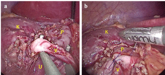
The approach of the right kidney began after opening Toldt's fascia in order to lower the right colon. The parietal peritoneum was opened at the level of the renal pelvis. In all cases, the mobilization of the inferior pole of the right kidney showed inferior polar vessels, which were clearly responsible for the obstruction (Figure 1a). In the left ureteropelvic junction obstruction, the opening of the mesocolon in the splenic angle gave access to the kidney. Subsequently, we dissected the pelvis, the ureter and the vessels, crossing in front of the Ureteropelvic Junction (UPJ). The lower pole vessels were then affixed in a cephalad position, at a suitable distance from the UPJ, by overlapping the vessels with the redundant pelvic anterior wall tissue, which was fixed with 2 absorbable sutures (Figure 1b). Diuretics were then intravenously injected to ensure the presence of peristaltic contractions of the ureter, without increasing the diameter of the pyelon.
In two cases, the entire procedure was performed with standard straight laparoscopic instruments.In the other cases, we also used the new JAIMY™ instrument (Endocontrol™), an articulated and motorized 5 mm needle holder, designed for laparoscopic surgery. It offers to surgeons an unlimited 360° rotation and a bidirectional controlled flexion for an improved triangulation during the suturing procedure.
Postoperative patient follow-up
Patients were assessed to a postoperative clinical and ultrasonographic evaluation 2 months after surgery, and to a control MAG3 scintigraphy performed at least 6 months after surgery. Then, they had an annual follow-up during the next three years.
Results
Patients were 2 boys and 2 girls, with a median age of 11.5 (5.7-12.9) years. The revealing factor of UPJO was pain in the case of 2 patients, an incidental ultrasound finding following a road accident in the third case, anda urinary tract infection that required ultrasonographic investigationsin the fourth case.
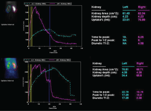
None of the patients had a prenatal history of renal dilation, and the right kidney was involved in 3 of 4 cases. A renal ultrasound was performed for all patients, which showed a median pyelocalyceal dilation of 22.5 mm (18-29 mm). For one patient, the crossing vessels were seen on renal ultrasound. Two patients required an additional angiography scan to confirm the existence of a polar artery crossing the junction, and the presence of crossing vessels was assessed during surgery for the last patient. The renal function of the dilated kidney on MAG3 scintigraphy was normal in two cases and impaired in the other two (23% and 27% of relative renal function, respectively), which also had renal cortical thinning on the ultrasound exam. Spontaneous urinary excretion was impaired in all cases, with a very limited response after furosemide injection (Figure 2).
Median operative time was 94 minutes (86-136 minutes) with negligible blood loss. In 2 cases, we had to use an additional 5 mm trocar that was inserted once in the subxiphoid region and once in the left iliac fossa to offer a better access and exposition of the renal pelvis.
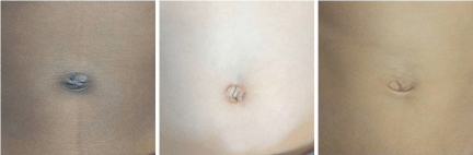
All postoperative courses were uneventful, with a visual analogue pain scale below 3/10 and an oral intakethat startedon the first postoperative day, which allowed the patients to be discharged. Only the first girl was required to stay in hospital until day 2. With a median delay of 16 months (12-72), there was no postoperative complication, and the ultrasound performed 2 months later showed the absence of pelvis dilation in all of the cases. Three patients underwent a postoperative MAG3 scintigraphy, with a median delay of 8 months (7-23) after surgery, which showed a good resolution of the obstruction (Figure 2). The last one was not submitted to this postoperative exam due to the complete resolution of preoperative symptoms and the disappearance of any pelvic dilation on ultrasound exams during the 23 months of follow-up. The umbilicus scar was almost invisible (Figure 3).
Discussion
The standard treatment of UPJO in children, regardless of etiology, has long been open dismembered pyeloplasty. Since the first reported experience of laparoscopic pyeloplasty in children in 1995 [2], this approach has gained wide acceptance as a minimally invasive procedure to correct UPJO. The overall incidence of UPJO caused by lower pole crossing renal vessels is approximately 18.5% in current literature [3]. However, among symptomatic children, the proportion of those with UPJO caused by extrinsic pressure from crossing renal vessels was significantly higher, reaching 30-50% [4,5]. In two of our cases, preoperative investigations only included ultrasounds and scintigraphy. In the other cases, we decided to assess the presence of lower pole crossing vessels by using an angiography scan due to the patients clinical history, with variable features of kidney dilation on serial ultrasounds and a preserved renal function, but an increase in the number of painful episodes, which meant that surgical indication was debatable.
Based on the original description in 1949 by Hellström, et al. [6], the vascular hitch seems an attractive surgical option compared to the standard dismembered pyeloplasty. Indeed, it avoids the need to violate the collecting system, thus preventing potential complications such as leakage or anastomotic strictures, or the use of drains or stents. The vessels are later enclosed in an overlapping tunnel of pelvic tissue, rather than affixed against the renal pelvis wall with perivascular adventitial sutures. The first vascular hitch by transperitoneal laparoscopy was reported by Godbole [7] on 13 patients. All underwent surgery involving three 5 mm trocars. The operating time was 92 minutes (74-120), with a short hospital stay of 24 to 36 hours. The success rate of this technique was more than 95%, with relief of pain, absence of complications and conversion, and improved data on ultrasound and scintigraphic exams after an average follow-up of 6 months. Its success led to the adoption of the technique by other surgeons (Table 1).
Nevertheless, identifying lower pole crossing vessels should not be the only determinant for performing vascular hitch because patients may have an associated intrinsic ureteral component of UPJO along with crossing vessels. Indeed, Janetscheck, et al. [8] found that, when the UPJ was routinely incised and inspected during dismembered pyeloplasty with associated crossing vessels, up to 33% of patients had an intrinsic abnormality. One of the difficulties of this intervention is therefore to ascertain that the obstruction is the direct result of a sole Inferior Polar Artery (IPA), without some part of an associated intrinsic obstruction of the UPJ. Many teams have recently described tools designed to find a potential intrinsic obstruction before performing the vascular hitch to avoid potential failure of the intervention. Consequently, Schneider, et al. [9] proposed that three different anatomical variations could be described, depending on where the IPA was placed with respect to the junction - above (type 1), in front of (type 2) or below (type 3) -, with the vascular hitch being suggested only for the third group. Parente, et al. [10] proposed to perform a calibration of the UPJ just before the pyeloplasty using a high-pressure inflation balloon, given that the radiological observation of a waist during balloon inflation is in favor of an associated intrinsic stenosis of the junction and bears the indication for pyeloplasty rather than a simple vascular hitch. Miranda, et al. [11] proposed to measure intra-pelvic pressure through the percutaneous insertion of a needle during surgery, allowing to directly assess the decrease of pressure when the IPA is mobilized, which may confirm the indication of simple vascular hitch instead of dismembered pyeloplasty. Nevertheless, such approaches require further confirmation. Our strategy was to carefully inspect the UPJ and to perform the vascular hitch in the absence of visible strictures, and then to use diuretics to confirm the presence of peristaltic contractions of the ureter, without increasing the diameter of the pyelon.
In recent years, the introduction of the LESS approach obscured the concept of an essential triangulation required in laparoscopy. Indeed, LESS requires more technical skills than conventional laparoscopy, but it might lead to faster and even more painless postoperative recovery, and may provide an obvious cosmetic advantage for the patient. Operative time in our first experience was comparable to that of reported standard laparoscopic series on comparable patients [12]. The increase of operative time in our second case was due to a longer time of suture, which can be explained by the fact that it was the first time we used the JAIMY™ needle-holder. Thereafter, operative time became similar to previous interventions, showing that the use of this new electronic needle holder at least did not impair surgical performance.
The management of UPJO using the LESS approach was described for the first time in 2011 by Tugcu, et al. [13] in a series of 11 pediatric patients, with a mean age of 10, comparable to our patients. In their study, they performed laparoscopic dismembered pyeloplasty in 11 children suffering from UPJO, among whom they found crossing lower pole vessels in 3 cases. However, no vascular hitch had been performed in their experience, the vessel being either simply dissected free or transposed posterior to the junction.
Conclusion
We have described here the first experience of vascular hitch using a LESS approach. The LESS procedure remains relatively cumbersome and further instrument refinement seems mandatory before the technique can achieve a level of standardization like conventional laparoscopy. Articulated instruments pave the way to such LESS standardization.
Disclosure Statement
This research did not receive any specific grant from funding agencies in the public, commercial, or not-for-profit sectors.
References
- Kaouk JH, Palmer JS (2008) Single-port laparoscopic surgery: Initial experience in children for varicocelectomy. BJU Int 102: 97-99.
- Peters CA, Schlussel RN, Retik AB (1995) Pediatric laparoscopic dismembered pyeloplasty. J Urol 153: 1962-1965.
- Rigas A, Karamanolakis D, Bogdanos I, et al. (2003) Pelvi-ureteric junction obstruction by crossing renal vessels: clinical and imaging features. BJU Int 92: 101-103.
- Ross JH, Kay R, Knipper NS, et al. (1998) The absence of crossing vessels in association with ureteropelvic junction obstruction detected by prenatal ultrasonography. J Urol 160: 973-975.
- Rooks VJ, Lebowitz RL (2001) Extrinsic ureteropelvic junction obstruction from a crossing renal vessel: demography and imaging. Pediatr Radiol 31: 120-124.
- Hellstrom J, Giertz G, Lindblom K (1951) Pathogenesis and treatment of hydronephrosis. J Belge Urol 20: 1-6.
- Godbole P, Mushtaq I, Wilcox DT, et al. (2006) Laparoscopic transposition of lower pole vessels-the 'vascular hitch': an alternative to dismembered pyeloplasty for pelvi-ureteric junction obstruction in children. J Pediatr Urol 2: 285-289.
- Janetschek G, Peschel R, Frauscher F (2000) Laparoscopic pyeloplasty. Urol Clin North Am 27: 695-704.
- Schneider A, Ferreira CG, Delay C, et al. (2013) Lower pole vessels in children with pelviureteric junction obstruction: laparoscopic vascular hitch or dismembered pyeloplasty? J Pediatr Urol 9: 419-423.
- Parente A, Angulo JM, Romero R, et al. (2016) High-pressure balloon assessment of pelviureteric junction prior to laparoscopic "vascular hitch". Int Braz J Urol 42: 154-159.
- Miranda ML, Pereira LH, Cavalaro MA, et al. (2015) Laparoscopic transposition of lower pole crossing vessels (Vascular Hitch) in children with pelviureteric junction obstruction: How to be sure of the success of the procedure? J Laparoendosc Adv Surg Tech A 25: 847-851.
- Gundeti MS, Reynolds WS, Duffy PG, et al. (2008) Further experience with the vascular hitch (laparoscopic transposition of lower pole crossing vessels): An alternate treatment for pediatric ureterovascular ureteropelvic junction obstruction. J Urol 180: 1832-1836.
- Tugcu V, Ilbey YO, Polat H, et al. (2011) Early experience with laparoendoscopic single-site pyeloplasty in children. J Pediatr Urol 7: 187-191.
- Simforoosh N, Javaherforooshzadeh A, Aminsharifi A, et al. (2010) Laparoscopic management of ureteropelvic junction obstruction in pediatric patients: a new approach to crossing vessels, crossing vein division, and upward transposition of the crossing artery. J Pediatr Urol 6: 161-165.
- Singh RR, Govindarajan KK, Chandran H (2010) Laparoscopic vascular relocation: alternative treatment for renovascular hydronephrosis in children. Pediatr Surg Int 26: 717-720.
- Assem A, Hashad MM, Badawy H (2013) Retroperitonoscopic pyelopexy for pelviureteral junction obstruction with crossing vessel in adolescents: Hellstrom principle revisited. J Pediatr Urol 9: 415-418.
- Abbo O, Patard PM, Mouttalib S, et al. (2015) Laparoscopic transposition of lower polar vessels for pyelo-ureteral junction obstruction: preliminary experience. Prog Urol 25: 96-100.
- Villemagne T, Fourcade L, Camby C, et al. (2015) Long-term results with the laparoscopic transposition of renal lower pole crossing vessels. J Pediatr Urol 11: 174.e1-174.e7.
- Chiarenza SF, Bleve C, Fasoli L, et al. (2016) Ureteropelvic junction obstruction in children by polar vessels. Is laparoscopic vascular hitching procedure a good solution? Single center experience on 35 consecutive patients. J Pediatr Surg 51: 310-314.
Corresponding Author
Françoise SCHMITT, MD, PhD, Pediatric Surgery Department, University Hospital of Angers, 4 Rue Larrey, 49933 Angers Cedex 9, France, Tel: +33-241-35- 42-90, Fax: +33-241-35-36-76.
Copyright
© 2017 Aurora M, et al. This is an open-access article distributed under the terms of the Creative Commons Attribution License, which permits unrestricted use, distribution, and reproduction in any medium, provided the original author and source are credited.