Reversible MRI Changes in the Splenium Related to Recent Cessation of Antiepileptic Medications
Case
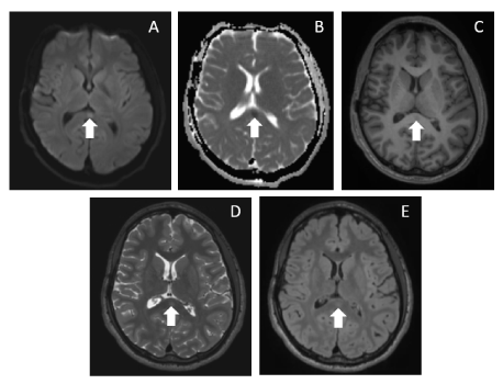
A 21-year-old male with history of refractory epilepsy presented for long-term video EEG monitoring. His home antiepileptic medications were weaned off over a seven-day period. On hospital day 4, he had a right temporal electro clinical seizure with secondary generalization and a second right temporal electrographic seizure. On hospital day 8, MRI brain identified an ovoid circumscribed lesion in the splenium of the corpus callosum which had not been present on prior scans (Figure 1). This characteristic lesion is thought to be induced by a rapid reduction of antiepileptic drugs, not with toxic drug effects or seizure frequency. It requires no further investigation [1].
References
Corresponding Author
John R McLaren, Department of Neurology, Massachusetts General Hospital, 175 Cambridge St, Suite 340, Boston, MA 02140, USA, Tel: 617-726-6540, Fax: 617-726-0230.
Copyright
© 2021 McLaren J. This is an open-access article distributed under the terms of the Creative Commons Attribution License, which permits unrestricted use, distribution, and reproduction in any medium, provided the original author and source are credited.