Cerebellar Vermian Dysplasia: The Tale of the Tail
Abstract
A recently described feature of cerebellar vermian dysplasia, the cerebellar "vermian tail", is a morphologically distinctive dysplasia of the nodular lobule of the posterior cerebellum that is appreciable on prenatal imaging. The vermian tail has been described in both human cases of Dandy-Walker malformation, as well as in a mouse model for Dandy-Walker malformation. However, the finding may also be seen in less severe anomalies of the posterior fossa, such as with isolated enlargement of the fourth ventricle. Attempting to predict the neurodevelopmental outcome of anomalies on this continuum presents a challenge. Normal histologic appearance of the cerebellar vermis is described, and the pathologic appearance of the posterior vermis that correlates with the finding of a "vermian tail" is explained. Lack of neurodevelopmental outcomes data to interpret the clinical significance of this finding is also discussed.
Keywords
Fetal MRI, Dandy-Walker variant, Dandy-Walker continuum, Vermian hypoplasia, Vermian dysplasia, Brain malformation
Introduction
Suspicion of an intracranial anatomic abnormality on prenatal ultrasound is a relatively common indication for prenatal MR imaging and for prenatal counseling to discuss the imaging implications for neurodevelopmental outcome. Posterior fossa cystic space enlargement is readily recognized by ultrasound, and a concern in this scenario is abnormal formation of the cerebellum [1-4].
For more than two decades, there has been discrepant use of terms to describe posterior fossa cystic space enlargement [4,5]. Several different classification schemes have been described [6-9], and the persistent lack of unified language creates continued confusion for diagnostic radiologists, high-risk maternal-fetal medicine physicians, genetic counselors, and pediatric neurodevelopmental subspecialists. The topic is made more complicated by the variable appearance of the cerebellar vermis and coverage of the fourth ventricle, attributable to ongoing maturation through the second and third trimesters of fetal growth [3,10-12].
A thorough editorial article written by Dr. A. Robinson discusses the confusing terminology regarding abnormal appearances of the cerebellar vermis, specifically addressing the term, "inferior vermian hypoplasia" [13]. In this article, Dr. Robinson cogently describes how studies evaluating cerebellar embryology, phylogeny, somatotopic mapping and functional MRI demonstrate that the cerebellar vermis arises from its own midline primordial tissue and it develops in a ventral to dorsal direction, rather than in a superior to inferior direction. Furthermore, in prenatal images, although one may interpret an abnormally developed vermis as inferiorly hypoplastic (in other words, small at its inferior aspect), in actuality, the cerebellar malformation may extend beyond the vermis. In an effort to emphasize that "there is no generic term which encompasses all the various etiologies that can cause a small vermis", Dr. Robinson argues that more appropriate terminology may be "vermian hypoplasia" or "vermian dysplasia" [13]. Accordingly, in this paper, an abnormally formed vermis perceived by prenatal ultrasound or MRI will be referred to as "vermian dysplasia."
In recent literature, a new feature of cerebellar vermian dysplasia has been described, with findings seen using both histopathologic assessment and with imaging. This finding is called the cerebellar "vermian tail", a morphologically distinctive dysplasia of the nodular lobule of the posterior cerebellum. In prenatal images of the cerebellar vermis in the midline sagittal plane, the cerebellar tail appears as a linear, posteriorly projecting extension of the posterior vermis. This pathologically-proven posterior vermian dysplasia with an imaging correlate has been described in both human cases of Dandy-Walker malformation, as well as in a mouse model for Dandy-Walker malformation [2,3,14,15]. The question of its clinical significance in the absence of more severe vermian malformation (for instance, classic Dandy-Walker malformation) remains unanswered, however. Furthermore, what constitutes a tail in prenatal images, exactly, has not been well established. Here, we present a discussion to raise concern for the potential over-interpretation of this finding in certain cases.
Posterior fossa cystic anomalies
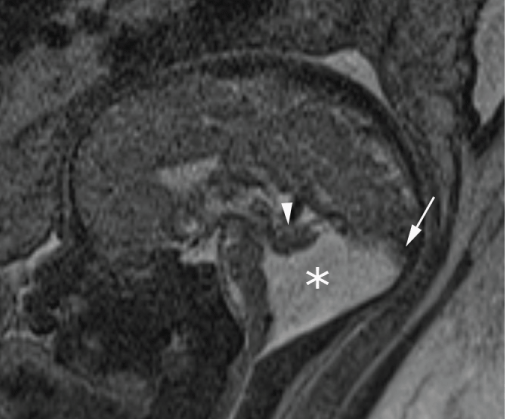
Among the more severe hindbrain malformations is the classic Dandy-Walker malformation (Figure 1), which refers to the following association: Large posterior fossa (abnormal elevation of the cerebellar vermis), a small and dysplastic cerebellar vermis, flattening of the fastigial point, and an enlarged fourth ventricle that communicates with a retrocerebellar cystic space [16-18]. Variations of this abnormal anatomic configuration have been assigned different terms, depending on the amount of apparent cerebellar vermian dysplasia and on the degree of fourth-ventricular enlargement. For instance, the term "Dandy-Walker variant" was established in a classification system to describe a small cerebellar vermis without posterior fossa enlargement [6], whereas "Dandy-Walker complex" has been used to describe varying degrees of vermian dysplasia, fourth-ventricle enlargement and normal-to-large posterior fossa [7].
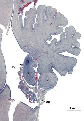
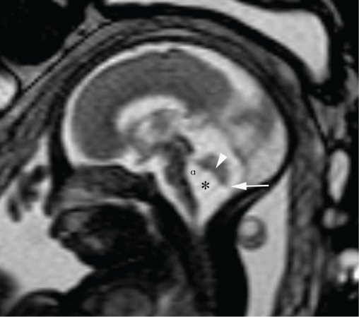
Additional posterior fossa cystic abnormalities, such as mega cisterna magna and Blake's pouch cyst, confound the uniform application of these terms in reporting. Interobserver disagreement between these categories is common [4,5,19,20]. With various methods of in situ imaging, mega cisterna magna refers to a fluid-filled space greater than 10 millimeters in the anterior-posterior dimension between the caudal-ventral surface of the fourth ventricle and the occiput, without enlargement of the rostral fourth ventricle or vermian malformation. In most instances, the measured space includes not only the cisterna magna proper (the space between the dorsal surface of the medulla oblongata and the occiput), but also the caudal fourth ventricle, as the posterior medullary velum ("membranous roof" of the fourth ventricle; "MR" in Figure 2), a thin membrane that separates the two, is not resolved with contemporary imaging. Blake's pouch cyst describes abnormal persistence of an embryological membrane arising from the roof of the fourth ventricle that results in a space-occupying enlargement of the fourth ventricle, which may deform the cerebellar vermis (Figure 3). Normally, there is transient evagination of the membranous fourth ventricular roof as an ependyma-lined diverticulum extending posteriorly and caudal to the cerebellum; communication with the subarachnoid space occurs variably between the 7th week and the 4th month of gestation, when medial and lateral foramina form in the ependymal membrane and the pouch relaxes into its mature flat configuration ventral to the fluid-filled intra-arachnoid space that is the cisterna magna proper [1,8,21,22]. According to one model [1], some cystic malformations in the posterior fossa can be explained based on aberrations in the completeness and/or timing of fenestration of Blake's pouch. s
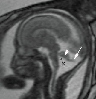
Over two decades ago, Strand, et al. presented their rationale for discarding the terms "Dandy-Walker variant" and "mega cisterna magna", and instead considered all cystic posterior fossa anomalies as being on a spectrum [5]. At one end of this spectrum, with normal to mild neurodevelopmental outcomes, is the Blake's pouch cyst, and at the opposite end of the spectrum, with more severe outcomes, is the classic Dandy-Walker malformation. The authors point out that the histological observations in the Dandy-Walker malformation match those of the Blake's pouch cyst. In both conditions, they argue, the cyst walls are composed of an inner layer of ependyma continuous with that of the fourth ventricle, an intermediate layer of attenuated neuroglial or astroglial tissue, and an outer layer of pia-arachnoid, not associated with characteristics typical of secondary arachnoid cysts as might be expected following hemorrhage or infection [5]. More recently, experts in the field have argued that cystic posterior fossa anomalies do indeed fall on a spectrum, and Dandy-Walker continuum is a more suitable term to capture variable appearances of cerebellar vermian abnormal growth and development, associated with or without fourth ventricle enlargement and with or without posterior fossa enlargement [8]. Attempting to predict the neurodevelopmental outcome along various points on this continuum is the challenge. Somewhere in the middle of the continuum is isolated cerebellar vermian dysplasia (referring to an underdeveloped, small appearance of the inferior cerebellar vermis). Some cases of isolated vermian underdevelopment may be accompanied by the appearance of a beak-like, elongated extension from the posterior vermis, which might be considered a cerebellar "vermian tail" (Figure 4). The team of care providers is then left with this question: Depending on the age of the fetus, does the suggestion of a cerebellar vermian tail indicate a change in prognosis?
Vermian development from the pathologist's perspective
Growth and elaboration of the cerebellar hemispheres and midline vermis continue through fetal development [3,10-12]. The cerebellar vermis is comprised of 10 lobules, and the most posterior portion of the cerebellar vermis includes three of these principal lobules: Pyramis, uvula, and nodulus. As previously summarized by Kapur, et al. the uvula and pyramis are nearly equivalent in length, and both extend to the inferior boundary of the vermis. The nodulus lies deep to the uvula and is not visible to the pathologist from the dorsal aspect of the cerebellum. The nodulus is ovoid, and through most of midgestation, the ventral superior portion contains densely packed neuroglial precursors clustered in the midline (the germinal zone). A normal fetal vermis is characterized by these six features (Figure 2): 1) A steep fastigium (indentation in the dorsal roof of the fourth ventricle immediately rostral to the nodulus); 2) The uvula extends inferior to the nodulus; 3) The nodulus is ovoid; 4) The ventral surface of the nodulus has folia; 5) The posterior medullary velum attaches to the inferior surface of the nodulus (approximately half-way between the fastigium and the caudal tip of this lobule); and, 6) The germinal zone is seen at the rostral end of the nodulus (rostral to insertion of the velum), at least through 2nd trimester [3].
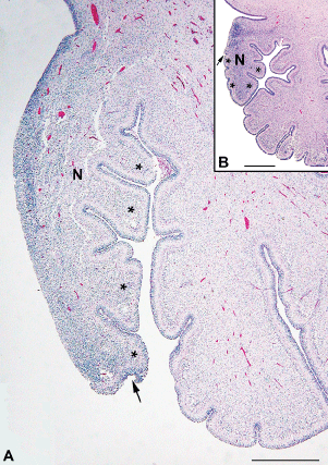
In our experience with prenatal imaging cases that show only mild enlargement of the fourth ventricle, rotation of the cerebellar vermis augmenting the tegmentovermian angle (the angle between the dorsal surface of the brainstem and the ventral surface of the vermis), and normal size of the posterior fossa (all imaging features typical of a Blake's pouch cyst) and subsequent termination of pregnancy, fetal neuropathological assessment sometimes reveals abnormalities of the posterior vermis that were not identified by imaging. Sagittal sections of the cerebellar vermis reveal abnormal development of the vermian lobular architecture (a "vermian tail") that does not follow the rules listed above: The nodulus extends inferior to the uvula and may appear elongated, as though the ventral surface of the vermis was pulled inferiorly in conjunction with distention of the membranous roof of the fourth ventricle, and the germinal zone is not restricted to the superior-dorsal aspect of the nodulus (Figure 5).
Outcome reflected by the tail remains a question
The significance of such vermian dysplasia evident under the microscope is unknown. Perhaps this degree of posterior vermian dysplasia would equate to a discernible neurodevelopmental deficiency using satisfactory assessment tools beyond infancy; or, perhaps the finding is clinically silent and bears no impact on future outcome. This is not known. Regardless, histopathologic evidence of abnormal cerebellar development is disconcerting, and the imaging correlate of nodular elongation in the sagittal plane causes the radiologist or maternal-fetal medicine physician to pause. Should the finding of a vermian tail raise concern for an abnormal neurodevelopmental outcome?
An important consideration is the fetal age - it might be that the appearance of the cerebellar vermian tail bears no meaning through the second trimester, but if present late in gestation, then concern for a neurodevelopmental deficit is more likely. To support this possibility, we also have seen cases with prenatal imaging showing a mild Dandy-Walker continuum versus Blake's pouch cyst plus a thin, beak-like posterior vermian extension (possible tail), in which the pregnancy continued without complication and postnatal brain MR imaging of the newborn infant showed normal appearance of the posterior fossa (Figure 6). Cases like these call into question whether the radiologic vermian tail is the same as that identified by the pathologist, as it seems unlikely that the malformation observed histologically could resolve into a normal nodulus. Regardless, a vermian tail identified by prenatal imaging may not persist and does not necessarily predict abnormal postnatal neurodevelopment. Currently, there is only one published paper [15] that includes postnatal imaging describing a vermian tail in infants with a deletion of chromosome 6p25 (FOXC1), which is associated with Dandy-Walker malformation, and this article shows what the authors perceive as a vermian tail - "a common extended and dysplastic posterior vermis with an indistinct choroid plexus".
It is conceivable that innumerable cases diagnosed as Blake's pouch cyst may have had the vermian tail finding, either by imaging or by pathology, or possibly both. Several published articles including cases diagnosed as Blake's pouch cyst show a cerebellar vermian tail-like structure on sagittal midline fetal brain T2-weighted (Robinson and Goldstein, 2007, figure 11B; Garel, 2010, figure 3; and, Guibaud, 2012, figure 4C) [1,23,24]. As an example of a published case wherein pathologic dysplasia associated with a Blake's pouch cyst went unrecognized, Robinson and Goldstein (2007, figure 12) show an example of a persistent Blake's pouch cyst detected on prenatal ultrasound that appeared to resolve by postnatal imaging; in retrospect, the sagittal histopathologic section from the fetal vermis in that case is reported as normal, but the published image (figure 12C) illustrates abnormal elongation of the nodulus and absence of ventral folia, which is the histologic equivalent of a cerebellar tail [1].
A discussion of prior publications reporting outcomes evidence for posterior fossa cystic anomalies is relevant here, and specifically outcomes data for Blake's pouch cyst cases. Available outcomes data for Blake's pouch cyst cases are reassuring. For instance, Colleoni Galdolfi, et al. published a series of 63 prenatal cases with a neurologic follow-up at 1-5 years available, and the authors demonstrated about 90% of fetuses with either Blake's pouch cyst or mega cisterna magna without associated anomalies had normal neurologic development compared with only 50% of those with Dandy-Walker malformation and vermian hypoplasia [25]. An excellent meta-analysis published by D'Antonio, et al. supports a favorable outcome for Blake's pouch cysts as well [26]. In this meta-analysis, the authors use the following classification system for posterior fossa anomalies: 1) Classic Dandy-Walker Malformation; 2) Mega cisterna magna (> 10 mm); 3) Blake's pouch cyst (rotated vermis, normal size of posterior fossa); and 4) Vermian hypoplasia (based only on size). There was no significant association between Blake's pouch cyst and the occurrence of abnormal neurodevelopmental delay, with a pooled proportions rate of 4.7% (95% CI, 0.7-12.1%) and range of 0-5%, compared with a prenatal diagnosis of Dandy-Walker malformation, showing a rate of 58.2% (95% CI, 21.8-90.0%). Interestingly, this study also discerned no statistically significant increased rate of abnormal neurodevelopmental outcome for vermian hypoplasia, although the authors comment that a small number of studies confounds this particular evaluation [26].
Assuming that these outcomes studies include infants, who had a prenatal diagnosis of a persistent Blake's pouch cyst may have shown the "tail sign", one might surmise that assigning significance to this imaging finding in the absence of associated brain anomalies could lead to overly cautious counseling and unnecessary pregnancy terminations. The currently published descriptions of the cerebellar tail in mouse and human studies show the tail as a thick, T2-weighted dark structure on fetal MRI [14,15], and perhaps thin beak-like tails should be assessed differently and more correctly interpreted as the roof of a Blake's pouch cyst. A retrospective search for the "tail sign" in prenatal imaging in cases of mild Dandy-Walker continuum at varying gestational ages with postnatal clinical follow-up is warranted to better understand the significance of this abnormality and to more carefully establish its diagnostic descriptors.
References
- Robinson AJ, Goldstein R (2007) The cisterna magna septa vestigial remnants of blake's pouch and a potential new marker for normal development of the rhombencephalon. J Ultrasound Med 26: 83-95.
- Russo R, Fallet-Bianco C (2007) Isolated posterior cerebellar vermal defect: A morphological study of midsagittal cerebellar vermis in 4 fetuses--early stage of Dandy-Walker continuum or new vermal dysgenesis? Journal of Child Neurology 22: 492-500.
- Kapur RP, Mahony BS, Finch L, et al. (2009) Normal and abnormal anatomy of the cerebellar vermis in midgestational human fetuses. Birth Defects Res A Clin Mol Teratol 85: 700-709.
- Limperopoulos C, Robertson RL, Khwaja OS, et al. (2008) How accurately does current fetal imaging identify posterior fossa anomalies? Am J Roentgenol 190: 1637-1643.
- Strand RD, Barnes PD, Poussaint TY, et al. (1933) Cystic retrocerebellar malformations: Unification of the Dandy-Walker complex and the Blake's pouch cyst. Pediatr Radiol 23: 258-260.
- Altman N, Naidich TP, Braffman BH (1992) Posterior fossa malformations. AJNR Am J Neuroradiol 13: 691-724.
- Barkovich AJ, Kjos BO, Norman D, et al. (1989) Revised classification of posterior fossa cysts and cyst-like malformations based on the results of multiplanar MR imaging. AJR Am J Roentgenol 153: 1289-1300.
- Robinson AJ, Ederies MA (2016) Diagnostic imaging of posterior fossa anomalies in the fetus. Seminars in Fetal Neonatal Medicine 21: 312-320.
- Siebert J, Kapur RP (2002) Rulers rule: Present and future applications of cerebellar morphometry. Pediatr Dev Pathol 5: 422-424.
- Langelaan J (1919) On the development of the external form of the human cerebellum. Brain 42: 130-170.
- Larsell O (1934) Morphogenesis and evolution of the cerebellum. Arch Neurol Psych 31: 373-395.
- Larsell O (1947) The development of the cerebellum in man in relation to its comparative anatomy. J Comp Neurol 87: 85-129.
- Robinson AJ (2014) Inferior vermian hypoplasia - Preconception, misconception. Ultrasound Obstet Gynecol 43: 123-136.
- Bernardo S, Vinci V, Saldari M, et al. (2015) Dandy-Walker Malformation: Is the "tail sign" the key sign? Prenat Diagn 35: 1358-1364.
- Haldipur P, Dang D, Aldinger KA, et al. (2017) Phenotypic outcomes in Mouse and Human Foxc1 dependent Dandy-Walker cerebellar malformation suggest shared mechanisms. Elife 6.
- Dandy WE (1921) The diagnosis and treatment of hydrocephalus due to occlusion of the foramina of Magendi and Luschka. Surg Gynecol Obs 32: 112.
- Walker AE (1944) A case of congenital atresia of the foramina of Luschka and Magendie. Journal of Neuropathology and Experimental Neurology 3: 368-373.
- Benda CE (1954) The Dandy-Walker syndrome or the so-called atresia of the foramen of Magendie. J Neuropathol Exp Neurol 13: 14-39.
- Limperopoulos C, du Plessis AJ (2006) Disorders of cerebellar growth and development. Curr Opin Pediatr 18: 621-627.
- Limperopoulo C, Robertson RL, Estroff JA, et al. (2006) Diagnosis of inferior vermian hypoplasia by fetal MRI: Potential pitfalls and neurodevelopmental outcome. Am J Obstet Gynecol 194: 1070-1076.
- Azab W, Shohoud S, Elmansoury T, et al. (2014) Blake's pouch cyst. Surg Neurol Int 5: 112.
- Calabrò F, Arcuri T, Jinkins JR (2000) Blake's pouch cyst: An entity within the Dandy-Walker continuum. Neuroradiology 42: 290-295.
- Garel C (2010) Posterior fossa malformations: Main features and limits in prenatal diagnosis. Pediatr Radiol 40: 1038-1045.
- Guibaud L, Larroque A, Ville D, et al. (2012) Prenatal diagnosis of "isolated" Dandy-Walker malformation: Imaging findings and prenatal counselling. Prenat Diagn 32: 185-193.
- Colleoni Gandolfi G, Contro E, Carletti A, et al. (2012) Prenatal diagnosis and outcome of fetal posterior fossa fluid collections. Ultrasound Obstet Gynecol 39: 625-631.
- D'Antonio F, Khalil A, Garel C, et al. (2016) Systematic review and meta-analysis of isolated posterior fossa malformations on prenatal imaging (part 2): Neurodevelopmental outcome. Ultrasound Obstet Gynecol 48: 28-37.
Corresponding Author
Teresa Chapman, Professor of Radiology, Department of Radiology, Mail Stop MA.07.220, Seattle Children's Hospital, University of Washington School of Medicine, 4800 Sand Point Way NE, Seattle, WA 98105, USA, Tel: 206-987-1577, Fax: 206-987-2341.
Copyright
© 2018 Chapman T, et al. This is an open-access article distributed under the terms of the Creative Commons Attribution License, which permits unrestricted use, distribution, and reproduction in any medium, provided the original author and source are credited.

