Intradiploic Skull Hematoma in Infants: An Underestimated Diagnosis
Abstract
Background
Intradiploic hematoma was first reported in 1934 by Chorbski and Davis. It usually occurs after a mild trauma where a small bleeding accumulates within the diploe in between the tabula interna and externa of the skull. It is to be differentiated from calcified or ossified cephalhematoma. The bleeding in cephalhematoma is subperiosteal and tends to resorb spontaneously within 3-6 months in comparison to intradiploic hematoma which needs a surgical intervention.
Description
We reported on 2 cases of intradiploic hematoma, where a hard swelling was noticed in the skull of the newborn shortly after a prolonged normal delivery. Surgical remodeling was done in both patients with very good cosmetic results.
Conclusion
An intradiploic hematoma in infants is usually misdiagnosed as cephalhematoma that is calcified or ossified later. Differentiation between these 2 different pathologies is mandatory. As for cephalhematoma, conservative methods are advised primarily except in case of ossification later. In case of huge intradiploic hematoma, surgery is advised from the start as there is no role for waiting because it will not resolve spontaneously. Neurosurgeons should be aware of these entities.
Keywords
Cephalhematoma, Intradiploic hematoma, Skull
Background
Chronic intradiploic hematoma was first reported in 1934 by Chorbski and Davis [1]. It usually occurs after a mild trauma where a minimal haemorrhage accumulates within the diploe in between the tabula interna and externa of the skull. It is to be differentiated from calcified or ossified cephalhematoma, a subperiosteal collection of blood which occurs in 0.2-3% of infants after birth [2]. Cephalhematomas usually develop in the parietal eminence and does not extend past the suture line. Hence, they can be differentiated from caput succedaneum and subgaleal hematomas [3].
Although cephalhematomas occur more frequently during difficult labor, they can also appear after normal delivery during the neonatal period [4]. The lesions generally spontaneously resorb within 1 month, but if the cephalhematoma persists beyond this time, it may progress towards ossification [5]. The incidence of ossified cephalhematoma is a rare clinical entity, and its pathogenesis is unclear. But there is another entity of hematoma which is immediately firm. These develop between the diploe and separate the inner and outer bone layers.
The presence of a benign chronic hematoma should be considered as part of the differential diagnosis for each intradiploic mass lesion. Alarming signs during history taking when a previous minor or major head trauma is reported. Due to the possibility of progressive growth, surgical excision of an intradiploic hematoma is recommended after radiological diagnosis of the condition [6].
Case presentation no. 1
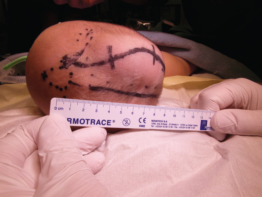
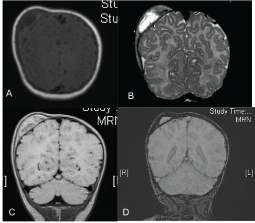
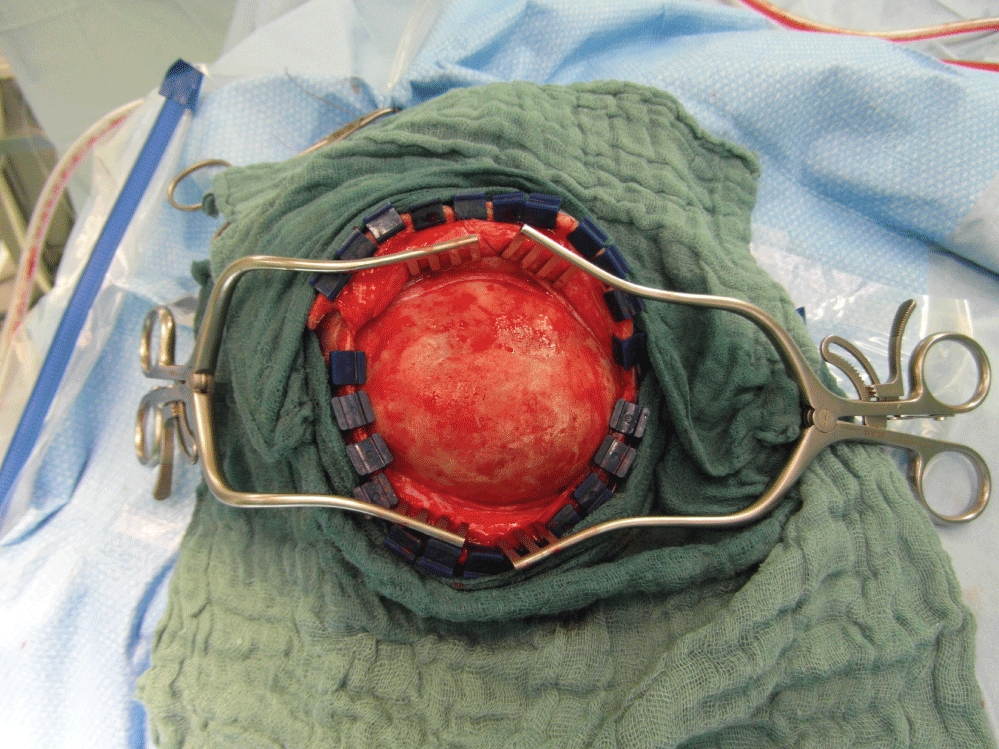
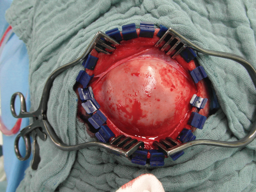
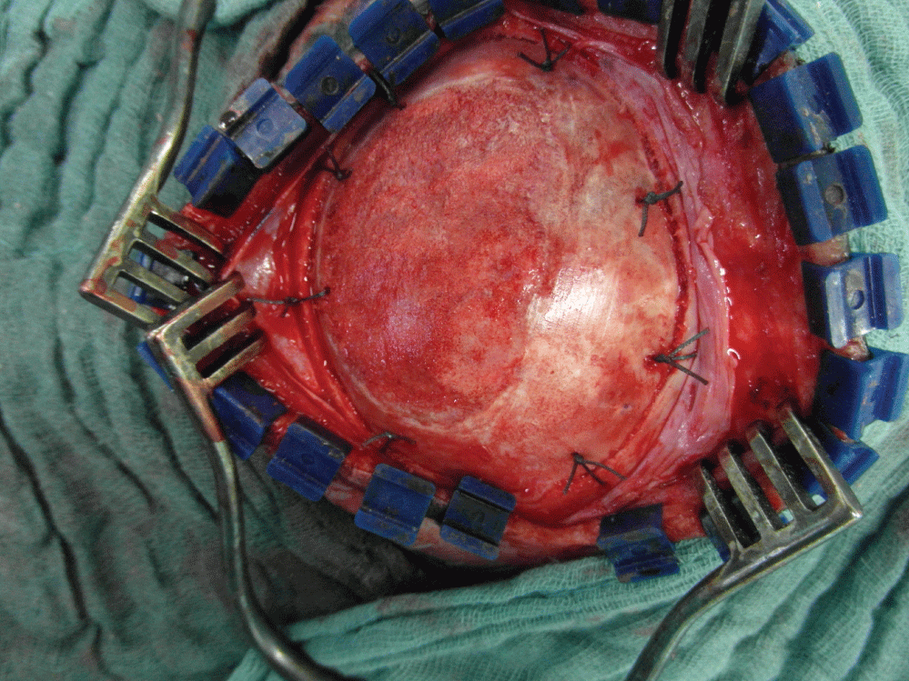
A 2-month-old boy presented with an asymmetrical skull swelling since birth in the right parieto-occipital region 10 × 7 cm in size (Figure 1). The patient had been born at full term by a prolonged normal vaginal delivery without the use of vacuum extraction. On examination, he had a head circumference of 38 cm. The mass was eggshell hard and fixed to the bone. The patient was neurologically intact. A magnetic resonance imaging (MRI) showed an intradiploic mass, without intracranial abnormalities (Figure 2). A decision was made to perform a surgical excision of the bony swelling completely to correct the skull deformity at the age of 2 months. A sigmoid incision was made in the parieto-occipital region parallel to the long axis of the mass. A smooth localized elevation of the bone was found (Figure 3). The whole mass was excised primarily after a burr hole was applied outside the lesion and finally a craniotomy all around the lesion. The bone flap was elevated, and the dura was intact. The outer calvarial bone was drilled to relieve a yellow pathological gel-like organized hematoma (Figure 4). The pathologic bone was as hard as an egg shell. After the outer pathologic bone, along with the consolidated old hematoma was excised, we found the inner calvarial bone very thin and uneven. The bone flap was remodelled using the high-speed drill and then replaced and fixed with 6 non-absorbable sutures to the surrounding skull bone. On histologic examination of the outer bone, well-formed mature bony trabeculae were seen, as well as the organized ossified hematoma. The postoperative course was uneventful, and follow-up examination after 8 months revealed a satisfactory good cosmetic result. Accordingly, control follow up has been ended.
Case presentation no. 2
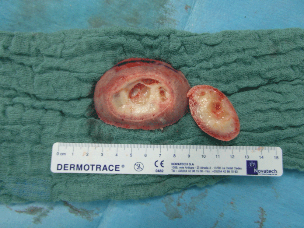
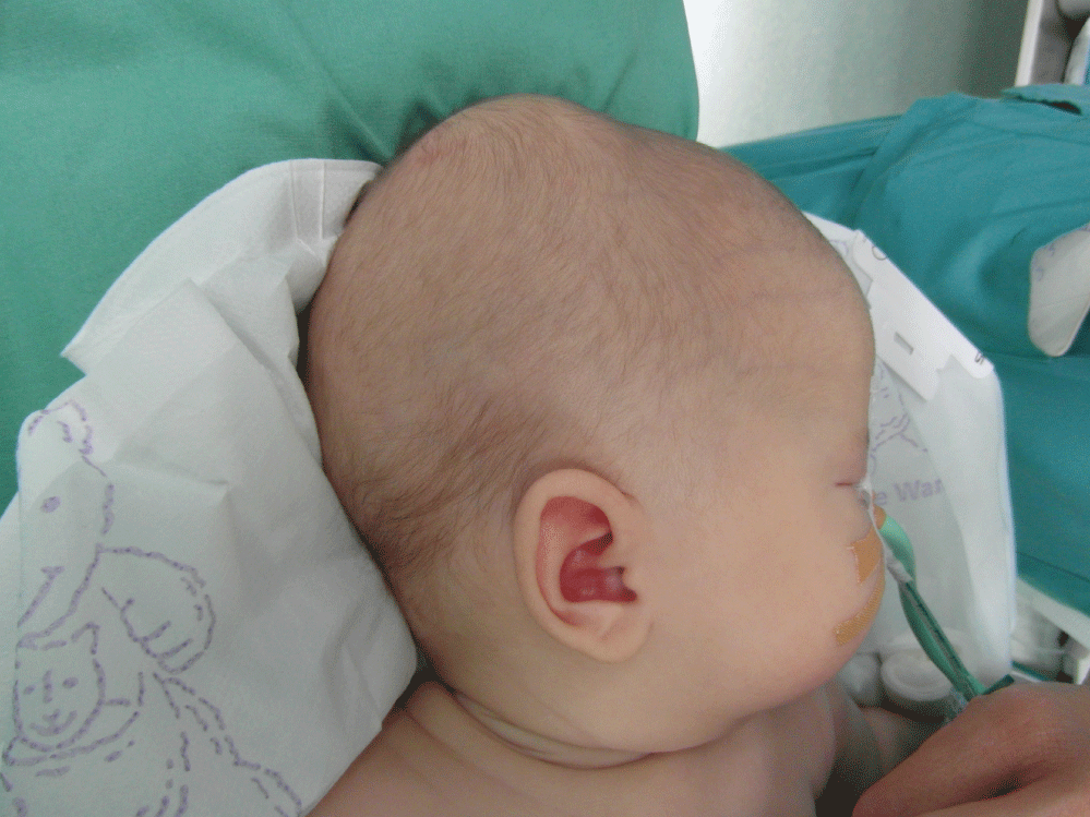
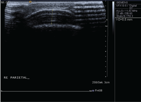
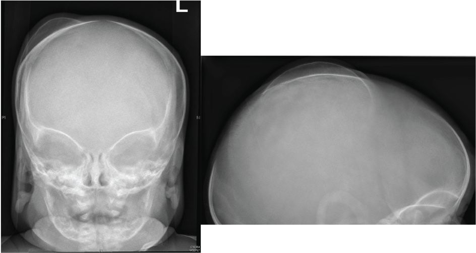
A 3-month-old boy presented with an asymmetrical skull swelling since birth in the right parieto-occipital region 8 × 6 cm in size (Figure 5). The patient had been born at full term by a normal vaginal delivery with the aid of vacuum extraction. The swelling was initially firm and 3 weeks later became egg-shell hard and stable in size and form. On examination, the mass was exactly as described earlier. An Ultrasonography (Figure 6) as well as skull X-rays (Figure 7) showed an intradiploic mass, without intracranial abnormalities. As discussed before, we decided also to perform a surgical excision of the bony swelling to correct the skull deformity. A sigmoid incision was made in the parieto-occipital region parallel to the long axis of the mass. A smooth localized elevation of the bone was found (Figure 8). The whole mass was excised as discussed before, handled in the same fashion as described by the previous patient and re-fixed to the surrounding bone using 8 non-absorbable sutures (Figure 9). The postoperative course was uneventful, and follow-up examination after 3 months revealed a satisfactory good cosmetic result. Accordingly, no more follow up visits were planned.
Discussion
Cephalhematoma is a collection of blood beneath the periosteum of the cranial vault bone. It occurs secondary to rupture of blood vessels between the skull and the periosteum. Most of the cases are reported in the newborns due to birth trauma and rarely following trauma or surgical interventions. The frequency of neonatal cephalhematoma is reported to be 0.2-2.5% of live births [7]. The exact mechanism underlying the formation of cephalhematomas is not known. Probably, subperiosteal blood vessels may be torn by sudden inward movement of the skull caused by sudden or prolonged compression of the skull. The shearing forces can also be elicited between the skull bone and periosteum, displacing the periosteum away from the underlying skull bone. The condition is self-limiting and resolves in more than 80% by gradual hemolysis, which turns the swelling to be fluctuant and finally resolving within 3-4 weeks [7]. When the hematoma does not resolve, it may get organized, and calcification [8-10] or ossification may be seen. There are only few published case reports in the literature reporting ossification in a cephalhematoma [8,11]. In such cases, the inner wall is formed from the original calvarial bone and the newly ossified bone forms the outer wall. The organized hematoma may still be seen in the intervening space between 2 bony walls. The newly laid outer ossified bone may even show hemopoietic tissue in the marrow spaces enclosed by the mature trabeculae of lamellar bone as mentioned by Gupta, et al. [7] in their case. They assumed that possibly the osteogenic progenitor cells in the periosteum along with the cytokines and growth factors in the hematoma play a role in the process of ossification similar to healing of a bony fracture.
We expect that all cephalhematomas are soft swellings in the beginning which tend to resorb or harden later. We describe here another entity in which the swelling from the beginning is hard. We assume that in such cases the bleeding occurs in between the bony lamellae from instance since the skull bone of infants is relatively softer. This is proved in our 2 cases as the intraoperative findings showed an expanded outer table of the skull but without bluish discoloration which is usually the case in cephalhematomas. Later, an organization of the hematoma as well as an ossification process of the residual hematoma takes place which was proved by the histological examination of the resected material in our 2 patients which showed trabecular bony tissue with osteoblasts and in between aggregates of lymphocytes, plasma cells, fatty tissue and blood residues.
Chronic intradiploic hematomas have been reported with the use of anticoagulants [12], birth trauma [11,13], and following shunt surgery [14]. Patients present with painless progressive skull swelling, which usually causes increased intracranial pressure. Radiological features are sometimes confusing and the lesion is often suspected to be neoplastic, but usually intradiploic hematomas can be diagnosed preoperatively. MRI scans typically show the intradiploic calvarial mass surrounded by bone, and not contrast-enhancing. These masses do expand within the diploid and cause separation of the outer and inner tables [12,13,15,16]. Hence, neurosurgeons should be able, after proper history taking, and using from laboratory and radiological data to differentiate between acute and chronic intradiploic skull hematomas.
The differentiation between intradiploic hematoma and cephalhematoma depends on the history taking regarding the nature of the swelling from the start as well as the radiological findings; in cephalhematoma the blood clot will be found completely outside the calvaria, i.e. no bone overlying hematoma, while in intradiploic hematoma the blood clot will be found covered by the tabula externa. Hence, the differentiation between intradiploic hematoma and ossified cephalhematoma will be very difficult radiologically except through the nature of the swelling from the start as the cephalhematoma tends to be soft to firm in consistency at the beginning while intradiploic hematoma is bony hard from the start.
In our opinion, differentiation between cephalhematoma and acute intradiploic hematoma is mandatory. With cephalhematoma, we adopt primarily conservative measures expecting the bleeding will be resorbed later. In cases when the cephalhematoma is calcified or ossified, we advise surgery for cosmetic reasons. But in cases of our described intradiploic hematoma, there is no role for conservative measures from the beginning and we advise an early surgical intervention.
Conclusion
An intradiploic hematoma in infants can be misdiagnosed as calcified or ossified cephalhematoma. We believe that neurosurgeons should differentiate early between these two different pathologies. As for cephalhematoma conservative methods are advised except in case of ossification later. In case of intradiploic hematoma, surgery is advised from the start as there is no role for waiting because it will not resolve spontaneously. Intradiploic skull hematoma is not an early cephalhematoma and should not be misdiagnosed. A surgical correction is the only option for treatment and the neurosurgeons should be able to differentiate between these entities.
Conflict of Interest
On behalf of all authors, the corresponding author states that there is no conflict of interest.
Financial Support
No financial support to be declared. The contents of this paper (or portion of it) has never been presented previously in any scientific meeting or conference.
References
- Mobbs RJ, Gollapudi PR, Fuller JW, et al. (2000) Intradiploic hematoma after skull fracture: Case report and literature review. Surg Neurol 54: 87-91.
- Yoon SD, Cho BM, Oh SM, et al. (2013) Spontaneous resorption of calcified cephalhematoma in a 9-month-old child: Case report. Childs Nerv Syst 29: 517-519.
- Liu L, Dong C, Chen L (2016) Surgical treatment of ossified cephalhematoma: A case report and review of the literature. World Neurosurg 96: 614.e7-614.e9.
- Jang DG, Kang SG, Lee SB, et al. (2007) Simple excision and periosteal reattachment for the treatment of calcified cephalhematoma. J Neurosurg Pediatr 106: 162-164.
- Chida K, Kubo N, Suzuki T, et al. (2008) Surgical treatment of ossified cephalhematoma: A case report. No Shinkei Geka 36: 529-533.
- Tokmak M, Ozek E, Iplikçioğlu C (2015) Chronic intradiploic hematomas of the skull without coagulopathy: Report of two cases. Neurocirugia (Astur) 26: 302-306.
- Gupta PK, Mathew GS, Malik AK, et al. (2007) Ossified cephalhematoma. Pediatr Neurosurg 43: 492-497.
- Chung HY, Chung JY, Lee DG, et al. (2004) Surgical treatment of ossified cephalhematoma. J Craniofac Surg 15: 774-779.
- Kaufman HH, Hochberg J, Anderson RP, et al. (1993) Treatment of calcified cephalohematoma. Neurosurgery 32: 1037-1039.
- Petersen JD, Becker DB, Fundakowski CE, et al. (2004) A novel management for calcifying cephalohematoma. Plast Reconstr Surg 113: 1404-1409.
- Yücesoy K, Mertol T, Ozer H, et al. (1999) An infantile intraosseous hematoma of the skull. Report of a case and review of the literature. Childs Nerv Syst 15: 69-72.
- Dange N, Mahore A, Avinash KM, et al. (2010) Chronic intradiploic hematoma in patients with coagulopathy. J Clin Neurosci 17: 1047-1049.
- Reeves A, Edwards Brown M (2000) Intraosseous hematoma in a newborn with factor VIII deficiency. AJNR Am J Neuroradiol 21: 308-309.
- Giammusso V, Strukelj S (1980) Intradiploic hematoma in a patient who underwent internal drainage of CSF for hydrocephalus. J Neurosurg Sci 24: 105-108.
- Sato K, Kubota T, Kawano H (1994) Chronic diploic hematoma of the parietal bone. Case report. J Neurosurg 80: 1112-1115.
- Yuasa H, Watanabe H, Uemura Y, et al. (1992) Intraosseous hematoma of the skull: Case report. Neurosurgery 30: 776-778.
Corresponding Author
Ahmed El Damaty, Consultant Neurosurgeon, Department of Neurosurgery, Heidelberg University Hospital, Im Neuenheimer Feld 400, D-69120 Heidelberg, Deutschland, Heidelberg, Germany, Tel: +49-6221-56-38089, Fax: +49-6221-56-5856.
Copyright
© 2018 El Damaty A, et al. This is an open-access article distributed under the terms of the Creative Commons Attribution License, which permits unrestricted use, distribution, and reproduction in any medium, provided the original author and source are credited.

