Stroke in a Child: The Role of Mycoplasma Pneumoniae and Coagulation Factor VIII
Abstract
A 6-year-old child was admitted for an episode of generalized seizures. Shortly after admission in our Emergency Room, he developed somnolence and meningismus (GCS score 3). A brain magnetic resonance imaging showed signs of stroke. No laboratory findings were relevant except for an increasing in the coagulation factor VIII levels, so a therapy with heparin sodium was instaured without benefit. An increased serum titer of IgM antibodies for Mycoplasma pneumonia prompted us to introduce a combined therapy with IV clarithromycin and methylprednisolone, leading to a great improvement of symptoms (GCS score 15). This case shows that in a rare case of juvenile stroke it is mandatory to search for Mycoplasma infection to avoid a misdiagnosis and compromising the patient's prognosis.
Keywords
Stroke, Mycoplasma pneumonia, Coagulation Factor VIII
Introduction
Mycoplasma pneumoniae (MP) is linked to many neurologic syndromes [1]. Epidemiological studies show that children are more affected by Central Nervous System (CNS) manifestations secondary to MP infections than adults [1].
The pathomechanisms for extrapulmonary manifestations of MP infections remain largely unknown [2]. MP-induced CNS diseases have been linked with three broad pathophysiologic mechanisms: 1) Systemic dissemination with direct infection of extrapulmonary organs; 2) Autoimmune or Immune complex-mediated injury; and 3) Vascular occlusion [3].
Stroke has been associated with MP pneumoniae infection both in children and adults [4]. The pathophysiologic mechanisms underlying MP-associated stroke still is not completely understood. A procoagulable state has been often touted [3]. A systemic or focal vasculitic process is supported by several case reports and animal models [1].
Increased factor VIII (FVIII) levels are considered a moderately high independent risk factor for ischemic heart disease, ischemic stroke, and venous thromboembolism [5].
Here we describe a case of a child with MP infection-associated stroke and increased FVIII levels.
Case Presentation
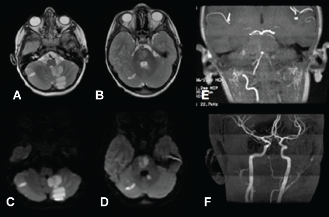
A 6-year-old child came to our Emergency Room for the onset of generalized seizures. Two days earlier he had fever (38 °C), nuchal-occipital throbbing headache, weakness in the right side of his body and dysarthria. Shortly after admission, he developed somnolence and meningismus A Brain Computer Tomography (CT) didn't show any acute injuries. After a lumbar puncture (normal chemical and microbiological findings), we started therapy with intravenous (i.v) ceftriaxone, acyclovir, mannitol and midazolam. The following day the patient showed a rapidly increasing lethargy and finally lapsed into a coma with Glasgow Coma Scale (GCS) Score of [3]. A brain Magnetic Resonance Imaging (MRI) performed in emergency showed evidence of ischemic stroke (Figure 1). He immediately started therapy with heparin sodium infusion (15000 U.I./24 h) without evident benefits on the symptoms. Chest radiograph, neck vessels Doppler, holter electrocardiogram and transthoracic echocardiogram were within normal ranges.
Laboratory investigation for the presence of inherited or acquired thrombophilia included measurement of antithrombin, free protein C, factor VIII (FVIII) functional activity, free protein S antigen, fasting homocysteine, factor V Leiden, prothrombin 20210A polymorphisms and antiphospholipid antibodies (i.e. lupus anticoagulant, anti-cardiolipin and anti-beta2-glycoprotein I antibodies) were within the normal ranges except for an increasing of the FVIII (262 IU/dl, n.v. 70-140).
Serum and cerebrospinal fluid analysis for the most common neurotropic viruses (HSV1, HSV2, VZV, CMV, EBV, Paramyxovirus, Rubivirus) and bacteria were unremarkable except an increased serum titer of IgM antibodies toward MP (14.3 UI/ml) with negative values of IgG (7.3 UI/ml) while a PCR for MP genome wasn't performed for lack of laboratory performance, so we started a therapy with iv clarithromycin (15 mg/kg/day 2 times/day for 7 days) and methylprednisolone (10 mg/kg for 5 days) with progressive improvement of the symptoms leading to a GCS score of 15.
After 15 days, a new MP titer test was performed showing a more than 4-fold increasing of the IgG antibodies (30.8 UI/ml) compatible with a seroconversion. We stopped the therapy with heparin sodium and started therapy with acetyl salicylic acid (100 mg/day).
Factor VIII levels were found consistently increased after three months (205 IU/dl) since the acute event, without correlation with the inflammatory markers such as C-reactive protein. FVIII levels were measured also in the patients' parents and we found a FVIII-increased titre in his mother (191 IU/dl and 175 IU/dl in two occasions within the space of three months, in the presence of ESR and C-reactive protein normal values). To date, the child shows only mild dysarthria.
Discussion
Although the mechanism of MP infection leading to stroke remains undetermined, an increasing number of evidences is in favour of a possible role of a direct MP infection role.
Stroke linked to MP infection has been reported in several cases, mainly in children. Chiang, et al. [6] found that patients with MP infection had a significantly higher incidence of stroke development than those without the infection. The bacteria have been found in atherosclerotic plaques and have been isolated from the cerebrovascular fluid of stroke patients [7].
The mechanisms by which MP can cause stroke could be: 1) Inducing directly the production of cytokines in blood vessels; 2) Inducing an autoimmune response through a mechanism of molecular mimicry causing local vasculitic and/or thrombotic vascular occlusion; 3) Inducing a systemic hypercoagulable state, which may occur through activation of chemical mediators such as complement factors [2].
Familial clustering has been demonstrated for elevated factor VIII levels, and individuals with high factor VIII levels were reported to be at increased risk for venous thromboembolism, myocardial infarction and peripheral artery disease (but not for ischemic stroke) as compared with their relatives with normal levels [8].
Two-thirds of the children with venous thromboembolism had elevated levels of FVIII, which remained persistently elevated in about half of the cases [9]. In a small series of children with ischemic stroke, those with cardioembolic stroke had a median FVIII level of 215 IU/dl (range 165-242) [10].
In our patient, the observed seroconversion for anti-MP antibodies and the good response to specific antibiotic therapy and steroids were compatible with the diagnosis of MP associated-stroke.
The increased factor VIII levels were demonstrated to be not inflammation-related, being consistent over time and showing a familial pattern. Therefore, we can suggest that the MP infection acted as a triggering event for the onset of thrombosis in a child with constitutional thrombophilia.
This case shows that in a rare case of juvenile stroke it is mandatory to search for MP and other viral or bacterial infections to avoid a misdiagnosis and compromising the patient's prognosis.
Acknowledgments
To Professor Anna Paola Batocchi, recently passed away, that helped us in solving this clinical case.
Declaration of Conflicting Interests
The authors declared no potential conflicts of interest with respect to the research, authorship, and/or publication of this article.
Funding
None of authors received an honorarium, grant, or other form of payment to produce the manuscript.
References
- Tsiodras S, Kelesidis I, Kelesidis T, et al. (2005) Central nervous system manifestations of Mycoplasma pneumoniae infections. J Infect 51: 343-354.
- Narita M (2010) Pathogenesis of extrapulmonary manifestations of Mycoplasma pneumoniae infection with special reference to pneumonia. Journal of Infection and Chemotherapy 16: 162-169.
- Narita M (2009) Pathogenesis of neurologic manifestations of Mycoplasma pneumoniae infection. Pediatr Neurol 41: 159-166.
- Perez C, Montes M (2002) Cutaneous leukocytoclastic vasculitis and encephalitis associated with Mycoplasma pneumoniae infection. Arch Intern Med 162: 352-354.
- Martinelli I (2005) Von Willebrand factor and factor VIII as risk factors for arterial and venous thrombosis. Semin Hematol 42: 49-55.
- Chiang CH, Huang CC, Chan WL, et al. (2011) Association Between Mycoplasma Pneumonia and Increased Risk of Ischemic Stroke: A Nationwide Study. Stroke 42: 2940-2943.
- Padovan CS, Pfister HW, Bense S, et al. (2001) Detection of Mycoplasma pneumoniae DNA in cerebrospinal fluid of a patient with M. pneumoniae infection- "associated" stroke. Clin Infect Dis 33: e119-e121.
- Bank I, Libourel EJ, Middeldorp S, et al. (2005) Elevated levels of FVIII:C within families are associated with an increased risk for venous and arterial thrombosis. J Thromb Haemost 3: 79-84.
- Goldenberg NA, Knapp-Clevenger R, Manco-Johnson MJ, et al. (2004) Elevated plasma factor VIII and D-dimer levels as predictors of poor outcomes of thrombosis in children. N Engl J Med 351: 1081-1088.
- Bernard TJ, Fenton LZ, Apkon SD, et al. (2010) Biomarkers of hypercoagulability and inflammation in childhood-onset arterial ischemic stroke. J Pediatr 156: 651-656.
Corresponding Author
Viviana Nociti, Department of Geriatrics, Neurosciences and Orthopedics, Institute of Neurology, Catholic University, Rome, Italy, Fax: +390635501909.
Copyright
© 2017 Mariotti P, et al. This is an open-access article distributed under the terms of the Creative Commons Attribution License, which permits unrestricted use, distribution, and reproduction in any medium, provided the original author and source are credited.