Unusual Nasal Foreign Body in a Cleft Palate Children: A Case Report
Keywords
Nasal foreign body, Cleft palate, Palatal fistulae, Van der Woude
Introduction
Nasal foreign bodies are frequent in children; nevertheless those who include palate and nasal cavity are rare. We present an unusual foreign body that affects the nose and hard palate, in patient with a partially corrected oronasal fistulae in a cleft palate syndrome.
This shows the importance of prevention and suspicion of this type of accidents in cleft palate patients, possible serious complications on the skull base, orbit, nasal mucosa and oral cavity could present depending of the entry mechanism and type of foreign body.
Case Presentation
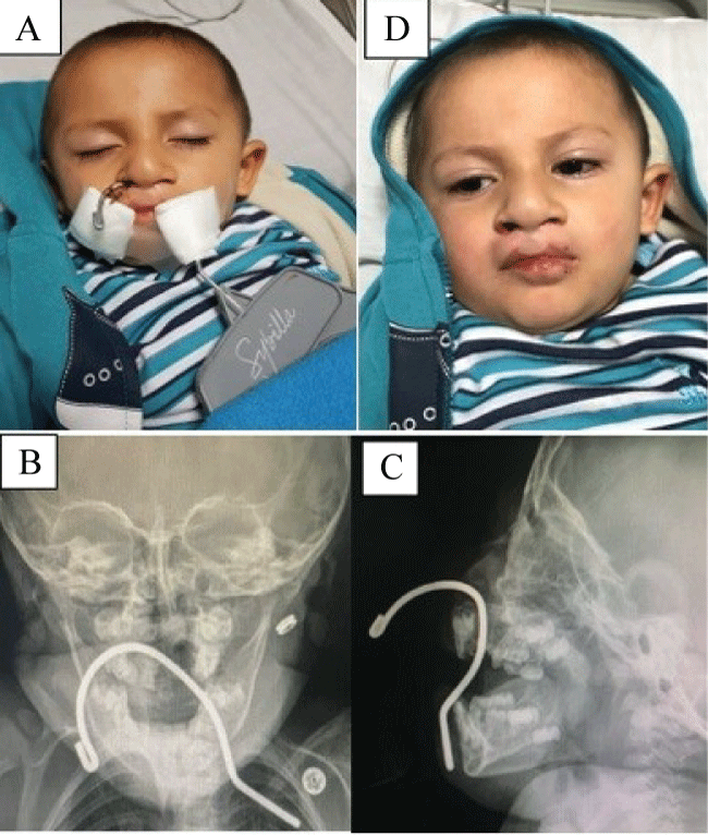
Male patient, 2-years-old, mestizo race arrives to the emergency room sent from a shopping precinct after introducing a clothes hanger through his mouth and came out through his nose. He had mild epistaxis which controlled spontaneously, no airway compromise.
He was previously diagnosed with Van der Woude syndrome and one year ago lip and palate were repaired.
At the emergency room, anterior rhinoscopy showed the tip of the hook coming out from the right nostril and it was followed to the floor of the right nasal cavity where it passed to the oral cavity through a palatal fistula. Figure 1A. There were no lesions on the turbinate's or the right lateral wall, no septal perforation, no purulent discharge (Figure 1B and Figure 1C). Face X ray was taking, without showing orbit, skull base or septal perforation.
Inspection of the oral cavity showed bilateral complete cleft of the secondary palate, scar tissue on the posterior primary palate and the proximal part of the hook that came out through the mouth. No lesions were found on the tongue, tooth or dental tissues.
The hook was removed manually under direct visualization in the emergency room by pulling the inferior end and directing the point in a retrograde way through the floor of the nose avoiding lesions in the septum, turbinates and orbit without other complications. There was no evidence of soft tissue laceration or strong bleeding. Patient was discharged and in the next controls no sequelae lesions where seen. Persistent palatal fistulae will be programed for surgical repair (Figure 1D).
Discussion
Cleft lip and palate is the most common congenital facial defect in the newborn, in Colombia the prevalence is reported from 1 in 500 to 1 in 1000 newborns [1].
Van der Woude syndrome is the most frequent form of syndromic cleft lip and palate [2] with an approximate incidence of 1:100,000 births, caused by a gene mutation that affects the interferon regulatory factor 6 protein. The main characteristics include cleft lip with or without cleft palate, isolated cleft palate, lower lip pits, hypodontia, epidermal and genital anomalies with normal intelligence. It has an autosomal dominant mode of transmission and a high degree of penetrance [3].
Foreign bodies are common in every emergency department. Analysis of the most common locations show esophageal in the first place followed by nasal, auricular and bronchial [4]. Children will find easy insertion through the mouth and the nose any object of appropriate size or shape [5]. Unilateral nasal foreign bodies are twice more common in the right side compared to the left [5].
Accidents are often domestic with objects present in the child's immediate environment [4]. In this case the patient was playing with a hook at a clothing store. He took the hook to his mouth and through the cleft palate advanced it until the right nostril where it came out.
Series of cases have described nonorganic compounds as the most common foreign bodies in the nose accounting for 72-80% of cases. 4 Organic come in second place with 36% and live insects are very rare. The most frequent type are spherical objects (beads, bead like toy fragments, dried vegetables, fruit seeds, nuts) followed by irregular soft objects (sponge, paper, leaf fragments) [5]. Button batteries are not uncommon and require an urgent management because of the risk of complications such as widespread necrosis and septal perforation [6].
Unilateral nasal discharge with a fetid odor are the two symptoms highly suggestive of foreign bodies. Nasal obstruction and epistaxis may also occur [7].
This case is a non-organic object with a particular shape that puts in risk vital structures like the cranial base or the orbits, with potential serious bleeding or infections. Also, it can perforate the septum, erode and damage the flaps or bony palate structures and fracture the teeth [8].
Few cases have reported foreign bodies in the palate and most of them were plastic objects impacted, the risk comes from the high frequency in which children place objects in their mouths [8].
He had previously cleft repair surgeries but residual oronasal communication. This commonly occur on the palate or in the alveolar ridge or labial vestibule and might become a potential source for the displacement of foreign bodies [9] that might end in palatal injuries and worsening of the velopharyngeal insufficiency [10].
The diagnosis is confirmed with the anterior rhinoscopy and the oral cavity examination. If the nasal foreign body is not seen, the ideal method is rigid or flexible nasal endoscopy [7]. In this case, the foreign body is directly visualized, adjacent lesions are discarded and fixation is made to avoid mobilization and further complications.
The treatment in this case was simple, quick and effective. Manual extraction was the best option; the key was direct visualization a careful handling. Historically, nasal foreign body removal is not associated with major complications like the outcome of this case [11]. Most frequent complications include local inflammation, ingestion of the foreign body and epistaxis. There is a low risk of aspiration [6].
A review of foreign bodies in an emergency setting reported a 65% success rate by the emergency department staff. 35% were referred to the ENT clinic and overall 10% were removed in the operating room [12].
Foreign bodies are frequent in cleft palate children, prevention is mandatory. One must teach parents what are the most frequent items for them to be aware, to recognize the symptoms and to avoid trying to pull it out because they might worsen the situation. Specialized care must be taken to remove it with the less complications.
References
- Otero L, Gutiérrez S, Cháves M, et al. (2007) Association of MSX1 with nonsyndromic cleft lip and palate in a Colombian population. Cleft Palate Craniofac J 44: 653-656.
- Tehranchi A, Behnia H, Nadjmi N, et al. (2017) Journal of Surgery Case Reports Multidisciplinary management of a patient with van der Woude syndrome: A case report. Int J Surg Case Rep 30: 142-147.
- Lam AK, David DJ, Townsend GC, et al. (2010) Van der Woude syndrome: dentofacial features and implications for clinical practice. Aust Dent J 55: 51-58.
- Abou-Elfadl M, Horra A, Abada RL, et al. (2015) Nasal foreign bodies: Results of a study of 260 cases. Eur Ann Otorhinolaryngol Head Neck Dis 132: 343-346.
- Cetinkaya EA, Arslan İB, Cukurova İ (2015) Nasal foreign bodies in children: Types, locations, complications and removal. Int J Pediatr Otorhinolaryngol 79: 1881-1885.
- Scholes MA, Jensen EL (2016) Presentation and management of nasal foreign bodies at a tertiary children's hospital in an American metro area. International Journal of Pediatric Otorhinolaryngology 88: 190-193.
- Figueiredo RR, Azevedo AA, Kós AO, et al. (2006) Nasal foreign bodies: description of types and complications in 420 cases. Braz J Otorhinolaryngol 72: 18-23.
- Rocha AC, Bernabé DG, Amato Filho G, et al. (2009) Foreign Body in the Hard Palate of Children and Risk of Misdiagnosis: Report of 3 Cases. J Oral Maxillofac Surg 67: 899-902.
- Kalan A, Tariq M (2000) Foreign bodies in the nasal cavities: a comprehensive review of the aetiology, diagnostic pointers, and therapeutic measures. Postgrad Med J 76: 484-487.
- Najadah IA, Sir D (2005) An unusual injury and partially-embedded foreign body mimicking a cleft palate in an infant. European Journal of Plastic Surgery 48: 253-257.
- Chan TC, Ufberg J, Harrigan RA, et al. (2004) Nasal foreign body removal. J Emerg Med 26: 441-445.
- Mackle T, Conlon B (2006) Foreign bodies of the nose and ears in children. Should these be managed in the accident and emergency setting. Int J Pediatr Otorhinolaryngol 70: 425-428.
Corresponding Author
Marrugo Pardo G, Titular Professor, Department of Otorhinolaryngology, Universidad Nacional de Colombia, Carrera 30 No. 45-03, Building 471 OFICCE 225, Bogotá, Colombia, Fax: 57-(1)-316-5145, Tel: +57-3153655228, Conmutador: 57-(1)-316-5000 Ext. 15161.
Copyright
© 2017 Pardo GEM, et al. This is an open-access article distributed under the terms of the Creative Commons Attribution License, which permits unrestricted use, distribution, and reproduction in any medium, provided the original author and source are credited.