Risk of External Ear Canal Skin Injury by an Unsheathed Straight Drill and Safety of a Sheathed Curved Drill for Transcanal Endoscopic Ear Surgery
Abstract
Objective
To investigate the potential risk of external ear canal injury using an unsheathed straight drill for bone dissection during transcanal ear surgery.
Methods
Twenty-two consecutive patients (13 men and 9 women, age range: 7-78 years) who underwent transcanal endoscopic tympanoplasty with atticotomy or atticoantrostomy for cholesteatoma. Eight patients underwent transcanal endoscopic ear surgery (TEES) using a conventional unsheathed straight drill and 14 patients underwent surgery using a sheathed curved drill. The condition of the skin in the cartilaginous portion of the external ear canal was assessed after TEES. The difference in condition between patients treated with an unsheathed straight drill and sheathed curved drill was investigated.
Results
After surgery, 50% (4 patients) of the 8patients treated using an unsheathed straight drill, demonstrated erosion on the cartilaginous portion of the external ear canal. This erosion cured 2-3 weeks after surgery. None of the 14 patients treated using a sheathed curved drill showed any skin damage in the cartilaginous portion of the external ear canal. The risk of erosive lesions on the cartilaginous external ear canal is significantly higher in TEES using an unsheathed straight drill than in TEES using a sheathed curved drill (p = 0.0096).
Conclusion
The use of an unsheathed straight drill in TEES risks damaging the skin of the external ear canal. Extensive attention is necessary when using this tool. A sheathed curved drill is the safer, more suitable tool for TEES.
Keywords
Endoscopic ear surgery, Sheathed curved drill, Unsheathed straight drill, External ear canal, Temporal bone dissection
Introduction
Ear surgery has commonly been performed under microscopy. The microscopic surgical view angle is straight; therefore, many important structures and spaces are hidden behind obstacles. However, endoscopy has a wide view angle, which enables surgeons to observe the previously hidden sides of the ear. Endoscopy was first used for observation of the middle ear by Mer, et al. in 1967 [1]. Nomura, used endoscopic photography for exploratory examination of the middle ear in 1982 [2]. However, the initial endoscopies were not performed with screens, and surgeons instead performed surgeries while looking into the eyepiece of an endoscope. At that time, endoscopic image resolution was lower than that of a microscope. Therefore, endoscopy was supportively used to examine the middle ear cavity. After the creation of charge-coupled device (CCD) camera technology, endoscopy images could be monitored on screens and magnified, and their resolution has improved. Thomassin, et al. used endoscopy combined with microscopic ear surgery in 1993 [3]. Tarabichi, used endoscopic ear surgery for chronic otitis media in 1999 [4], and acquired cholesteatoma in 1997 [5]. Kakehata, also developed endoscopic transtympanic tympanoplasty for conductive hearing loss with decreased invasiveness in 2006 [6]. The recent further development of digital technology has enabled transcanal endoscopic ear surgery (TEES) to be performed without a microscope. Along with these changes of surgical approach, various surgical tools need to be developed.
We focus in the present study on the use of surgical drills for bony dissection. We assessed the condition of the cartilaginous external ear canal after TEES with bone dissection for cholesteatoma using either an unsheathed straight drill or sheathed curved drill.
Patients and Methods
We retrospectively reviewed the medical records of 22 consecutive patients (13 men and 9 women, median age: 57 years, age range: 7-78 years) who underwent transcanal endoscopic tympanoplasty and atticoantrostomy for cholesteatoma at the Department of Otolaryngology at our institution between June 2013 and October 2014. Endoscopic ear surgery was performed using an endoscope (Visera Elite, Olympus Corporation). Two types of endoscopes were used: their visual directions were 0 and 30 degrees, respectively, and their view angles were 71 and 68 degrees, respectively (A70960A and A70961A, Olympus Corporation, Tokyo, Japan). Each endoscope had a diameter of 2.7 mm, with an effective length of 158 mm and a total length of 231 mm, and was fitted with a full Hi-Vision 3CCD camera (CH-S190-XZ-E; Olympus Corporation, Tokyo, Japan).
When bony dissection was necessary, either a common unsheathed straight drill (0.5-2 mm) or sheathed curved drill (0.5-2 mm) (Visao, Medtronic, US) was used. The condition of the cartilaginous external ear canal during and after the surgery was monitored and compared between surgeries using an unsheathed straight drill and those using a sheathed curved drill. Lesions caused by the surgical incision of the external ear canal were excluded. These surgeries were performed by a single surgeon who had 16 years of experience in ear surgery. He performed TEES using an unsheathed straight drill from June 2013 to December 2013 and using a sheathed curved drill from December 2013 to October 2014.
The study protocol was approved by the ethics committee of Saitama Medical Center and performed in accordance with the ethical standards of the Helsinki Declaration (JAMA2000; 284: 3043-3049). The requirement for informed consent was waived. The statistical tests (Fisher's exact probability test for assessments of differences between two groups; all reported p-values are two-tailed) were performed using Ekuseru-Toukei ,2012 software (Social Survey Research Information Co. Ltd, Tokyo, Japan).
Results
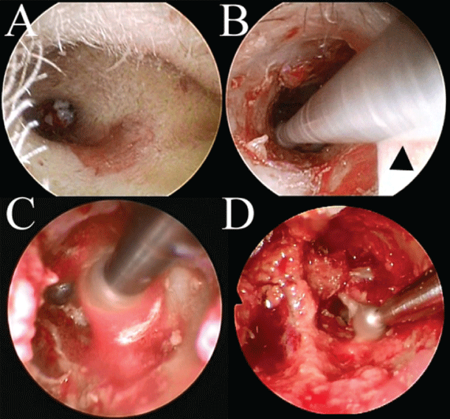
Eight patients (4 men and 4 women; median age, 57 years; age range, 18-73 years) underwent TEES using an unsheathed straight drill, while 14 patients (9 men and 5 women; median age, 47.5 years; age range, 7-78 years) underwent surgery using a sheathed curved drill. After TEES using an unsheathed straight drill, 50% (4 of 8) patients suffered from erosion (i.e. the superficial destruction of a surface of epidermis) on the posterior surface of the cartilaginous external ear canal (Figure 1A). After the first experience of erosion on the external ear canal, the surgeon tried less stress or less contact of the drill on the external ear canal. Nevertheless, the other subsequent patients suffered from erosion. However, TEES using a sheathed curved drill did not cause erosion. Of the patients for which the unsheathed straight drill was used, 2 underwent tympanoplasty with atticotomy, and 6 underwent tympanoplasty with atticoantrostomy. Of the patients for which the sheathed curved drill was used, 7 underwent tympanoplasty with atticotomy, and 7 underwent tympanoplasty with atticoantrostomy (Table 1).
The erosive lesions of the four patients were cured in 2-3 weeks. The risk of erosive lesions on the cartilaginous external ear canal was significantly higher for TEES using an unsheathed straight drill than for TEES using a sheathed curved drill (p = 0.0096).
Discussion
In TEES, an unsheathed straight drill caused temporal external ear canal lesions in 50% of patients, while a sheathed curved drill caused no lesions. In microscopic transcanal ear surgery, an unsheathed straight drill has been commonly used but has not generally caused external ear canal lesions.
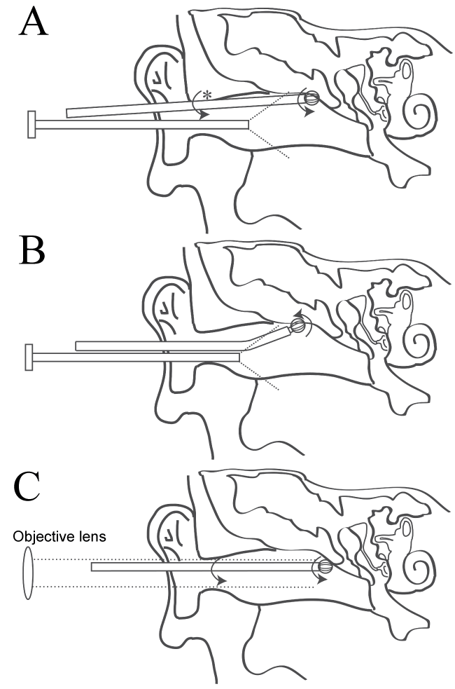
The rotating shaft of an unsheathed straight drill may damage the skin of the external ear canal (Figure 1B and Figure 2A). When using an endoscope, a surgeon cannot notice what happens behind the endoscope tip (Figure 1C) because this area is completely blind (Figure 1A). Conversely, when using a microscope, a surgeon can notice the situation in the external ear canal because the area is located between the objective lens and the objects, although the visual field for this region is out of focus and appears blurry (Figure 2C). Surgeons can thus pay attention to the external ear canal during bone dissection. Moreover, under microscopic surgery, the surgical view angle is straight and areas to the side are invisible. Therefore, surgeons do not try to dissect bone from these side areas. However, endoscopy enables surgeons to see these side areas. Surgeons may then try to dissect bone from these areas seven though they are not freely accessible to a straight drill. The rotating shaft of a drill stresses, pushes, and damages the surface of the external ear canal (Figure 1B). In contrast to a straight drill, a curved drill has a greater ability to access side areas (Figure 2B). Additionally, a sheathed curved drill does not damage through its rotation (Figure 1D and Figure 2B).
All the erosive lesions by an unsheathed straight drill were fully recovered without sequelae. Therefore, these erosions may be clinically insignificant. However, surgeons should try to diminish the potential risks. Unless sheathed curved drills are equipped, a speculum might prevent damage of the external ear canal. Another device, the ultrasonic bone dissector (Sonopet, Stryker, US), has been examined for bone dissection by Ito, et al. [7] and Kakehata, et al. [8]. This device ultrasonically vibrates its tip longitudinally and oscillates it torsionally, but it does not rotate like a drill. Therefore, the device does not catch soft tissue such as the tympanomeatal flap and the skin of the external ear canal. It can dissect bony tissue and aspirate the fine fragments. However, the size of the tip is more than 1.5 mm in width and this device may not be suitable for bone dissection adjacent to the ossicles. In these cases, a sheathed curved drill with a small sized tip (i.e. less than 1 mm in width) is much more suitable. Another alternate way to protect external ear canal is to use paper or silastic sheet to protect ear canal skin.
The other common tympanomeatal injury is involution of the tympanomeatal flap by the rotating drill although it did not happen to the patients in this study. This type of injury is possible with both an unsheathed straight drill and a sheathed curved drill. Paper or silastic sheet is a useful tool to protect tympanomeatal flap, and the ultrasonic bone dissector is safe device as well.
Endoscope resolution depends on the image sensing technology, monitor, and image processing technology. Because image sensing and processing technologies are rapidly progressing, endoscopy resolution is also expected to increase. To keep up with these technologies, further suitable devices for bone dissection and other maneuvers in TEES must be developed.
Finally, although this was a small study, it demonstrated the potential risk of external ear canal skin injury by an unsheathed straight drill and the alternate measures. This study has the limitation with respect to retrospective study, potential bias, and small number of patients. Because this study is focusing on the risk of external ear canal skin injury, prospective randomized controlled study is not suitable ethically. The attention bias to prevent damage intentionally cannot remove totally. However, after we experienced the external ear canal skin injury, the surgeon tried not damage the external ear canal. But skin injury inevitably occurred consequently. We believe that there is not attention bias at all. A study in larger number of patients with different similarly experienced surgeons is ideal. After a sheathed curved drill was equipped, the surgeon used it in TEES. Therefore the number is small but we believe that it is instructive.
Summary
The use of an unsheathed straight drill for TEES can damage the skin of the external ear canal. Extensive attention is necessary when using this tool. A sheathed curved drill is the safer and more suitable tool for TEES. Further devices for safe endoscopic surgery are expected to be developed.
Conflict of Interest
None.
Financial Disclosure
None.
References
- Mer SB, Derbyshire AJ, Brushenko A, et al. (1967) Fiberoptic endotoscopes for examining the middle ear. Arch Otolaryngol 85: 387-393.
- Nomura Y (1982) Effective photography in otolaryngology-head and neck surgery: endoscopic photography of the middle ear. Otolaryngol Head Neck Surg 90: 395-398.
- Thomassin JM, Korchia D, Doris JM (1993) Endoscopic-guided otosurgery in the prevention of residual cholesteatomas. Laryngoscope 103: 939-943.
- Tarabichi M (1999) Endoscopic middle ear surgery. Ann Otol Rhinol Laryngol 108: 39-46.
- Tarabichi M (1997) Endoscopic management of acquired cholesteatoma. Am J Otol 18: 544-549.
- Kakehata S, Futai K, Sasaki A, et al. (2006) Endoscopic transtympanic tympanoplasty in the treatment of conductive hearing loss: early results. Otol Neurotol 27: 14-19.
- Ito T, Mochizuki H, Watanabe T, et al. (2014) Safety of ultrasonic bone curette in ear surgery by measuring skull bone vibrations. Otol Neurotol 35: e135-e139.
- Kakehata S, Watanabe T, Ito T, et al. (2014) Extension of indications for transcanal endoscopic ear surgery using an ultrasonic bone curette for cholesteatomas. Otol Neurotol 35: 101-107.
Corresponding Author
Masafumi Ohki, MD, Department of Otolaryngology, Saitama Medical Center, Saitama Medical University, 1981 Kamoda, Kawagoe-shi, Saitama 350-8550, Japan, Tel: +81-49-228-3685, Fax: +81-49-225-6312.
Copyright
© 2017 Ohki M, et al. This is an open-access article distributed under the terms of the Creative Commons Attribution License, which permits unrestricted use, distribution, and reproduction in any medium, provided the original author and source are credited.


