Knee Loading due to Varus and External Rotation in Gait Supports Medial Compartment Wear in Total Knee Arthroplasty
Abstract
Malalignment of the lower extremity may be a cause for gonarthrosis and failure of total knee prosthetic components. Neutral lower limb alignment is considered straight alignment of the axes formed from the center of the femoral head to the center of the knee and from the center of the knee to the center of the ankle joint (the mechanical axes of the thigh and shank). Malalignment of the mechanical axis (measured by δv, the angle between the femoral and the tibial mechanical axes) occurs when varus moves the line of action farther medially from the knee joint center. Malalignment due to varus results in increases in medial compartment loading and has been attributed to medial compartment articular cartilage degeneration. External rotation of the hip and knee compared to the direction of gait is another form of malalignment. In external rotation, the knee displaces from the midline at stance phase. Knee loading and the consequences of external rotation on cartilage degeneration are not well understood. The purpose of our study was to develop a mathematical model that would calculate forces and moments in knees for gait and study how they vary with varus and external rotation malalignment. An additional objective was to develop a finite element model of total knee replacement to study stress patterns on the polyethylene insert during malaligned gait. We hypothesized that medial compartment loads would be increased by both varus and external rotation alignment of the knee compared to the direction of gait. We also hypothesize that stress patterns on total knee replacement (TKR) inserts under conditions related to malaligned gait will correlate with wear patterns observed in retrieval studies. It was found that for varus and external rotation, there is a shift towards adduction moment, which resulted in an increase in medial compartment loads, supporting our hypothesis. It was also found that the malalignment in gait results in alterations in tibial tray load magnitudes and load distribution that support elevated wear in the medial compartment as observed clinically.
Keywords
Varus, External rotation, Total knee replacement, Wear, Malalignment
Introduction
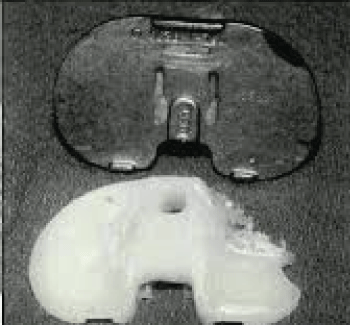
Total knee replacement (TKR) is a common treatment for gonarthrosis and has seen increasing use over the last 20 years. Malalignment of the lower extremity may be not only a cause for gonarthrosis but a precursor for failure of total knee prosthetic components [1-3]. Polyethylene loss in total knee replacement is evident and commonly reported in numerous retrieval studies as one cause of TKR failure [4-6]. In particular, wear in the anterior medial compartment is frequently observed (Figure 1) [7]. Malalignment of the TKR (e.g. varus or external rotation vs. neutral alignment) may be a contributing factor in accelerating medial compartment wear. Neutral lower limb alignment is considered straight alignment of the axes formed from the center of the femoral head to the knee center and from the center of the knee to the center of the ankle joint (the mechanical axes of the thigh and shank). Malalignment of the mechanical axis (measured by δv, the angle between the femoral and the tibial mechanical axes) occurs when varus moves the line of action farther medially from the knee joint center increasing the medial load [8]. External rotation of the knee compared to the direction of gait exacerbates medial loading by further displacing the knee from the midline at stance [9].
Previous work has shown that in varus malalignment, the line of the force shifts farther medially from the knee joint center increasing the load on the medial compartment of the knee [10-19] resulting in a joint reaction force that is approximately three and a half times the lateral compartment loads [8]. Moyer, et al. [20] showed that the mechanical axis angle accounted for 32%-54% of the explained variation in knee adduction moment, yielding a 3.2 N increase for every 1° increase in varus. Knees that visually demonstrate varus thrust, have been shown to have greater knee adduction moment compared to knees without varus thrust [21-23] and have been reported to be at least four times more likely to have pain during weight bearing activity [24].
The purpose of our study was to develop a mathematical model that would calculate forces and moments in knees for gait and study how they vary with varus and external rotation malalignment. An additional objective was to develop a finite element model of total knee replacement to study stress patterns on the polyethylene insert during malaligned gait. We hypothesized that medial compartment loads would be increased by both varus and external rotation alignment of the knee compared to the direction of gait. We also hypothesize that stress patterns on TKR inserts under conditions related to malaligned gait would correlate with wear patterns observed in retrieval studies.
Methods
Applied knee joint forces
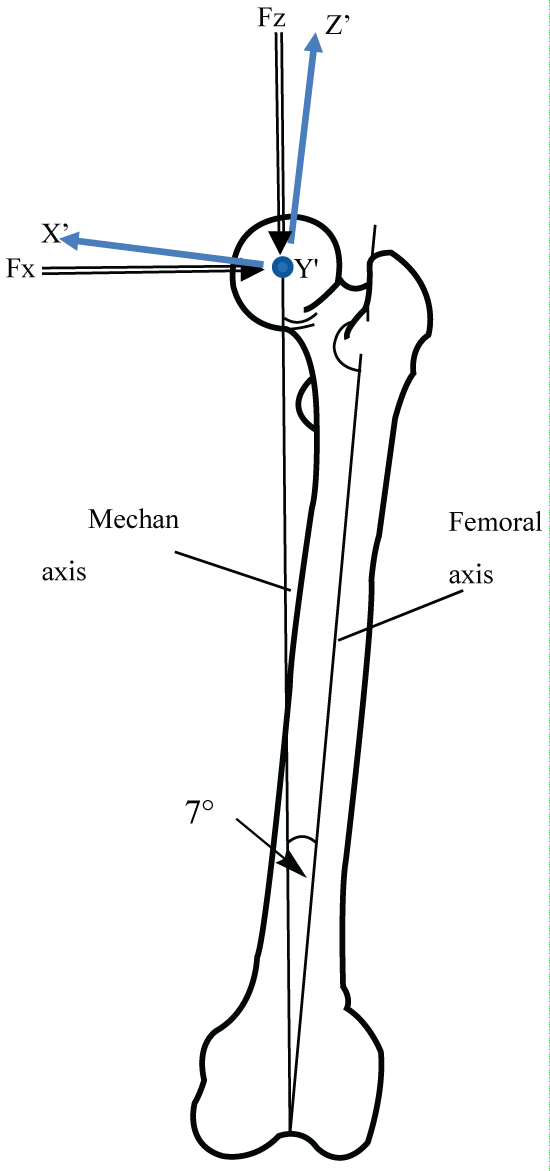
In the approach to define applied loads at the knee, results from clinical gait lab experiments were used [25]. Several full sets of hip joint load data as well as the angle at which these loads are applied is included in the referenced study. The reference includes information on several different loading scenarios including walking, climbing and descending stairs, sitting down, standing up, knee bending, standing on one leg, and stumbling. The current studied considered maximum loads while walking. An equilibrium approach was applied to the hip joint loads to find the overall forces acting on the knee. This involved taking "snapshots" of the data at each location of the leg at each increment in gait and imposing equilibrium at each of these positions. The hip forces were imposed on a simplified "stick-figure" model as done previously [26,27]. Our axes were a global anatomical axis system (x',y',z') as well as a local axis system (x,y,z) which places the z-axis along the mechanical axis of the femur (Figure 2). The mechanical axis is the axis from the center of the femoral head to the center of the knee. The data from Bergman, et al. [25] was offset seven degrees in the x-z plane from the mechanical axis. Therefore, the forces on the femoral head were redefined by a rotation about the y,y' axis of seven degrees. Several articles provided ranges for the angular orientation of the femur in the body to reorient the loads [28-30]. The forces at the knee where then determined using a force balance after positioning the femur and applying experimentally determined femoral loads as described below. The knee loads that define the "neutral" gait case are defined when the femur and the tibia are aligned according to the mechanical axis. This alignment assumes that the hip-knee-ankle angle is 180°, although reports indicate an angle of slightly less than 180° depending on the patient [31,32].
A unit vector, N, which establishes the orientation of the resultant force F from Bergmann, et al. [25] was determined by taking the force vector and dividing it by the magnitude of the force
The unit vector in the form of direction cosines is defined a
where
In equation (3) above, θx, θy, θz are the angles between the resultant force and the x, y and z axes, respectively. These direction cosines can be defined by the product of two cosines multiplied together. One direction cosine is defined by the angle between the force vector and the x-y plane (Φxy) or the y-z plane (Φyz). The second direction cosine is defined by the angle between the projected force on the x-y and y-z planes and the x (δx), y (δy) or z (δz) axes. The resulting direction cosines can be rewritten as
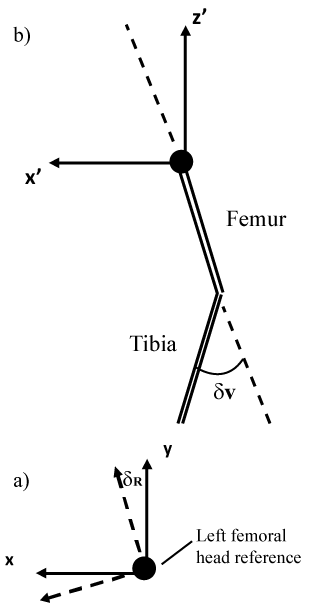
External rotation (δR) of the knee is defined as a positive rotation of the hip joint such that the hip and knee joint are rotated through the same angle about the z-axis (Figure 3a) similar in orientation to the previously defined δx. This results in the knee pointing outward. Taking this into consideration in addition to recognizing the relationship between substituting δx and δy i.e.
we arrive at the direction cosines for external rotation
Equation (6) defines the directions cosines of the resultant force corresponding to an internal rotation of δR.
For varus of the knee, the direction cosines are determined by adding the desired angle of varus, δv (Figure 3b), to the direction cosines defined previously. Recognizing the relationship between δz and Φy similar to eq. (5), and substituting that relation into eq. (4), we arrive at
Equation (7) defines the directions cosines of the resultant force corresponding to an varus rotation of δv.
Multiplying unit vectors by the magnitude of the force yields the force vectors for each case (normal gait, external rotation of the femur, and varus of the knee), i.e.
After loading to the femoral head is determined for each case (i.e. neutral, external rotation or varus) in its properly oriented directions, equilibrium is imposed on the femur in order to determine knee loads. For each load case, the sum of forces, F, is expressed as
Where f refer to the femoral head, k refers to knee forces are equ the knee and i refers to the three mutually perpendicular directions x, y or z. Thus, equilibrium ensures that the al in magnitude but opposite in directions to the applied femoral head forces such that
The x and y moments at the knee, Mx and My, respectively, are determined from the product of the components of the femoral head loads times the length of the femur
The moment along the mechanical axis, Mz, is zero consistent with femoral head loads applied to the mechanical axis. The Forces and moments are subsequently geometrically rotated to align with knee axis (Figure 4). This results in modifications to the forces and moments calculated in (10) and (11) resulting in a nonzero torsional moment, Mz. Subsequent calculations using a matlab based program were performed to determine applied knee loadings. To illustrate the extent of the knee loading for moderate to extreme varus and external rotation, both angles were varied for 5, 10 and 25 degrees of malalignment. The algorithm output gave force and moment across the gait cycle from which the critical locations were identified. The analysis showed the maximum force occurred 17% of the gait cycle which is shortly after the opposite leg is leaving the ground and the foot flat on the ground. Forces and moments at the knee were then used to calculate femoral shaft loads using equilibrium. The femoral shaft loads were applied in the finite element analysis described below.
Finite element model
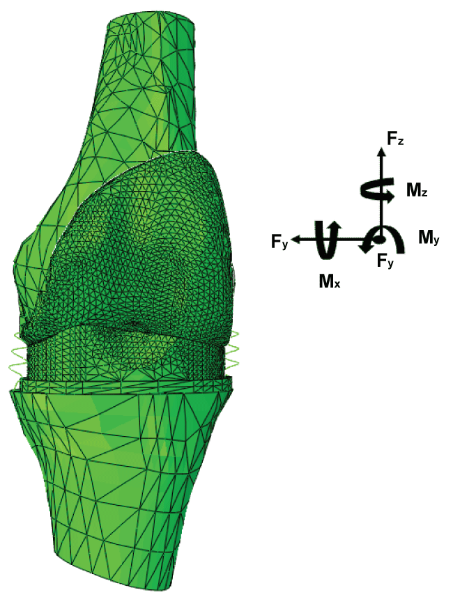
The finite element analysis was modeled after the ADVANCE® Medial-Pivot Knee. A physical model of the implant was scanned using the NextEngine 3D scanner (Santa Monica, CA), which gave the contact surface of both the tibial plastic and the femoral component. RapidWorks (Santa Monica, CA) was used to convert the pointcloud scanned image into a solid surface. RapidWorks was used to solidify the pointcloud into a solid surface by simply using the solidify feature. The two objects were then transferred into SolidWorks (Dassault Systems, Waltham, MA) where the implant model was attached to models of the femur and tibia. The implant was appropriately scaled for the size of the femur and tibia models and was placed in the proper orientation. There were two primary parts that were needed to properly position the knee. The first part is the set of angles that define the knee at a given position for gait. The second part is the contact points between the tibial insert and the femoral condyles in the implant. Gaudreault, et al. [33] was used to obtain flexion-extension, abduction-adduction and internal-external rotation angles of the knee throughout gait. At maximum loading corresponding to 17% gait position, the orientation of the femur to the tibia for normal or neutral gait was found to be 14 degrees of flexion, 0.4 degrees of adduction, and 1 degree of internal rotation. The femur was repositioned for varus and external rotation type gait. When 5 degrees of external rotation was added to the femur, the angle of rotation became 4 degrees. The contact points between the femoral and tibial components were defined at 17% gait [34]. The neutral position was modified for varus and external rotation forms of gait. For varus, an angle of 5 degrees was assumed based on the literature. Varus positioning resulted in condylar liftoff or separation in the lateral condyle of the knee. For external rotation of the knee, our positioning was based on clinical experience of one of the investigators of this paper (JDB) who is a practicing orthopaedic surgeon. In external rotation the point about which the knee moves in the rotation is located outside of the knee on the lateral side. Therefore, both condyles move anteriorly in the implant, but the medial side moves further anterior than the lateral side of the knee. To find the angle of external rotation, the normal gait model was subjected to an angle of external rotation, keeping the contact points along a prescribed path until the contact was made on the anterior portion of the medial side of the knee. This angle was chosen as 5 degrees. The angle was then used in the rest of the models for the augmented knee replacements. All three models were positioned in SolidWorks and imported into ABAQUS (Dassault Systems, Waltham, MA) finite element software. The model consisted of a tibial tray, tibial plastic, femoral component, and a portion of a left femur (Figure 4). The elastic moduli of the components were 20.0 GPa for the cortical bone, 230 GPa for the cobalt-chromium tibial tray and femoral component and 0.689 GPa for the ultra-high molecular weight polyethylene (UHMWPE). Partial tibia and femur were used to limit the size of the model to approximately 100,000 nodes. The applied loads were calculated as discussed previously and were applied to the femur while the tibia was fixed. The final important task was to define the interactions between the tibial plastic and the femoral component. We used finite sliding, a surface to surface discretization method, and a slave adjustment only to remove overclosure. We applied linear springs to either side to replicate the medial and lateral collateral ligaments. To aid with convergence, a tie constraint was used between the surfaces of the femoral component and the tibial plateau.
Results
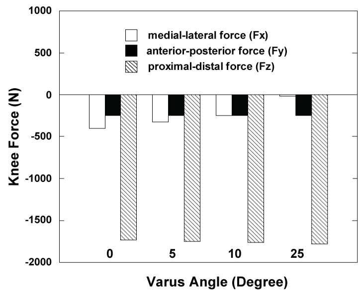
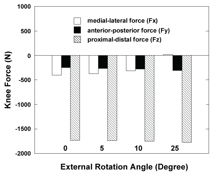
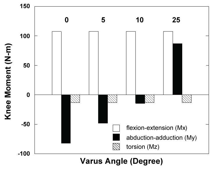
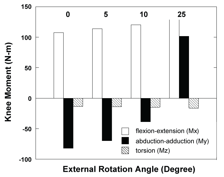
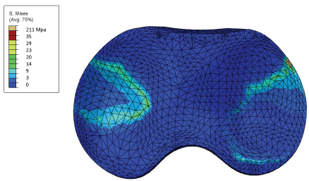
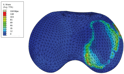
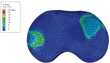
The knee force components for 0 (neutral), 5, 10 and 25 degrees are shown in Table 1 and Figure 5 for varus malalignment and in Table 2 and Figure 6 for external rotation malalignment. Results are reported as applied to the tibial plastic. Anterior-posterior (Fy) and proximal-distal (Fz) forces remain nearly unchanged with varus and external rotation. However, medial-lateral directed force (Fx) changes significantly from -403 N laterally directed force to near zero in magnitude as varus and external rotation angles increase. Thus, malalignment of either form results in forces applied to the knee that shift from medially directed to less medially directed and achieve a small laterally directed force for the case of 25 degrees of external rotation. The knee moment components for 0, 5, 10 and 25 degrees are shown in Table 1 and Figure 7 for varus malalignment and in Table 2 and Figure 8 for external rotation malalignment. Results are reported as applied to the tibial plastic. Flexion-extension moment (Mx) and rotation moment (Mz) also experience small changes with varus and external rotation angle. Abduction-Adduction moment (My) however experiences significant changes with increases in varus and external rotation angles. For both types of malalignment, the moment changes from an abduction moment (negative, away from the body center) to adduction moment (positive, towards the body center) as the malalignment angle increase. The adduction moment results in applied loads to the knee that increase the tibial tray medial compartment loads. These results are reflective in finite element simulations. Finite element results of the tibial tray demonstrate both medial and lateral tibial tray compartment loading near implant center for neutral alignment (Figure 9). For five degrees of varus malalignment (Figure 10), finite element results show concentrated medial compartment loading with elevated stress results compared to neutral alignment. Five degrees of external rotation (Figure 11) resulted in tibial tray loading in the anterior region of the medial compartment and the lateral-posterior region of the lateral compartment. Stresses in the contact region for neutral and external rotation where both condyles are in contact with the tibial tray result in von Mises stresses of approximately 29 MPa. For varus malalignment where only one condyle is in contact, von Mises stresses in the contact region are significantly higher, on the order of approximately 81-138 MPa.
Discussion
Results of this study show that knee malalignment had a significant effect on the forces and moments at the knee. In particular, the medial-lateral force and the abduction-adduction moment were most significantly affected. Adduction moments are responsible for higher loads in the medial compartment of the knee. Varus and external rotation during gait resulted in a large shift towards adduction moment. The compressive force on the medial compartment due to adduction moment in varus malalignment combined with the compressive normal force (Fz) along the mechanical axis resulted in an overall greater medial compressive compartment load compared to neutral alignment as observed in finite element results. Results for varus malalignment are consistent with previous studies that show higher adduction moment. The higher stresses concentrated in the medial compartment would results in higher wear rates as observed clinically. For a five degree external rotation, finite element results indicated a change in stress distribution rather than an increase in stress magnitude compared to neutral alignment. It is expected that the stress magnitudes would increase with additional external rotation. In addition, other types of activity not considered here may result in increased stress. This study investigated maximum loading while walking. Other load cases such as running or climbing stairs would result in higher applied loads and consequently higher tibial tray stresses than reported in the current study.
The importance of lower limb alignment in the preservation of femoral-tibial joint health has been shown in many previous studies. Knee adduction moment in subjects with moderate to severe OA ranged from 3.6% x body weight x height to 6.9% x body weight x height, respectively [35]. There is also a higher incidence of Knee Osteoarthritis (OA) in the medial than lateral compartment of the knee [1-3]. Patients having a knee with OA had greater peak knee adduction moment and frontal plane lever arm (perpendicular distance between the ground reaction force and the knee joint center) but less ground reaction force [36]. Bennell, et al. [37] reported that knee loading may be a risk factor for loss of medial tibial cartilage volume. There is also evidence in numerous radiographic, MRI and clinical studies that suggest that knee adduction moment is a strong risk factor the progression of medial compartment articular cartilage degeneration [8,37-39]. Varus malalignment has been associated with incident of cartilage damage in the medial compartment supporting the idea that varus increases the risk of initial development of osteoarthritis (OA). Increased medical loading due to varus malalignment has been correlated with subsequent disease progression as well [20,36,40-42]. Varus deformity leads to a perpetuating cycle of a decrease in medial joint space and even greater loads on the medical compartment [43].
Knee loading under external rotation is not well understood. Our findings suggest that the Posterior-anterior directed moments also change to more to anteriorly directed moments for external rotation, although not as significantly as varus malalignment. As stated previously, anteriorly directed applied moments to the knee corresponds to adduction moments and are responsible for higher loads in the medical compartment. External rotation resulted in a shift in the applied loads to the tibial tray, causing more load transfer in the anterior portion of the medial compartment reminiscent of implant wear as described by Knezevich, et al. [7]. Our finding suggests that individuals that externally rotate during gait could experience a higher medial compartment compressive loads and perhaps higher incidence of knee medial compartment articular cartilage degeneration.
The mathematical model developed in this study was written to calculate knee loads during neutral, externally rotated and varus gait cases based on static equilibrium using hip loads at the femoral head. The model is limited by the omission of hip and knee soft tissue loads. The omission of soft tissue loads would result in alterations to forces and moments at the knee. It is also responsible for the loss of a significant torsional moment along the mechanical axis. It can be seen from our result that the abduction-adduction directed moment (My) for normal gait is posteriorly directed, which is an abduction moment. From previous work we know that this moment should be anteriorly directed (i.e. Adduction moment), for normal gait. Our simplifying assumptions at the hip including the omission of hip abductor loading are likely responsible for the difference observed here as hip abduction for would result in a larger adduction moment at the knee. However, the model is descriptive in the sense that we can see that externally rotated and varus gaits shift the knee moments towards adduction moment, at peak load and throughout the gait cycle.
Results of this study found that for both types of misaligned gait there is a shift towards adduction moment. Increased adduction moment results in an increase in medial condyle loading, supporting our hypothesis. In addition, finite element simulations of misaligned gait demonstrate shifts in load magnitude and load distribution supporting elevated wear in the medial compartment as observed clinically. Therefore, for both types of misaligned gait considered in this study, joint health and post total knee replacement success needs to be more carefully examined.
References
- Ledingham J, Regan M, Jones A, et al. (1993) Radiographic patterns and associations of osteoarthritis of the knee in patients referred to hospital. Ann Rheum Dis 52: 520-526.
- Dearborn JT, Eakin CL, Skinner HB (1996) Medial compartment arthrosis of the knee. Am J Orthop (Belle Mead NJ) 25: 18-26.
- Bartel DL (1992) Unicompartment arthritis: biomechanics and treatment alternatives. Instr Course Lect 41: 73-76.
- Vandekerckhove PT, Teeter MG, Naudie DD, et al. (2017) The impact of coronal plane alignment on polyethlylene wear and damage in total knee arthroplasty: A retrieval study. J Arthroplasty.
- Van der list JP, Zuiderbaan HA, Pearle AD (2016) Why do medial unicompartmental knee arthroplasties fail today? J Arthroplasty 31: 1016-1021.
- Gupta SK, Chu A, Ranawat AS, et al. (2007) Osteolysis after total knee arthroplasty. J Arthroplasty 22: 787-799.
- Knezevich S, Vaughn BK, Lombardi AW, et al. (1993) Failure of the polyethylene tibial component of the TKR associated with aseptic loosening secondary to polyethylene and metallic wear debris. Orthopedics 16: 1136-1140.
- Schipplein OD, Andriacchi TP (1991) Interaction between active and passive knee stabilizers during level walking. J Orthop Res 9: 113-119.
- Abelson Brian (2010) Kinetic Health - Calgary.
- Andriacchi TP (1994) Dynamics of knee malalignment. Orthop Clin North Am 25: 395-403.
- Birmingham TB, Hunt MA, Jones IC, et al. (2007) Test-retest reliability of the peak knee adduction moment during walking in patients with medial compartment knee osteoarthritis. Arthritis Rheum 57: 1012-1017.
- Hurwitz DE, Ryals AB, Case JP, et al. (2002) The knee adduction moment during gait in subjects with knee osteoarthritis is more closely correlated with static alignment than radiographic disease severity, toe out angle and pain. J Orthopaedic Res 20: 101-107.
- Baliunas AJ, Hurwitz DE, Ryals AB, et al. (2002) Increased knee joint loads during walking are present in subjects with knee osteoarthritis. Osteoarthritis Cartilage 10: 573-579.
- Hilding MB, Lanshammar H, Ryd L (1995) A relationship between dynamic and static assessments of knee joint load. Gait analysis and radiography before and after knee replacement in 45 patients. Acta Orthop Scand 66: 317-320.
- Wada M, Maezawa Y, Baba H, et al. (2001) Relationships among bone mineral densities, static alignment and dynamic load in patients with medial compartment knee osteoarthritis. Rheumatology (Oxford) 40: 499-505.
- Wada M, Imura S, Nagatani K, et al. (1998) Relationship between gait and clinical results after high tibial osteotomy. Clin Orthop Relat Res 180-188.
- Andrew M, Noyes FR, Hewett TE, et al. (1996) Lower limb alignment and foot angle are related to stance phase knee adduction in normal subjects: a critical analysis of the reliability of gait analysis data. J Orthop Res 14: 289-295.
- Catani F, Marcacci M, Benedetti MG, et al. (1998) The influence of clinical and biomechanical factors on the results of valgus high tibial osteotomy. Chir Organi Mov 83: 249-262.
- Sharma L, Hurwitz DE, Thonar EJ, et al. (1998) Knee adduction moment, serum hyaluronan level, and disease severity in the medical tibiofemoral osteoarthritis. Arthritis Rheum 41: 1233-1240.
- Moyer RF, Birmingham TB, Chesworth BM, et al. (2010) Alignment, body mass and their interaction on dynamic knee joint load in patients with knee osteoarthritis. Osteoarthr Cartil 18: 888-893.
- Chang A, Hayes K, Dunlop D, et al. (2004) Thrust during ambulation and the progression of knee osteoarthritis. Arthritis Rheum 50: 3897-3903.
- Hunt MA, Schache AG, Hinman RS, et al. (2011) Varus thrust in medial knee osteoarthritis: quantification and effects of different gait-related interventions using a single case study. Arthritis Care Res (Hoboken) 63: 293-297.
- Willams DS, Isom W (2012) Decreased frontal plane hip joint moments in runners with excessive varus excursion at the knee. J Appl Biomech 28: 120-126.
- Lo GH, Harvey WF, McAlindon TE (2012) Associations of varus thrust and alignment with pain in knee osteoarthritis. Arthritis Rheum 64: 2252-2259.
- Bergmann GF, Graichen A, Rohlmann A, et al. (2010) Realistic loads for testing hip implants. Biomed Mater Eng 20: 65-75.
- Bobbert MF, Yeadon MR, Nigg BM (1992) Mechanical analysis of the landing phase in heel-toe running. Journal of Biomechanics 25: 223-234.
- Dasgupta S (2009) Biomechanical Modeling of Materials Lifting Activities for Exploring Relation between Low Back and Knee Loading leading to Low Back Pain and Knee Arthritis. Indian Journal of Biomechanics 180-185.
- Mizuno Y, Kumagai M, Mattessich SM, et al. (2001) Q-angle Influences Tibiofemoral And Patellofemoral Kinematics. J Orthop Res 19: 834-840.
- Gilroy AM, MacPherson BR, Ross LM (2008) Atlas of anatomy. Stuttgart: Thieme.
- Solomin LN, Shchepkina E, Kulesh P, et al. (2010) Determination of reference lines and angles of the long bones (guide for the doctors). R.R. Vreden Russian Research Institute of Traumatology and Orthopedics 48.
- Paley D (2002) Principles of Deformity Corrections. Springer-Verlag Berlin Heidelberg.
- Cherian JJ, Kapadia BH, Banerjee S, et al. (2014) Mechanical, anatomical, and kinematic axis is TKA: Concepts and practical applications. Curr Rev Musculoskeletal Med 7: 89-95.
- Gaudreault N, Fuentes A, Mezghani N, et al. (2013) Relationship between knee walking kinematics and muscle flexibility in runners. J Sport Rehabil 22: 279-287.
- Pinskerova V, Johal P, Nakagawa S, et al. (2004) Does the femur roll back with flexion? J Bone Joint Surg Br 86: 925-931.
- Kuroyanagi Y, Nagura T, Kiriyama Y, et al. (2012) A quantitative assessment of varus thrust in patients with medical knee osteoarthritis. Knee 19: 130-134.
- Hunt MA, Birmingham TB, Giffin JR, et al. (2006) Associations among knee adduction moment, frontal plane ground reaction force, and lever arm during walking in patients with knee osteoarthritis. J Biomech 39: 2213-2220.
- Bennell KL, Bowles KA, Wang Y, et al. (2011) Higher dynamic medial knee load predicts greater cartilage loss over 12 months in medial knee osteoarthritis. Ann Rheum Dis 70: 1770-1774.
- Miyazaki T, Wada M, Kawahara H, et al. (2002) Dynamic load at baseline can predict radiographic disease progression in medial compartment knee osteoarthritis. Ann Rheum Dis 61: 617-622.
- Wu DD, Burr DB, Boyd RD, et al. (1990) Bone and cartrilage changes following experimental varus or valgus tibial angulation. J Orhtop Res 8: 572-585.
- Crema MD, Roemer FW, Felson DT, et al. (2012) Factors associated with meniscal extrusion in knees with or at risk for osteoarthritis: The Multicenter Osteoarthritis Study. Radiology 264: 494-503.
- Sharma L, Chmiel JS, Almagor O, et al. (2013) The role of varus and valgus alignment in the initial development of knee cartilage damage by MRI: the MOST study. Ann Rheum Dis 72: 235-240.
- Sharma L, Lou C, Cahue S, et al. (2000) The mechanism of the effect of obesity in knee osteoarthritis: the mediating role of malalignment. Arthritis Rheum 43: 568-575.
- McNamara I, Birminghan TB, Fowler PJ, et al. (2013) High tibial Osteotomy: Evolution of research and clinical applications - a Canadian Experience. Knee Surg Sports Traumatol Arthrosc 21: 23-31.
Corresponding Author
Timothy L Norman, Professor of Mechanical and Biomedical Engineering, School of Engineering and Computer Science, Cedarville University, Cedarville, OH 45314, USA.
Copyright
© 2017 Norman TL, et al. This is an open-access article distributed under the terms of the Creative Commons Attribution License, which permits unrestricted use, distribution, and reproduction in any medium, provided the original author and source are credited.