Labial Frenum Incision to Extraction of Transmigrated Impacted Mandibular Canine
Abstract
Impacted tooth is one that, after its formation period and normal eruption time, is still inside the bone tissue. This article shows the surgical access technique for extraction of the impacted canine in the mandible, having the vertical access through the labial frenum. We believe that this access offers an excellent surgical field, without causing sequelae in the anterior region of the chin and without causing damage to the periodontal structures and in the mentonian nerve bundle. This approach is also used for harvesting a chin block graft, for bone reconstruction for rehabilitation with dental implants and also in chin repositioning (mentoplasty) in orthognathic surgeries.
Keywords
Oral surgery, Impacted teeth, Labial frenum, Mandible
Introduction
An impacted tooth is a tooth that at the end of its development period, still remains within the bone tissue and is not positioned in the dental arch.
Thus, imaging diagnosis is essential for the success of orthodontic and surgical procedures, minimizing complications and providing patients with more effective treatment.
According to Hupp, et al. [1] the removal of impacted teeth can be difficult or relatively easy. The main factor in determining the difficulty of removal is the ease of access and its visualization. Accessibility is determined by the exposure, via ostectomy and the formatting of the exit path of the whole or sectioned tooth. The literature defined that the unerupted canines should be used orthodontically or if this is not possible, they should be extracted from the arch [2].
Yavuz, et al. stated that the incidence of impacted mandibular canines was 1.29% in their study, and extraction was the most common treatment performed [3].
We present a clinical case of the use of a vertical incision in the labial frenum for surgical access and extraction of a transmigrated impacted lower canine.
Case Study
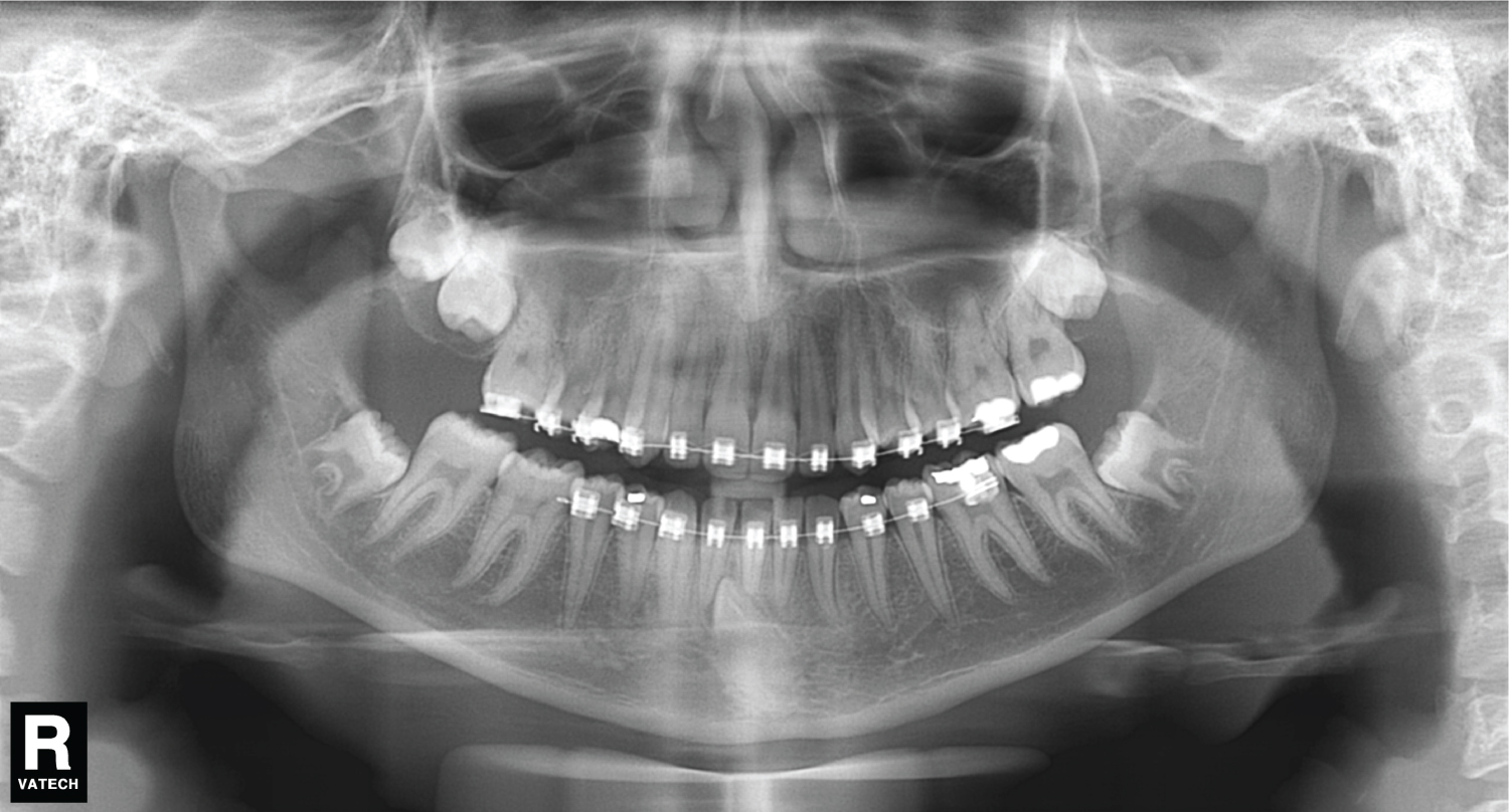
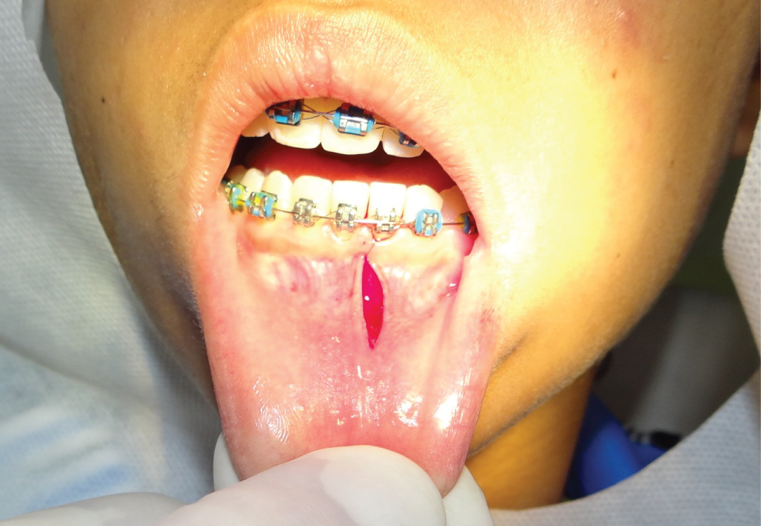
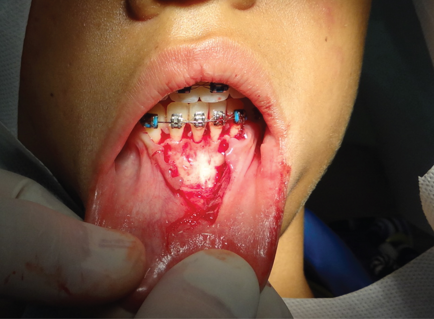
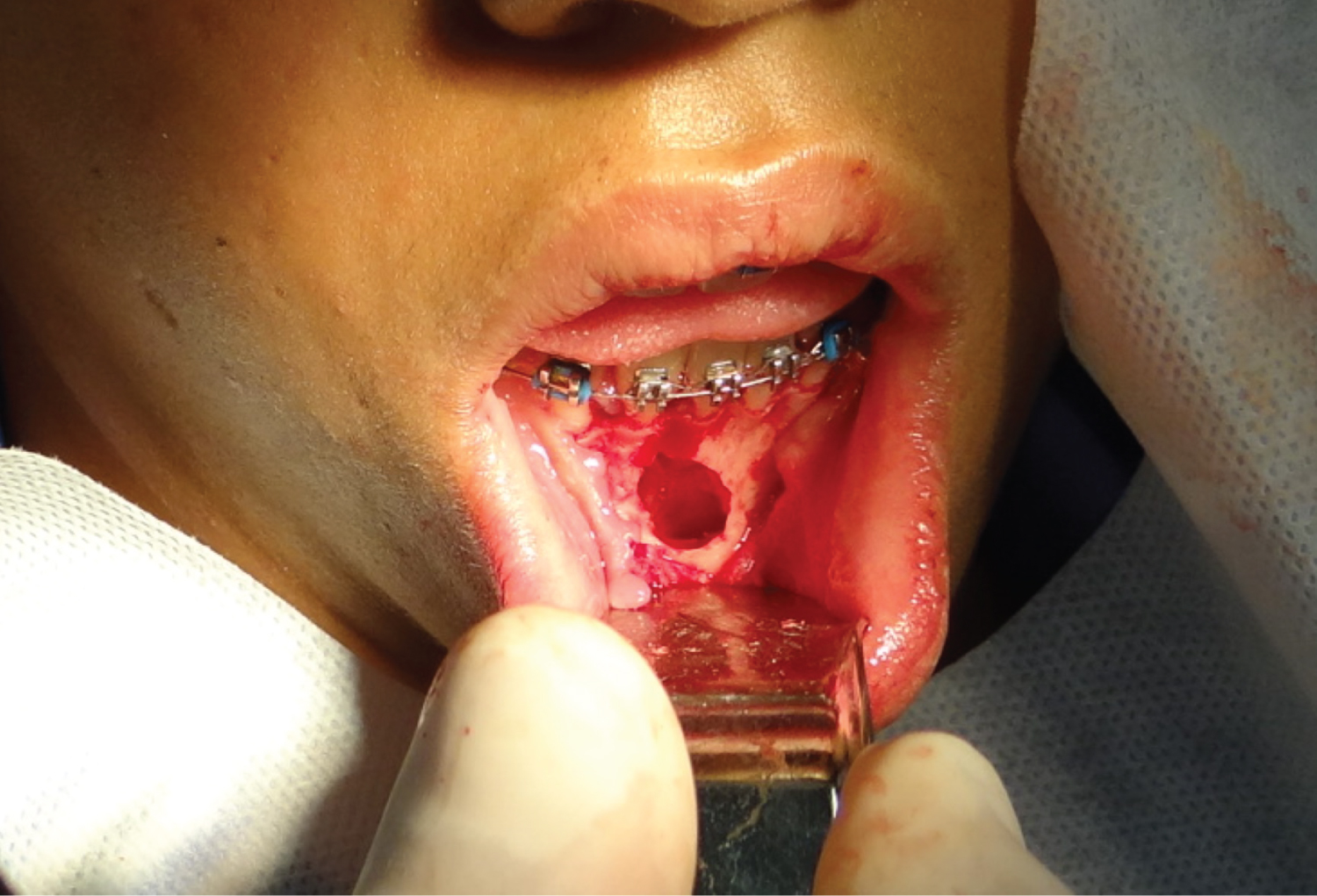
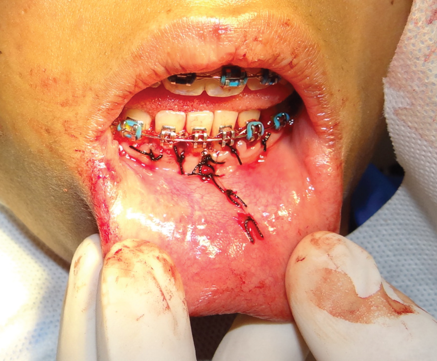
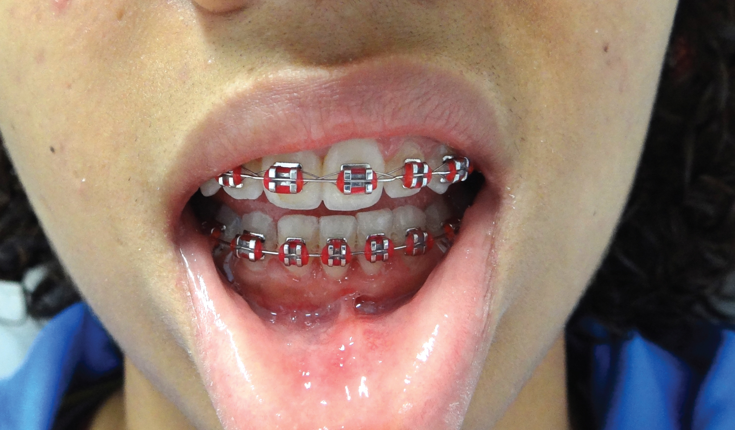
Female patient, 16-year-old, was referred for extraction of left mandibular impacted canine (FDI- 33) by orthodontic treatment reasons. During clinical examination an unerupted tooth was detected in alveolar position near the sagittal plane, causing a slight bulging of the alveolar sulcus, confirmed on panoramic radiograph (Figure 1). After planning and informed consent by the patient`s mother, a vertical access was performed through the lower labial frenum (Figure 2), a mucoperiosteal flap was elevated (Figure 3), the unerupted tooth was exposed via ostectomy with a high speed drill, dental sectioning and removal was performed (Figure 4). The surgical site was irrigated with saline solution and sutured (Figure 5). Repair occurred normally and the mucosal suture was removed after 7 days. Control after 3 weeks show the completed mucosal repair without changes in the labial frenum (Figure 6). Postoperative controls were performed until 2 months and the patient was discharged.
Discussion
The traditional surgical access would be the horizontal approach at the vestibular fold. This procedure causes greater surgical trauma and poses risk to the sensitivity of the chin because it reaches the mental nerve terminations. The vertical access in the labial frenum was chosen, as it offers an excellent surgical field in the sagittal region of the mandible. And the mucoperiosteal flap protects the mentonian nerve bundle.
This approach is also used for harvesting a chin block graft, for bone reconstruction for rehabilitation with dental implants and also in chin repositioning (mentoplasty) in orthognathic surgeries. We indicate its use because of its ease, low risk of neurological sequelae, and absence of complications.
References
- Hupp J, Ellis E, Tucker MR (2015) Cirurgia Oral e Maxilofacial Contemporânea. (6th edn), Rio de Janeiro: Elsevier.
- Verma SL, Sharma VP, Singh GP (2012) Management of a transmigrated mandibular canine. J Orthod Sci 1: 23-28.
- Yavuz MS, Aras MH, Büyükkurt MC, et al. (2007) Impacted mandibular canines. J Contemp Dent Pract 8: 78-85.
Corresponding Author
Elio H Shinohara, DDS, PhD, Assistant Surgeon, OMF Surgery Department, Hospital Regional de Osasco SUS/SP, Brazil.
Copyright
© 2023 Tedeschi GK, et al. This is an open-access article distributed under the terms of the Creative Commons Attribution License, which permits unrestricted use, distribution, and reproduction in any medium, provided the original author and source are credited.