A Hidden Oral Lesion
Abstract
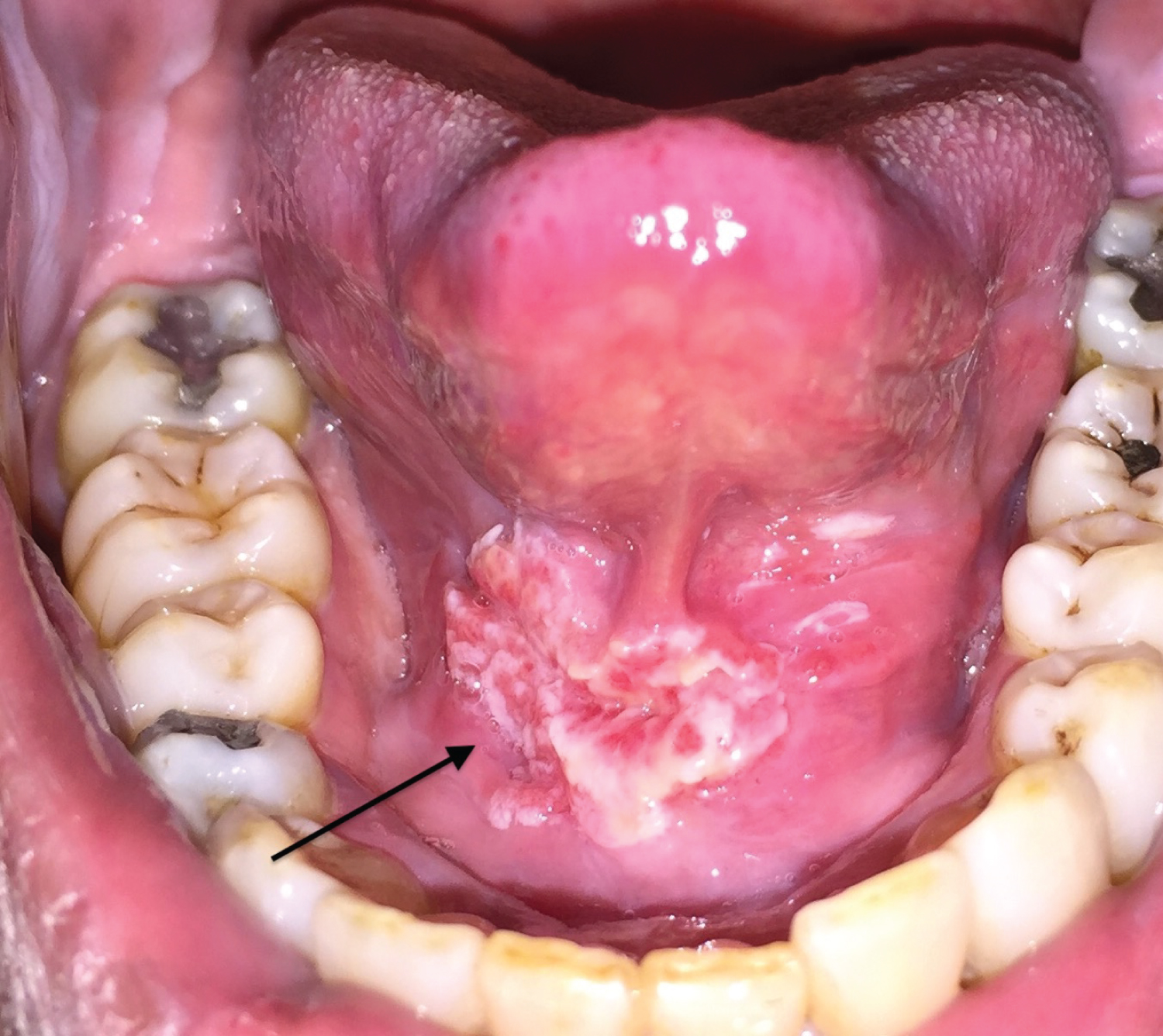
A 60-year-old man with a 10-pack-year smoking history was seen in the Oral surgery clinic for pain upon tongue movement especially with eating. A physical examination revealed a 1 cm by 3 cm exophytic mixed red and white lesion in the floor of the mouth and the ventral surface of the tongue extending posteriorly on the right side (Figure 1). On palpation, this was tender, firm, fixed, sessile with raised areas and rolled margins. Differential diagnoses included a squamous cell carcinoma or benign lesions such as verruca vulgaris or a squamous papilloma. Histopathology demonstrated a well-differentiated squamous cell carcinoma with islands of malignant squamous epithelium invading into the lamina propria with keratin pearl formation. Patient underwent surgical resection followed by chemo-radiation with an uneventful post-treatment course. There remains a need for public education on oral manifestations of tobacco and the need for oral examinations.
Corresponding Author
Sagar Khanna, BDS, DDS, Oral and Maxillofacial Surgery, Cleveland Clinic Foundation, Cleveland, OH, NY, USA, Tel: +1-507-206-1498.
Copyright
© 2022 Khanna S. This is an open-access article distributed under the terms of the Creative Commons Attribution License, which permits unrestricted use, distribution, and reproduction in any medium, provided the original author and source are credited.