Pituitary Adenoma with Evidence of Intracranial Extramedullary Haematopoiesis
Abstract
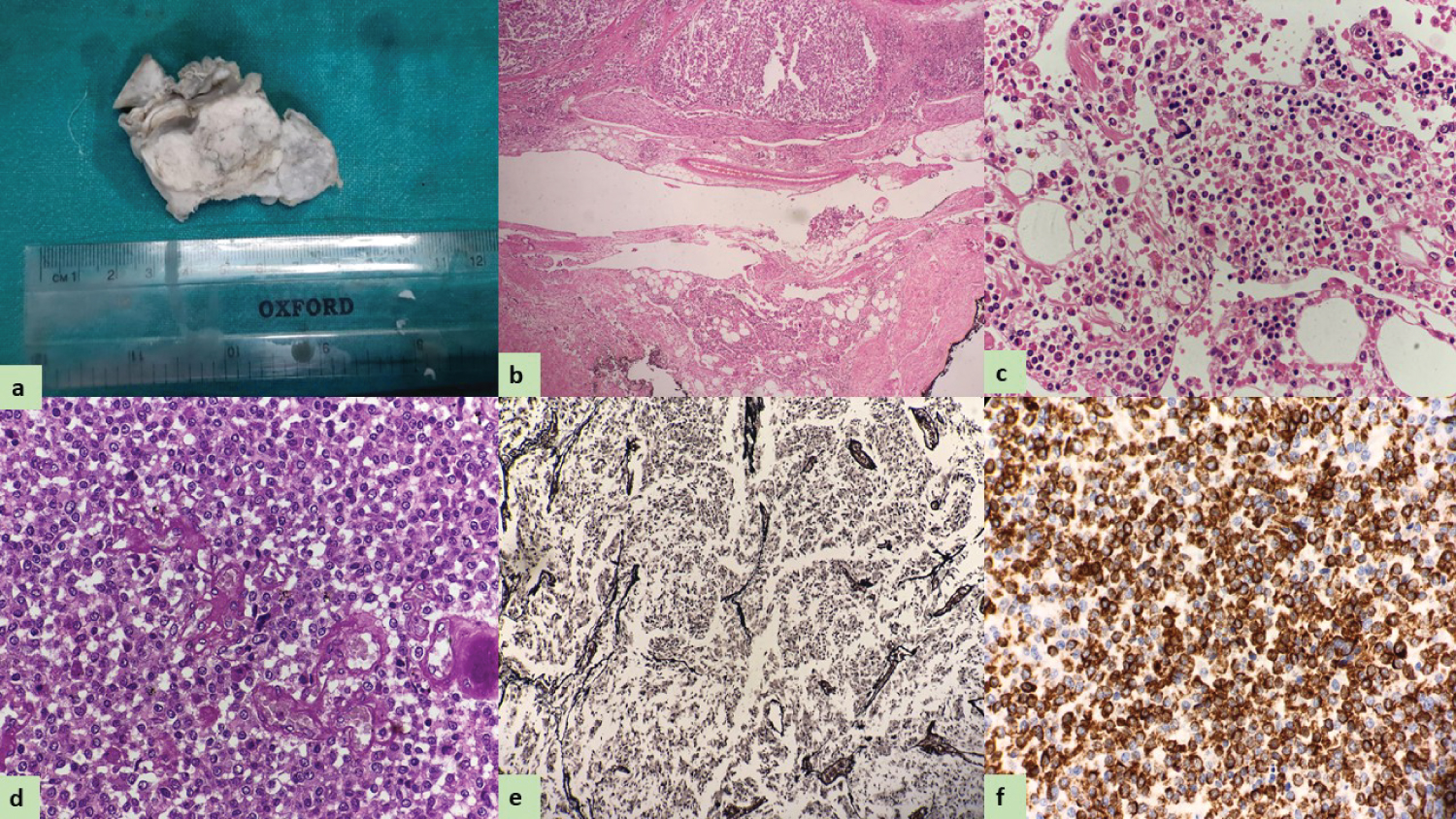
A pituitary mass and other organs of a deceased female who died of ingesting some poisonous substance was received. The pituitary mass occupied the almost whole of the anterior lobe [Figure 1a]. The mass was enclosed on all sides by a narrow rim of the anterior lobe which has almost disappeared over the lateral aspects of the tumour. On histopathological examination tumor cells are monomorphic with well-defined cell borders, eosinophilic granular cytoplasm & nuclei showing a salt and pepper appearance. The focus of extramedullary haematopoiesis was present [Figure 1b, Figure 1c and Figure 1d]. Reticulin stain showed loss of reticulin between the tumor cells [Figure 1e]. The tumour cells are immunoreactive to synaptophysin [Figure f].
Pituitary adenomas are benign, slow-growing tumors of the anterior pituitary. Pituitary adenoma can be classified based on size as microadenoma, macroadenoma, and giant tumors. Microadenoma is tumors less than 10 mm, while macroadenoma includes tumors larger than 10 mm. Giant pituitary tumors are more 40 mm. In a meta-analysis from 32 autopsy series, the pituitary adenomas frequency was 10.5%. Imaging studies, like MRI head, revealed 10% to 38% of pituitary incidentaloma [1]. Based on hormonal secretion adenomas can be divided into functional and non-functional. Approximately 50% of macroadenomas if untreated will enlarge and patient experience worsening of vision. Non-functioning pituitary adenomas are the second most common type of pituitary tumor after prolactin producing tumors. Surgical resection is considered the optimal first line therapy for symptomatic non-functioning macroadenoma [2]. Extramedullary haematopoiesis (EMH) is a compensatory effort when body is unable to meet body demand by orthotopic intramedullary bone marrow which under normal physiological condition are responsible for production of erythrocyte, myelocyte, and megakaryocyte lineages within the medullary cavity of bones. So, EMH has been associated with various hematological disorder. The ectopic site which have been reported other than liver, spleen, and lymph nodes are skin, breast, gastrointestinal tract, thyroid gland, middle ear, lung, paranasal sinuses, urinary tract, around the spinal cord, and rare reports of intracranial EMH [3]. Biopsy and Tc99m labelled sulphur colloid are good alternative for detecting orthotopic and heterotopic bone marrow as sulphur colloid particles are taken up by cells of the reticuloendothelial system [4]. EMH in a malignant meningioma has been reported by Gregorios JB et al in 1983. Sorsdahl and Saleeby believed that origin of EMH is by metaplasia or heterotopia of totipotent connective tissue cells transforming into hematopoietic tissue [5]. Intracranial EMH also reported in association with other CNS tumor includes hemangioblastomas, intracranial lipomas, and a pilocytic astrocytoma [6]. Treatment option available are transfusion therapy, cytotoxic drugs, and low dose radiotherapy. Surgery less commonly preferred due to its invasive nature and recurrence [3].
References
- Russ S, Shafiq I (2020) Pituitary adenoma. Stat Pearls, Treasure Island, Stat Pearls Publishing.
- Wass JA, Karavitaki N (2009) Non-functioning pituitary adenomas: The Oxford experience. Nat Rev Endocrinol 5: 519-522.
- Singer A, Quencer R (2014) Intracranial extramedullary haematopoiesis: A rare cause of headaches. J Neuroimaging 24: 524-527.
- Ohta Y, Shichinohe H, Nagashima K (2002) Spinal cord compression due to extra medullary haematopoiesis associated with polycythemia vera –case report. Neurol Med Chir (Tokyo) 42: 40-43.
- Gregorios JB, Bay JW, Dudley AW Jr (1983) Extramedullary hematopoiesis in a malignant meningioma. Neurosurgery 13: 447-451.
- Beckner ME, Lee JK. Schochet SS, et al. (2003) Intracranial extramedullary hematopoiesis associated with pilocytic astrocytoma: A case report. Acta Neuropathol 106: 584-587.
Corresponding Author
Jyotsna Naresh Bharti, Associate Professor, Department of Pathology, All India Institute of Medical Science, Jodhpur, Rajasthan, India
Copyright
© 2022 Bharti JN. This is an open-access article distributed under the terms of the Creative Commons Attribution License, which permits unrestricted use, distribution, and reproduction in any medium, provided the original author and source are credited.