Small Bowel Myxoma Inciting Ileo-Ileal Intussusception: The First Reported Case Managed Laparoscopically
Abstract
This case describes the clinical and pathologic findings as well as the surgical management of a patient diagnosed with a rare small bowel myxoma. This is only the ninth reported case of this condition, and the first to be managed laparoscopically. The patient presented with an ileo-ileal intussusception with resultant small bowel obstruction, which was managed with minimally invasive surgical resection with intracorporeal anastomosis. Since the disease does not carry a risk of seeding or recurrence, we feel that laparoscopy is an advantageous approach to managing the disease in the hands of a skilled minimally invasive surgeon.
Keywords
Myxoma, Intussusception, Ileo-ileal, Small bowel, Laparoscopy, Myxoid
Introduction
Small bowel myxoma represents an extremely rare tumor of mixed primitive mesenchymal origin [1]. On gross examination, they appear gelatinous, shiny, and white-tan [2,3]. They often have superficial mucosal ulceration with a thin hemorrhagic layer beneath and small, mature capillaries [3]. Pathologically, these tumors are somewhat of a diagnosis of exclusion. Some distinct microscopic characteristics are a composition of spindle cells loosely arranged in mucoid stroma associated with reticulin fibers [1]. Myxomatous tumors stain positive for vimentin, however are almost always negative for $100, smooth muscle actin, desmin, and c-KIT [3]. These tumors are typically associated with minimal mitotic activity [4]. Myxomas also rarely have vascular involvement [3]. Given these characteristics, it is exquisitely rare to see associated metastatic disease [4]. In fact, the only documented cases of myxomatous tumors presenting with metastases are those with cardiac myxomas [3,4]. Patients with small bowel myxomas typically present with constipation, nausea, and vomiting, consistent with small bowel obstructions, as our patient did [1]. The danger in leaving these tumors untreated is typically secondary to erosion or compression of local vital structures [4].
Case Presentation
The patient is a 56-year-old male who presented to John F Kennedy Hospital in Washington Township, New Jersey with complaints of abdominal pain that had been episodic over a period of three days. He reported peri-umbilical pain associated with constipation, nausea, and vomiting, as well as anorexia. His medical history was significant for diabetes and hypertension. The patient had no previous abdominal surgery.
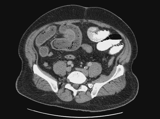
A CT scan of the abdomen and pelvis was performed which demonstrated a small bowel obstruction secondary to an ileo-ileal intussusception (Figure 1). A transition point of dilated to decompressed bowel was identified at the location of the intussusception.
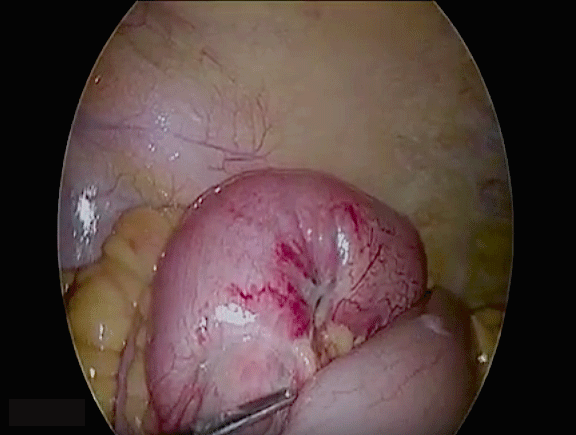
Due to the patient's clinical and radiographic findings, the decision was made to take the patient to the operating room for a laparoscopic exploration. The abdomen was entered left of the umbilicus using a direct visualization 5 mm trocar and was insufflated. The decision was made to enter left of the midline due to CT scan findings of a right lateral lesion. Upon inspection of the bowel, an area of hyperemic and dilated bowel was identified in the distal ileum with a palpable thickening/mass. No external extension of tumor or erosion was identified. Two additional 5 mm port sites were inserted into the left lower quadrant and the bowel was traced from the cecum, proximally. Approximately 20 cm proximal to the terminal ileum located in the right lower quadrant, the intussusception was noted (Figure 2). An additional 5 mm trocar was inserted in the left upper abdomen and the peri-umbilical port was converted to a 12 mm trocar. The bowel was transected proximal and distal to the intussusception using a 60 mm laparoscopic stapling device in usual fashion. The small bowel was then aligned in an isoperistaltic side-to-side orientation and three 2-0 silk sutures were placed intra-corporeally along the bowel to maintain orientation. The lumen of the anastomosis was created in usual fashion using a 60 mm laparoscopic stapling device through small enterotomies created at the end of the anastomotic site. The defect that was created for the stapling device was then closed intra-corporeally using a two-layer closure with 2-0 vicryl running suture. The mesenteric defect was then closed using 2-0 silk suture. The abdomen was adequately suctioned and irrigated and the specimen was removed through the largest port in an endoscopic retrieval bag. All port sites were closed appropriately and the specimen was sent for pathology. Post-operatively, the patient was managed with nasogastric decompression until bowel function resumed. Nasogastric tube was removed on post-operative day three and he was tolerating a diet. He underwent cardiac evaluation for chest pain and was found to have a non-ST elevation myocardial infarction. Cardiac catheterization identified severe multi-vessel disease and he subsequently endured a robotic assisted off-pump 2-vessel coronary artery bypass graft on post-operative day 13 from his bowel resection and percutaneous stenting of remaining cardiac disease. Of note, during his cardiac workup echocardiogram did not reveal any cardiac lesions that would suggest concomitant myxomatous disease. He was referred to a medical oncologist after the results of his pathology returned to ensure that no further treatment or surveillance were required.
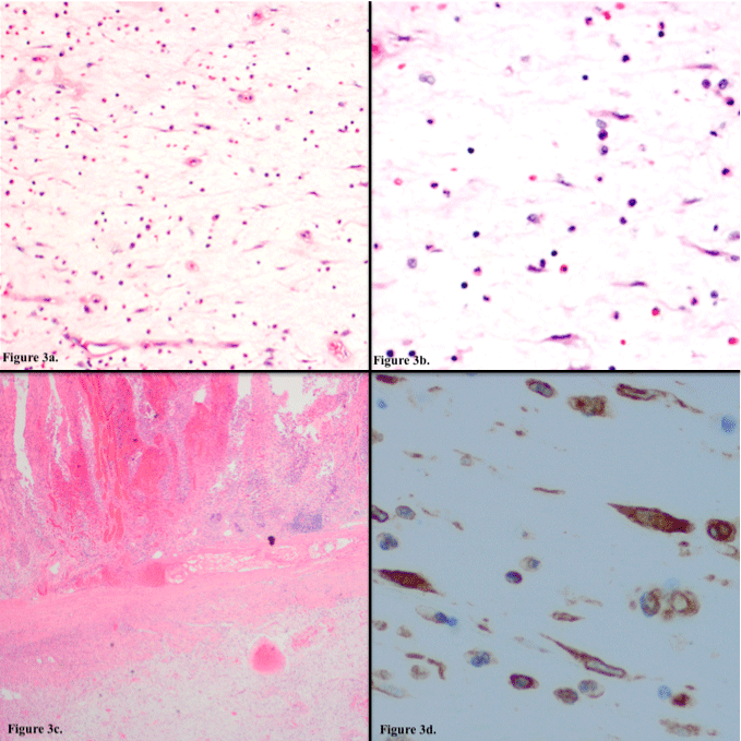
Gross evaluation of the specimen revealed a 25.5 cm section of small intestine. Once opened, a semi-firm pink and black intraluminal pedunculated polypoid lesion measuring 5.0 × 2.5 × 2.5 cm with a homogeneous and gelatinous cut surface was identified. This was located 11.5 and 12.0 cm from proximal and distal resection margins respectively. No evidence of deep invasion was identified. Sections of the lesion were submitted for microscopic examination which showed cytologically bland-appearing spindle cells present in a myxoid and hemorrhagic stroma associated with small, mature capillaries and scattered inflammatory cells (Figure 3a and Figure 3b). The mucosa overlying the lesion was ulcerated with hemorrhagic ischemic changes (Figure 3c). Immunohistochemistry showed these bland-appearing spindle cells to be positive for vimentin (Figure 3d) with lack of staining for c-kit (CD117), CD31, CD34, S-100, desmin and smooth muscle actin. These gross, histopathologic and immunophenotypic findings are strongly indicative of an ileal myxoma.
Discussion
Myxomas are associated with elongated periods of slow growth followed by bursts of rapid growth, which can lead to compression of surrounding structures. In our case, this would explain the nature of the patient's acute presentation. Of the eight existing reported cases, seven were in the ileum. It is unknown why myxomas occur more frequently in the ileum than the jejunum, and there have been no statistical studies to prove a true partiality given the limited number of reported cases. Across all organ systems, myxomatous tumors typically occur in the atrium of the heart. After echocardiogram, cardiac catheterization and bypass grafting, no indication of concomitant atrial myxoma was identified in this patient. These lesions can be solitary or associated with Carney Complex [3]. Carney Complex is a condition comprised of myxomas, lentiginosis, endocrine disorders, and endocrine or non-endocrine tumors; however, our patient was not found to have this condition. Our patient did not have any concomitant myxomatous lesions that have been identified to date.
In the era of minimally invasive surgery, we feel that attempting to perform a diagnostic evaluation of the abdomen in the setting of adult intussusception laparoscopically should be the preferred approach when safe. In this case, the patient had no prior abdominal surgery, minimizing the suspect of intra-abdominal adhesions and thus decreasing our risk of injury upon entering the abdomen. Considerations for choosing laparoscopy in surgical management of small bowel obstruction include shorter hospital length of stay and decreased morbidity [5,6]. When small bowel obstruction is the result of intussusception in adults, this could be due to an intraluminal, extraluminal or mural lead point. These processes can include tumors, suture or staple lines, lymphoid hyperplasia or local inflammation [7]. In the setting of this case, laparoscopic examination was performed in order to gain understanding of the process that was occurring. Once the intussuscepted segment was encountered, we felt that this could be managed safely under completely laparoscopic conditions.
Conclusion
The last noted case of small bowel myxoma reported in literature was in 2013. Both patients presented with an ileo-ileal intussusception with resultant small bowel obstruction. Management of this disease is not well described as the cases are so few. We present the ninth reported case of small bowel myxoma, and the first to be managed with minimally invasive surgical resection with intracorporeal anastomosis. When diagnostic laparoscopy does not indicate any surrounding invasion or compression of vital structures, we feel that completing resection laparoscopically is an appropriate management strategy. Since this tumor carries a very low risk of seeding or recurrence, we present this as an advantageous approach to managing the disease in the hands of a skilled minimally invasive surgeon.
References
- Weinberger HA (1956) Segmental myxomata of the small intestine with intussusception. Ann Surg 144: 263-270.
- Varsamis N, Tavlaridis T, Lostoridis E, et al. (2013) Myxoma of the small intestine complicated by ileo-ileal intussusception: Report of an extremely rare case. Int J Surg Case Rep 4: 609-612.
- Wang Y, Sharkey FE (2003) Myxoma of the small bowel in a 47-year-old woman with a left atrial myxoma. Arch Pathol Lab Med 127: 481-484.
- Stout AP (1948) Myxoma, the tumor of primitive mesenchyme. Ann Surg 127: 706-719.
- Nordin N, Freedman J (2016) Laparoscopic versus open surgical management of small bowel obstruction: an analysis of clinical outcomes. Surg Endosc 30: 4454-4463.
- O'Connor DB, Winter DC (2012) The role of laparoscopy in the management of acute small bowel obstruction: a review of over 2,000 cases. Surg Endosc 26: 12-17.
- Huang BY, Warshauer DM (2003) Adult intussusception: diagnosis and clinical relevance. Radiol Clin North Am 41: 1137-1151.
Corresponding Author
Mara Piltin, Department of General Surgery, Rowan University School of Osteopathic Medicine, Stratford, NJ, 08084, USA.
Copyright
© 2018 Piltin M, et al. This is an open-access article distributed under the terms of the Creative Commons Attribution License, which permits unrestricted use, distribution, and reproduction in any medium, provided the original author and source are credited.


