Anterior Pelvic Quadrangular External Fixation Frame: A Simple Modification of the Anterior External Fixator to Allow an Anterior Approach to the Pelvis
Abstract
Unstable and displaced anterior pelvic ring traumatic injuries can be provisionally and sometimes definitively stabilized using anterior pelvic external fixation. For some patients with severe instability, it is useful to maintain the reduction achieved by the initial anterior pelvic external fixation device during the definitive open reduction and internal fixation of the anterior pelvic injury. Unfortunately, conventional anterior pelvic external fixation bar configurations usually obstruct surgical access for an open anterior approach, clamp application, and fixation. We present a simple technique using a quadrangular frame consisting of 4 carbon fiber bars connected to standard anterior pelvic external fixation pins. This quadrangular frame construct provides the surgeon and assistants a much less obstructed surgical access to the anterior pelvis while maintaining the important stabilizing effect of the external fixator.
Keywords
Pelvis, External, Fixation, Frame, Anterior, Quadrangular
Introduction
Surgical treatment of unstable, displaced pelvic ring disruptions has become the standard of care to prevent long term sequelae of pelvic malunion [1]. Multiple techniques exist to stabilize an unstable pelvic ring, ranging from external fixation to various types ofinternal fixation [2-7]. Anterior pelvic external fixation is a useful option as a temporary or definitive treatment in complex anterior ring injuries and is often used as an adjunct to posterior pelvic ring fixation [2,8].
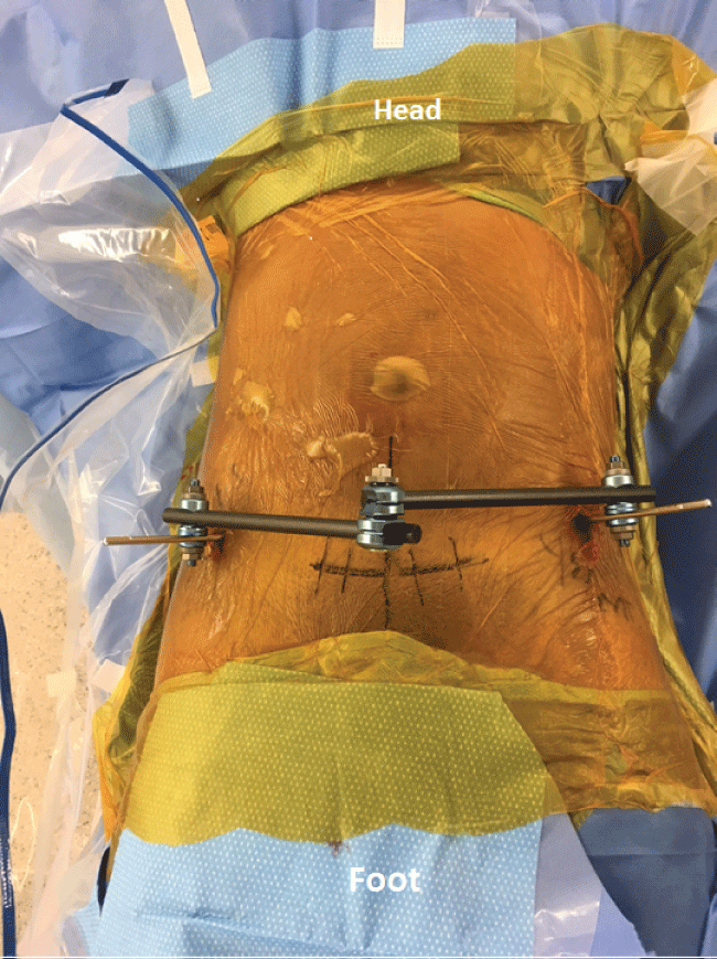
In hemodynamically unstable patients or patients with severe anterior pelvic ring instability, it is advantageous to maintain the external fixation frame during Open Reduction and Internal Fixation (ORIF) of the anterior pelvis. Unfortunately, the standard two bar anterior pelvic frame construct (Figure 1A and Figure 1B) obstructs the surgical approach and surgical access, and is therefore commonly removed prior to conversion to ORIF. Any stabilizing reduction afforded by the external fixator is thus lost. Additionally, in cases of severe pelvic instability, it may be advantageous to maintain the pelvic frame to aid in reduction and/or reduce the force required of pelvic reduction clamps. The purpose of the present report is to describe a simple quadrangular anterior pelvic frame modification that maintains the frame and its reduction while not obstructing anterior pelvic open surgical access.
Surgical Technique
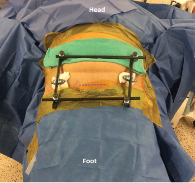
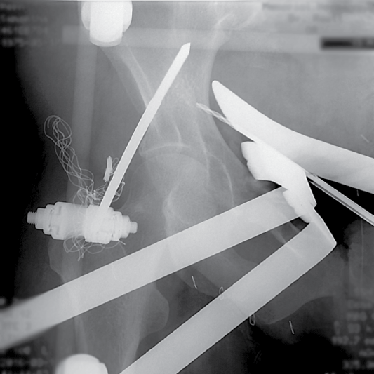
This technique has been used most commonly in patients sustaining complex anterior pelvic ring injuries, typically with bilateral pubic ramus fractures that are unstable and/or comminuted with or without an associated pubic symphyseal disruption. The quadrangular frame (Figure 2A and Figure 2B) can be placed at the index surgery when a subsequent open approach to the anterior pelvis is anticipated. Alternatively, a routine anterior pelvic external fixation frame can be converted to a quadrangular frame at the time of definitive ORIF of the anterior ring.
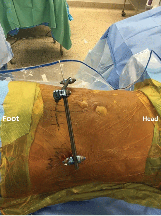
The quadrangular frame is assembled after 5 mm supra-acetabular external fixation half-pins have been placed into each ilia using a previously described technique [9]. The proposed skin incision for an anterior approach (Pfannenstiel, anterior intrapelvic, low pubic midline) is drawn on the skin with a marker to allow assembly of the frame around it so as not to block subsequent visualization and instrumentation during ORIF. Pin-bar clamps are used to place an 11 mm carbon fiber bar onto each pin oriented in the vertical axis, typically 150-200 mm in length (Figure 2A and Figure 2B). These vertical bars are centered on the iliac pins so that horizontal bars can next be connected to the vertical bars at both the cranial and caudal ends of the vertical bars using bar-bar clamps (Figure 2A and Figure 2B).
Once the quadrangular frame has been assembled, the initial obstructing two bar anterior frame can be removed without significant change in the initial reduction. If the surgeon opts to remove the initial obstructing frame prior to assembling the quadrangular frame, the initial reduction is maintained using two sponges wrapped around each pin and then clamped together prior to loosening the initial frame. Once the quadrangular frame is constructed, the clamp is released and the sponges are removed. If the initial reduction needs revision or adjustment, it is performed using fluoroscopy if necessary and then the quadrangular frame pin-bar and bar-bar clamps are terminally tightened.
Surgical Case Example
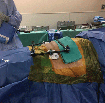
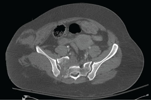
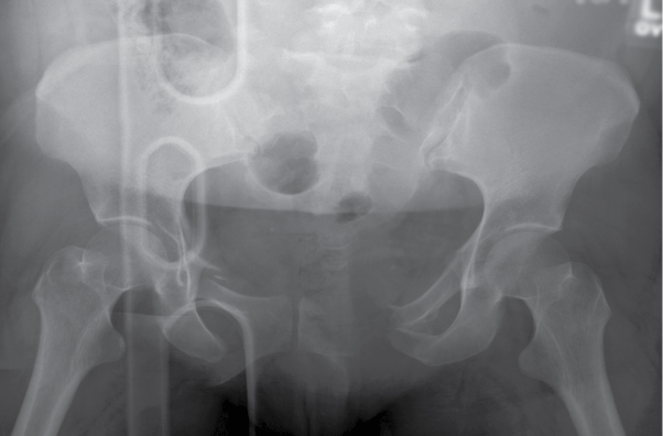
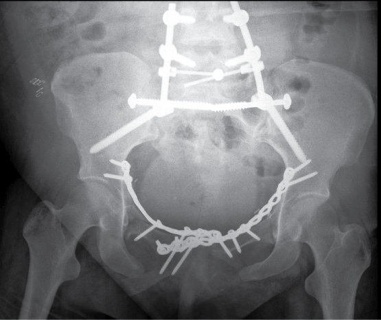
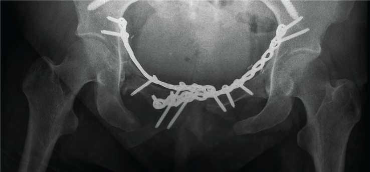
We present a case example of a 41-years-old female who was crushed beneath a car causing significant pelvic instability due to bilateral pubic ramus fractures, a left sided sacroiliac joint disruption, and a highly comminuted right sacral fracture (Figure 3A and Figure 3B). The patient was initially evaluated and resuscitated routinely according to Advanced Trauma Life Support (ATLS) protocols. During her initial resuscitation, she underwent closed reduction and anterior pelvic external fixator placement, along with percutaneous stabilization of her left sacroiliac joint disruption. Once her clinical situation stabilized on post-injury day five, she was returned to the operating room for definitive pelvic management using ORIF for both her anterior and posterior pelvic injury sites. The anterior pelvic ORIF was performed first with the patient positioned supine. The initial anterior pelvic external fixation frame had maintained a satisfactory overall pubic region reduction, but it obstructed the planned surgical site. The initial frame was therefore converted to a quadrangular frame. The quadrangular frame was constructed onto the two pins, and then the initial frame was removed. A Pfannenstiel exposure was next used to expose, accurately reduce, and then stabilize the fractures. The quadrangular frame was positioned to not crush her protuberant abdomen. The quadrangular frame did not interfere with the exposure, retraction, reductions, or fixations (Figure 4). Following internal fixation of the anterior pelvic ring, the quadrangular frame was removed and she was repositioned prone for the sacral fracture ORIF. Iliosacral screws and lumbopelvic fixation were used to stabilize the fracture (Figure 5A and Figure 5B).
Discussion
Use of pelvic external fixation for displaced and unstable anterior pelvic ring injuries is common in orthopedic trauma [10]. A variety of pin sites, pin sizes and numbers, and frame complexities have been described, mostly in an attempt to achieve maximal pelvic ring stability in order to use the frame as definitive treatment [11-14]. Other reports have used oblique pins and rods to allow compression and distraction along an oblique axis to achieve a pelvic ring reduction [15]. We describe a simple frame design modification that allows both the frame and reduction to be maintained without obstructing the anterior surgical exposure to the pelvic ring. This technique is most useful with a severely unstable anterior ring injury to provide additional stability, but can be used with pubic symphyseal disruptions in which the frame provides an additional reduction aid.
On occasion, patients with external fixation pin sites that begin to show signs of superficial infection or progressive skin tears due to the forces across the pins, the quadrangular frame would allow for maintenance of the initial pelvic ring reduction while converting to plate and screw fixation. Similarly, for polytraumatized patients that are temporized in an external anterior frame that need to be positioned prone for later procedures, the quadrangular frame can be used to maintain a reduction while converting to plate and screw fixation. This allows for anterior frame removal to avoid potential issues with frame impingement between the patient's soft tissues, frame, and operating table.
Because of its design and bar locations, the quadrangular frame cannot be used as a definitive frame or left on for a prolonged period of time, as difficulty with impingement of the bars on the abdomen or thighs will occur with sitting. The quadrangular frame is not intended to be used definitively and does not change recovery from the pelvic surgery. Constructing the frame may add to the overall surgical time but in our experience generally takes less than 5 minutes. This study was not intended to compare the biomechanical stability of the quadrangular frame to a standard 2 bar external fixation frame. The quadrangular frame may be more costly because it requires four bars and six clamps when compared to the two bars and three clamps needed for routine anterior pelvic frames. With inappropriate surgical planning, the quadrangular frame can block drill bit or clamp application needed for open reduction. In addition, percutaneous anterior column screws and screws from anterior to posterior in the supracetabular area can be obstructed by use of the quadrangular frame. In some patients with extreme obesity and in patients being positioned prone, the bars may impinge on the soft tissue preventing safe use of the frame. In cases in which it is desirable to maintain anterior pelvic external fixation after anterior pelvic internal fixation, we convert back to a standard 2-bar frame prior to leaving the operating room.
In conclusion, the anterior pelvic quadrangular frame is a simple modification of the anterior pelvic external fixator which may provide pelvic stability during an anterior open surgical approach for ORIF.
References
- Dujardin FH, Hossenbaccus M, Duparc F, et al. (1998) Long-term functional prognosis of posterior injuries in high-energy pelvic disruption. J Orthop Trauma 12: 145-150.
- Bellabarba C, Ricci WM, Bolhofner BR (2000) Distraction External Fixation in Lateral Compression Pelvic Fractures. J Orthop Trauma 14: 475-482.
- Tucker MC, Nork SE, Simonian PT, et al. (2000) Simple anterior pelvic external fixation. J Trauma Acute Care Surgery 49: 989-994.
- Gardner MJ, Nork SE (2007) Stabilization of unstable pelvic fractures with supraacetabular compression external fixation. J Orthop Trauma 21: 269-273.
- Gardner MJ, Mehta S, Mirza A, et al. (2012) Anterior pelvic reduction and fixation using a subcutaneous internal fixator. J Orthop Trauma 26: 314-321.
- Cole PA, Gauger EM, Anavian J, et al. (2012) Anterior pelvic external fixator versus subcutaneous internal fixator in the treatment of anterior ring pelvic fractures. J Orthop Trauma 26: 269-277.
- Haidukewych GJ, Kumar S, Prpa B (2003) Placement of Half-Pins for Supra-acetabular External Fixation: An Anatomic Study. Clin Orthop Relat Res 411: 269-273.
- Mitchell PM, Corrigan CM, Patel NA, et al. (2016) 13-Year experience in external fixation of the pelvis: complications, reduction and removal. Eur J Trauma Emerg Surg 42: 91-96.
- Calafi LA, Routt MLC (2013) Anterior pelvic external fixation: is there an optimal placement for the supra-acetabular pin? Am J Orthop 42: E125-E127.
- Poka A, Libby EP (1996) Indications and techniques for external fixation of the pelvis. Clin Orthop Relat Res 329: 54-59.
- Ponsen KJ, Joosse P, Van Dijke GA, et al. (2007) External fixation of the pelvic ring: an experimental study on the role of pin diameter, pin position, and parasymphyseal fixator pins. Acta Orthop Oct 78: 648-653.
- Eagan M, Kim H, Manson TT, et al. (2015) Internal anterior fixators for pelvic ring injuries: Do monaxial pedicle screws provide more stiffness than polyaxial pedicle screws? Injury 46: 996-1000.
- Sellei RM, Kobbe P, Dadgar A, et al. (2015) External fixation design evolution enhances biomechanical frame performance. Injury 46: S23-S26.
- Kim WY, Hearn TC, Seleem O, et al. (1999) Effect of pin location on stability of pelvic external fixation. Clin Orthop Relat Res 361: 237-244.
- Evans AR, Routt MLC, Nork SE, et al. (2012) Oblique distraction external pelvic fixation. J Orthop Trauma 26: 322-326.
Corresponding Author
Bryce A Cunningham, Department of Orthopedic Surgery, University of Texas Health Science Center, Texas Medical Center, 6400 Fannin Street, Suite 1700 Houston, TX 77030, USA, Tel: 314-780-8188.
Copyright
© 2017 Cunningham BA, et al. This is an open-access article distributed under the terms of the Creative Commons Attribution License, which permits unrestricted use, distribution, and reproduction in any medium, provided the original author and source are credited.