A Rare Involvement Region in a Patient with B-Cell Chronic Lymphocytic Leukemia: Skin
Abstract
Skin involvement in Chronic Lymphocytic Leukemia (CLL) is rare and can present with very different lesions. Our case was a 51-year-old woman who had been followed without medication for 2 years with the diagnosis of CLL in an external center. Papillomatous lesions have developed in different parts of her face for about 1 year. Skin biopsy was performed 1 month ago and chronic lymphocytic leukemia infiltration was detected; so she was referred to our clinic. The patient with Rai stage II CLL refused to take chemotherapy for these lesions and was followed up regularly. In this case report, we emphasized that skin lesions which can present with very different forms should be kept in mind in terms of skin involvement in CLL patients, and also the alternative treatments were reviewed.
Keywords
CLL, Skin involvement, B-cell leukemia
Introduction
Chronic Lymphocytic Leukemia (CLL) is the most common form of leukemia with an incidence of 4.2/100,000/year. The median age at diagnosis is between 67-72 years. 10% of CLL patients are younger than 55 years [1]. Skin involvement is seen in 4-20% of patients, and there is limited information in the literature on CLL patients with skin involvement [2]. The reported lesions are usually nodule, papule, plaque, ulcer, and exfoliative erythroderma in the head and neck region [3].
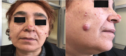
In this article, we reported a 51-year-old woman who was diagnosed with CLL for 2 years and who had papillomatous skin lesions (the largest one was 1.5 cm in diameter) on the nose and cheeks.
Case Presentation
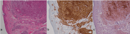
A 51-year-old woman was admitted to the hospital due to papillomatous lesions on her face for a year. Her past medical history was unremarkable except for that she was diagnosed with Chronic Lymphocytic Leukemia (CLL) in October 2014 (which was 2 years before our admission) and imaging tests for staging at the time of diagnosis showed as Rai stage 0. About 1 year after diagnosis; in September 2015, milimetric nodular lesions developed on the cheeks and nose of the patient. Lymph nodes in the neck and abdomen developed 8 months after skin findings. Radiological examinations have been reperformed in May 2016 and showed as Rai stage II. Biopsy was performed from the lesion on the left cheek in October 2016 due to the progression of skin lesions. Upon that the result of biopsy was consistent with chronic lymphocytic leukemia infiltration, she was referred to our clinic (Figure 1).
In physical examination which was made in admission to the hospital in December 2016, there were multiple papillomatous lesions (the largest one was 1.5 cm in diameter) on the nose and cheeks (clustered on the nose ridge) (Picture 1), a 1.5 cm right submental lymph node, a 2.5 cm right supraclavicular lymph node and bilateral cervical lymph nodes (the largest one was 2 cm in diameter). Moreover, the liver was palpable 2-3 cm below the costal margin and the traube's space was closed. In laboratory examination, hemoglobin level was 15.5 g/dL, leukocyte count was 36.1 × 10⁹/L, absolute neutrophil count was 3.9 × 10⁹/L, absolute lymphocyte count was 27.9 × 10⁹/L, absolute monocyte count was 3.4 × 10⁹/L, platelet count was 190 × 109/L, LDH level was 353 U/L and other biochemical parameters were normal. The patient's bone marrow biopsy made in 2014 revealed that CD20, PAX-5, CD5, CD23 and Bcl2 were diffusely positive, CD3-positive T cells were rare, and Cyclin D1 showed staining in the vascular endothelium. It was reported as "B cell lymphocytic infiltration ; consistent with small lymphocytic lymphoma/chronic lymphocytic leukemia". The classic cytogenetic results of bone marrow made in 2014 were found as 46, XX. Computerized Tomography (CT) of neck, thorax, abdomen and pelvis was performed for staging. In the neck CT scan,there were multiple pathological lymph nodes (the largest one was 2.5 cm in diameter on the right side) in the cervical region that showed occasionally conglomerate, and soft-tissue masses (the largest one was 1.5 cm in diameter on the right side) at both buccal levels that were localized in skin-subcutaneous tissue and exhibited an exophytic growth pattern. In the thoracic CT scan, there were multiple mediastinal lymph nodes (the largest one was 13.5 mm in diameter) and lymph nodes (the largest one was 3 cm in diameter on the left side) in both axillary areas. In the abdominal CT scan, the liver measured 17 cm, the spleen measured 13 cm, and there were multiple pathological paraaortic lymph nodes (the largest one was 25 mm in diameter). In the pelvic CT scan, there were bilateral inguinal lymph nodes (the largest one was 18 mm diameter). Chemotherapy was planned due to skin involvement in the patient with Rai stage II CLL, but the patient was followed up clinically because she did not accept chemotherapy.
Discussion
CLL is a low grade monoclonal disorder characterized by B cell lymphoproliferation. Leukemic skin involvement is seen in 4-20% of patients [2]. Although the mean disease duration until detection of skin lesions has been reported up to 39 months, skin lesions may also present as the first symptom of CLL in 16.7% of patients [4]. In our patient, the skin lesions developed 23 months after CLL was diagnosed. While Fullen, et al. reported that the patient had advanced-stage CLL (Rai stage IV) when skin lesions were detected [5], Sokumbi, et al. reported that the two patients had early-stage CLL (Rai stages I and II) when skin lesions were detected [6]. Our patient had Rai stage 0 CLL when skin lesions occured but skin biopsies were made about 1 year after lesions were developed. When the lesions were confirmed to be CLL skin involvement by biopsy, re-staging was performed and evaluated as Rai stage II.
Although the mechanism of skin involvement in CLL is unclear, it is considered to be associated with upregulation of Intracellular Adhesion Molecule-1 (ICAM-1) and Lymphocyte Function-Associated Antigen-1 (LFA-1) [7]. There is conflicting information in the literature on the prognostic effect of skin involvement. It has been reported that when skin involvement is detected, it is usually identified as Richter's syndrome or T-CLL and also indicates a poor prognosis [8,9]. However, in a retrospective study evaluating 42 CLL patients with skin infiltration, it was found that skin lesions did not affect the prognosis, and the 5-year survival rate was reported as 66.6% [4]. In a study of Kaddu, et al. which included 27 patients, it was indicated that skin findings in CLL were associated with a favorable prognosis and did not require specific treatment [10].
As treatment is recommended to be planned according to hematologic findings, it has been reported that surgical excision can be performed for small solitary or clustered lesions and radiotherapy can be used for larger lesions [11]. In a study of Dimeo, et al., the involved area was irradiated at a dose of 36 Gy in 18 fractions, and it was reported that complete regression was observed in the lesion [2]. Systemic chemotherapy has also been offered as an option in cases with aesthetic anxiety due to common, large lesions and it was reported that prednisone and chlorambucil or rituximab gave good results [12]. Although alkylating agents are generally used to treat disseminated leukemia cutis in B-CLL [13,14], the current data shows that skin involvement is controlled by nucleoside analogues such as fludarabine and cladribine [13,15]. In our patient, skin lesions were not suitable for radiotherapy because they were in different parts of the face. Despite the aesthetic problems, the patient refused chemotherapy because she hesitated the side effects of systemic chemotherapy, and therefore the follow-up was planned.
As a result, skin lesions that occur in different forms in CLL patients should be considered in terms of skin involvement of CLL, and skin biopsy should be performed. The need for surgery, chemotherapy, radiotherapy or follow-up in skin involvement of CLL should be assessed on a patient-by-patient basis. More research is needed to determine the impact of skin involvement on CLL prognosis and to improve follow-up standards.
References
- Turkish Hematology Association (2016) National daignosis and therapy guide, Chronic Lymphocytic Leukemia.
- diMeo N, Giuseppe S, Giusto T (2013) Cutaneous B-cell chronic lymphocytic leukaemia resembling a granulomatous rosacea. Dermatol Online J 19: 20033.
- Lu C, Li L, Qiao Q, et al. (2015) Cutaneous manifestations in a patient with chronic lymphocytic leukemia involving the head, neck and distal extremities. Exp Ther Med 9: 877-879.
- Cerroni L, Zenahlik P, Hofler G, et al. (1996) Specific cutaneous infiltrates of B-cell chronic lymphocytic leukemia: a clinicopathologic and prognostic study of 42 patients. Am J Surg Pathol 20: 1000-1010.
- Fullen DR, Jacobson SN, Valdez R, et al. (2003) Granuloma annulare-like infiltrates with concomitant cutaneous involvement by B-cell non-Hodgkin's lymphoma: report of a case. Am J Dermatopathol 25: 57-61.
- Sokumbi O, Gibson LE, Comfere NI, et al. (2012) Granuloma annulare-like eruption associated with B-cell chronic lymphocytic leukemia. J Cutan Pathol 39: 996-1003.
- Uccini S, Ruco LP, Monardo F, et al. (1993) Molecular mechanisms involved in intraepithelial lymphocyt emigration: A comparative study in skin and tonsil. J Pathol 169: 413-419.
- Giles FJ, O'Brien SM, Keating MJ (1998) Chronic lymphocytic leukemia in (Richter's) transformation. Semin Oncol 25: 117-125.
- Hoyer JD, Ross CW, Li CY, et al. (1995) True T-cell chronic lymphocytic leukemia: A morphologic and immunophenotypic study of 25 cases. Blood 86: 1163-1169.
- Kaddu S, Smolle J, Cerroni L, et al. (1996) Prognostic evaluation of specific cutaneous infiltrates in B-chronic lymphocytic leukemia. J Cutan Pathol 23: 487-494.
- Cerroni L, Kerl H, Gatter K (2004) An illustrated guide to skin lymphoma. Second edition, Oxford, Blackwell Science, 121-122.
- Aviv B, Hana F, Pietro Q, et al. (2012) Cutaneous B-cell neoplasms mimicking granulomatous rosacea or rhinophyma. Arch Dermatol 148: 824-831.
- Robak E, Robak T, Biernat W, et al. (2002) Successful treatment of leukaemia cutis with cladribine in a patient with B-cell chronic lymphocytic leukaemia. Br J Dermatol 147: 775-780.
- Robak E, Robak T (2007) Skin lesions in chronic lymphocytic leukemia. Leuk Lymphoma 48: 855-865.
- Ural AU, Kubar A, Avcu F, et al. (2007) Oral fludarabine treatment of cutaneous infiltration in a patient with B-cell chronic lymphocytic leukaemia. Clin Exp Dermatol 32: 151-154.
Corresponding Author
Tuba Bulduk, Department of Hematology, Faculty of Medicine, Eskişehir Osmangazi University, ESOGÜ Meşelik Yerleşkesi 26480 ESKİŞEHİR, Turkey, Tel: +905062512433, Fax: +90-222-239-37-80.
Copyright
© 2017 Bulduk T, et al. This is an open-access article distributed under the terms of the Creative Commons Attribution License, which permits unrestricted use, distribution, and reproduction in any medium, provided the original author and source are credited.