Association of a Persistent Ductus Arteriosus and a Persistent Left Superior Vena Cava: A Case Report and Review of the Literature
Abstract
Introduction/Purpose: The persistence of a left superior vena cava (LSVC) is a rare malformation characterized by an absence of involution of this one. We report a syncope revealing a persistent left superior vena cava.
Clinical observation: A 27-year-old patient with no cardiovascular or thromboembolic risk factors was admitted for syncope with rest dyspnoea. The clinical evaluation showed desaturation to 90% on room air, good haemodynamic stability, an arrhythmia with a continuous left latero-sternal systolo-diastolic murmur of intensity 4/6thassociated with a systolic murmur at the tricuspid focus.
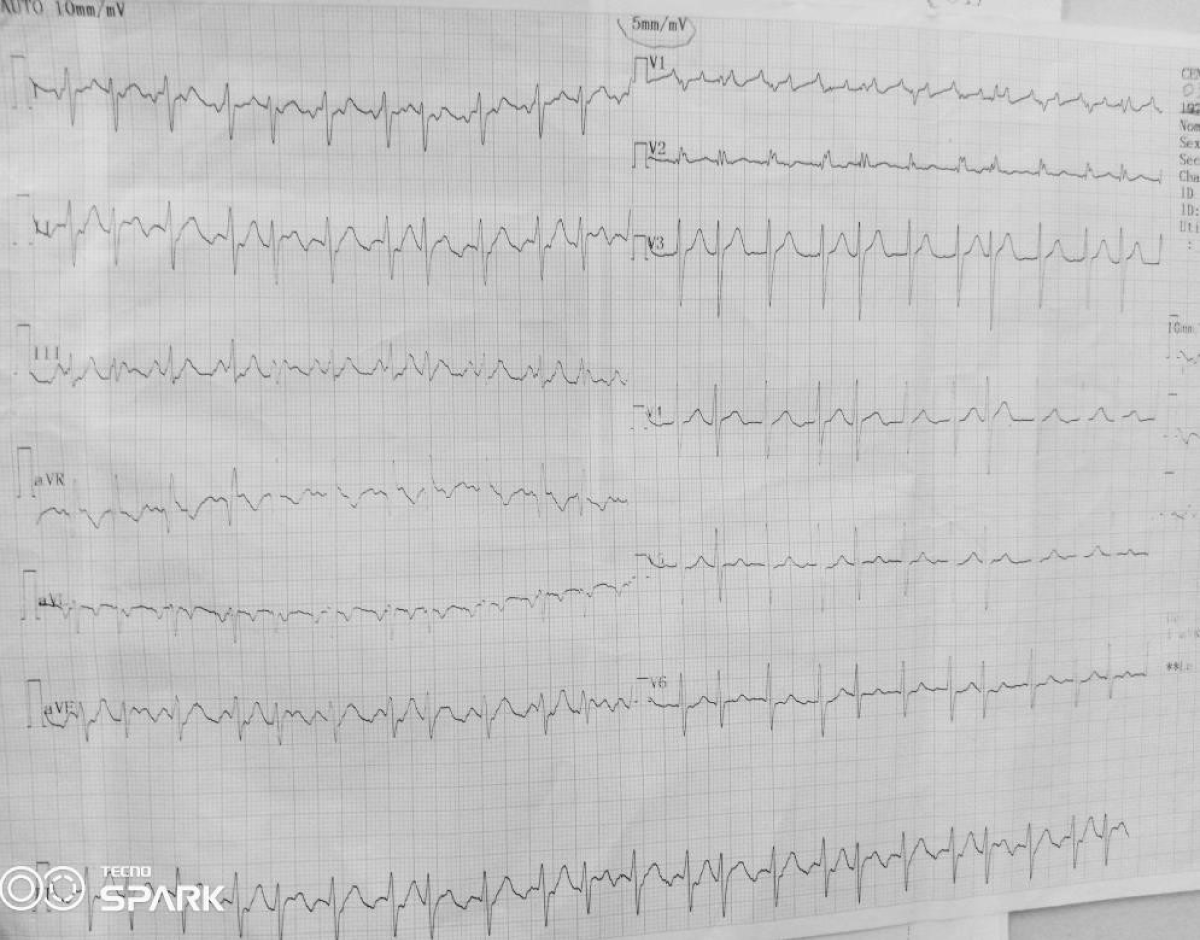
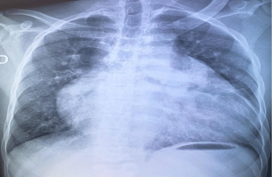
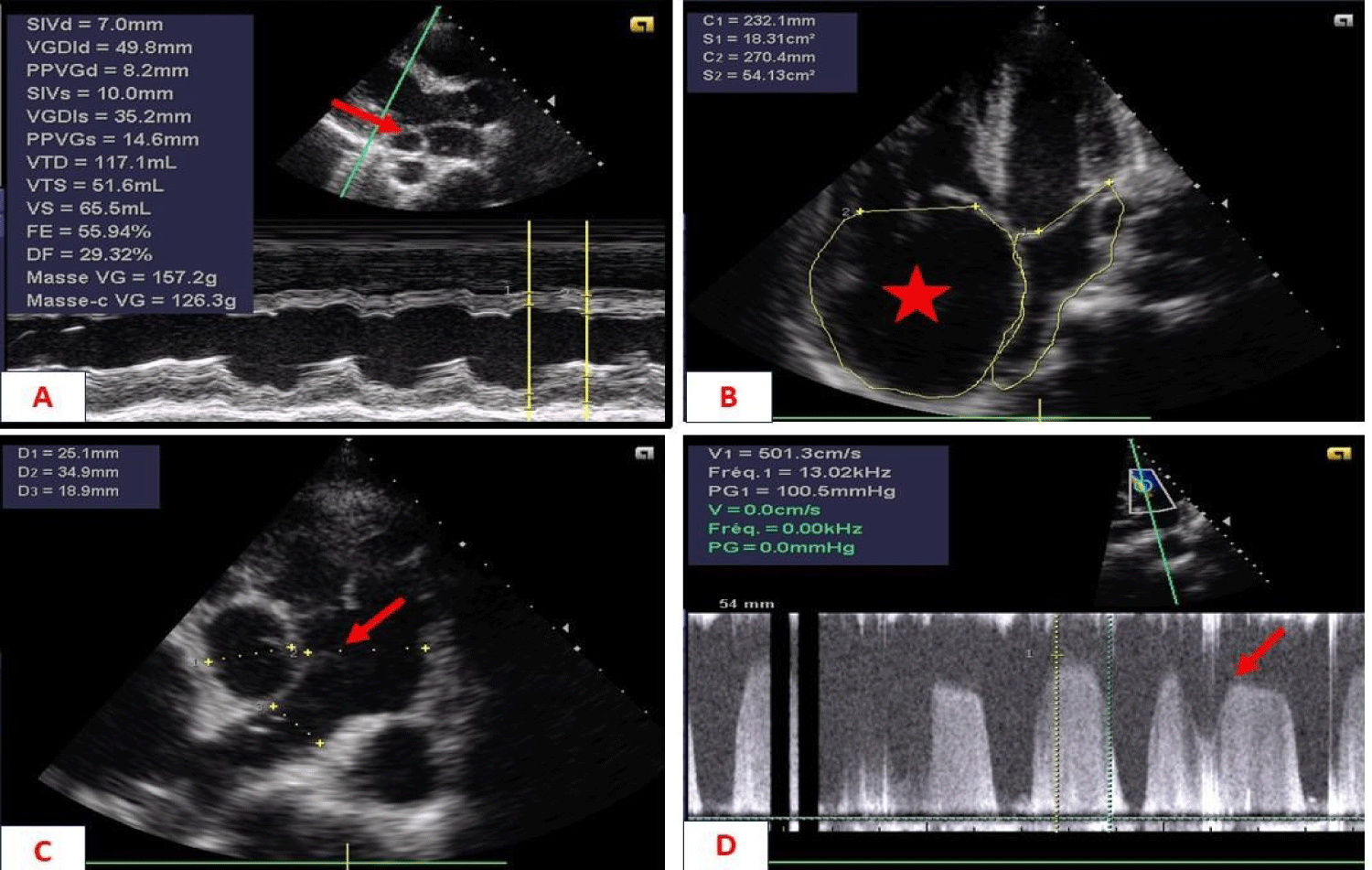
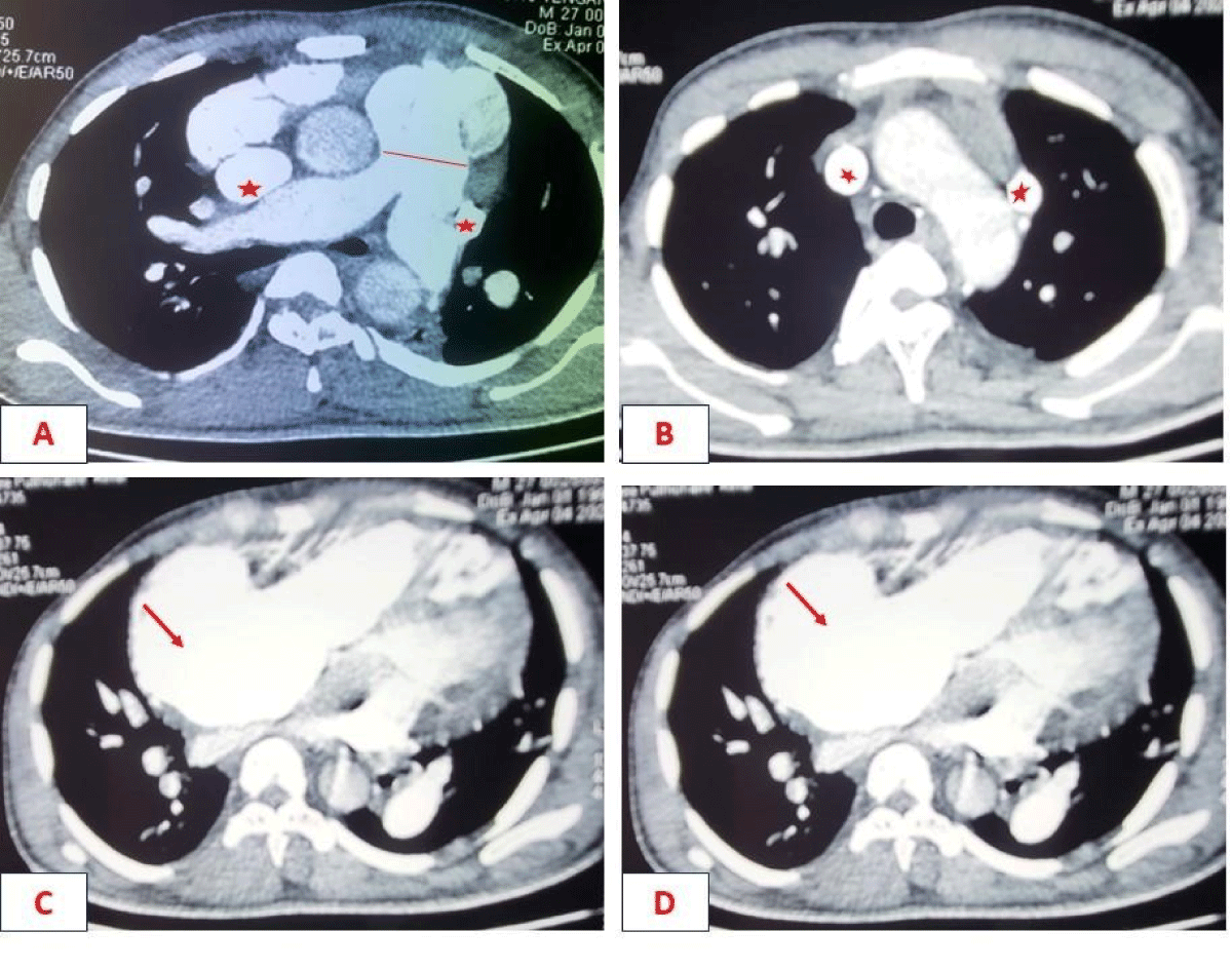
Biological tests showed polycythemia of 7 million/ul and mild anaemia of 11g/dL. The electrocardiogram showed atrial fibrillation (AF) with rapid ventricular response, while the X-ray showed significant cardiomegaly. Echocardiography showed a short-axis parasternal slice centred on the PA, with a tripod image and a left-right shunt between the aorta and PA. Continuous Doppler showed systolic flow in this shunt with diastolic extension without return to the isoelectric line. There was a passage of air bubbles into the left cavities (the patient had a venous line), dilatation of the coronary sinus, dilatation of the right cavities, and severe PAH at 90 mmHg. Echocardiography concluded that the patient had PCA with severe right-sided dilatation and severe PH associated with abnormal venous return. Chest CT showed the existence of two superior vena cava, one on the left and the other on the right, draining into the left and right atria respectively.
The diagnosis was persistent patent ductus arteriosus and SCAV complicated by AF and syncope. Treatment included oxygen therapy, curative anticoagulation with Enoxparin and an antiarrhythmic drug (Amiodarone). The patient developed a cardiovascular collapse resistant to vasoactive amines and died after 7 days in hospital.
Conclusion: The persistence of a ductus arteriosus associated with a left superior vena cava in an adult who has been asymptomatic for a long time is very rare. PCA would have made it possible to reach adulthood by protecting the left ventricle through the return of blood from the SVCG to the PA, hence the significant impact on the right cavities due to pulmonary hyperflow. This clinical presentation requires early medical and, above all, appropriate surgical management to avoid a fatal outcome.
Keywords
Patent ductus arteriosus, Left superior vena cava, Literature review, Burkina Faso
Introduction
Persistent left superior vena cava (PLSVC) is a congenital malformation in which the left superior vena cava persists instead of being reabsorbed during embryonic development [1]. This anomaly, which is generally asymptomatic and often discovered accidentally, can also lead to a variety of clinical symptoms, ranging from minor signs to more severe manifestations, such as syncope [2,3]. Persistent SCV is often associated with other cardiac and vascular anomalies such as septal defects, tetralogy of Fallot, patent ductus arteriosus, or coarctation of the aorta, making the clinical diagnosis complex [4]. The pathophysiological mechanisms underlying syncope in these cases are mainly related to compression of the left superior vena cava, which disrupts venous return to the heart, leading to cerebral hypoperfusion [5]. Despite the rarity of this condition, treatment must be rapid and appropriate in order to prevent serious complications [5]. In this report, we detail the clinical case of a 27-year-old adult with a persistent left superior vena cava associated with a patent ductus arteriosus. The diagnostic aspects, strategies and therapeutic implications of this anomaly will be examined in this clinical case while highlighting the challenges encountered during its management.
Clinical observation
The patient was a 27-year-old mechanic with no known cardiovascular risk factors. He was admitted for syncope of sudden onset with no prodromal symptoms or associated signs. The patient had been suffering from permanent palpitations and exertional dyspnoea for several months. On physical examination, consciousness was clear, with blood pressure at 124/74 mmHg, tachycardia at 133 beats per minute and xiphoid tingling. Auscultation revealed irregular heart sounds with a tricuspid insufficiency murmur of intensity 4/6 and an aortic insufficiency murmur of intensity 3/6. Examination of the other equipment was normal. The electrocardiogram showed coarse-mesh atrial fibrillation with a rapid ventricular response of 140 cycles per minute (Figure 1). The chest X-ray showed cardiomegaly (cardiothoracic index = 0.75), a right inferior arch protrusion (dilatation of the right atrium), and a convex middle arch with an outward point (Figure 2). On Doppler echocardiography, there were visible bullae in the left cavities, significant dilatation of the right cavities, persistence of the ductus arteriosus, pulmonary hypertension (PAPS= 90 mmHg) and major dilatation of the coronary sinus in favour (Figure 3). The CT-scan also showed dilation of the right cavities and confirmed the presence of two superior vena cava, one on the right draining into the right atrium and the other on the left draining into the coronary sinus (Figure 4). The biology work-up showed polycythemia at 7 million/ul and mild anaemia at 11g/dL. The diagnosis was persistent CA and SCAV complicated by AF. The patient was treated with a curative dose of Enoxaparin followed by Rivaroxaban and Amiodarone. The course was marked by the onset of cardiovascular collapse refractory to vasopressive amides, followed by death on the seventh day of hospitalisation.
Review of the literature - Discussion
The literature was searched using PubMed and Google Scholar. This search was carried out using the following three equations: "association of a left superior vena cava and a persistent ductus arteriosus", "persistent left superior vena cava" and "persistent ductus arteriosus" and "persistent left superior vena cava", "persistent ductus arteriosus". In total we obtained six articles [6-11] which are summarised in Table 1. Generally speaking, this association appears to be rare in the literature and is mainly documented in Asian countries [6-11]. Although there are asymptomatic forms known as aged forms, it can be responsible for the rapid development of fixed pulmonary hypertension, leading to the death of children from an early age.
The persistence of the left superior vena cava is a rare congenital anomaly, observed in approximately 0.3% to 0.5% of cases in the normal population and 10% in subjects with congenital heart disease [2]. Although the majority of patients are asymptomatic, in some cases persistent SCV may be responsible for severe symptoms, including syncope, mainly related to disruption of venous return [12]. Persistent CSVG can lead to compression of the coronary sinus and right superior vena cava, reducing venous return to the heart. This impaired blood flow to the right atrium and right ventricle can lead to cerebral hypoperfusion, responsible for the transient loss of consciousness seen in syncope [3].
The clinical symptoms of persistent SCSV vary according to the severity of compression and the presence of other associated malformations. However, in asymptomatic forms, persistent CSBV is often discovered incidentally during routine examinations [13]. Other symptoms may include signs of venous congestion or cardiac symptoms such as dyspnoea, chest pain or palpitations associated with rhythm disorders, malaise or even syncope [3].
Diagnosis of persistent SCV is based on imaging. Transthoracic and transoesophageal echocardiography allow direct visualisation of the left superior vena cava and assessment of the impact on blood flow [14]. They can be combined with cardiac MRI, which offers better resolution of vascular structures and can confirm the persistence of the LVES [15]. In addition, a chest CT scan may be useful to assess the topography and anatomical relationships of the SCCV in relation to other vascular and cardiac structures [16]. These techniques are essential for assessing the extent of the malformation and its impact on the circulatory system.
Treatment of persistent SCV is often conservative in asymptomatic cases. However, in cases where syncope or other cardiovascular complications occur, more active treatment may be required. Treatment options include endovascular ablation or ligation of the persistent SVC in some severe cases, particularly if the compression causes significant symptoms [17]. In other cases, medical treatment may be considered. This treatment aims to alleviate the clinical symptoms and includes heart failure drugs and anti-arrhythmic drugs, in particular amiodarone. In the case of severe conductive disorders, pacemaker implantation is the treatment of choice [18]. Surgery is often reserved for patients with serious complications or who are refractory to medical treatment [19].
The prognosis for persistent SCV is generally favourable, especially in asymptomatic forms and particularly in the case of early surgical management [8-10,20]. However, when associated with a persistent ductus arteriosus, the evolution and prognosis may be life threatening, especially if discovered late at the stage of fixed pulmonary hypertension. In these situations, signs of tissue hypoperfusion, particularly in the brain, such as syncope, may occur and require more intensive treatment. The prognosis of patients who undergo surgery, although generally better, depends on the nature of the malformation and the response to treatment [20]. Early surgery can improve patients' quality of life by reducing the frequency of syncope and associated symptoms.
Conclusion
Persistent left superior vena cava is a rare but clinically significant malformation. Although often asymptomatic, it can cause severe symptoms, such as syncope, due to disruption of venous return. Diagnosis is based primarily on echocardiographic and CT imaging, while treatment ranges from monitoring to surgical intervention in severe forms. Early and appropriate treatment generally results in a favourable prognosis, with targeted interventions depending on the severity of symptoms and complications.
Conflicts of interest
We have no conflicts of interest
Consent
We have obtained the patient's consent for publication of these results.
References
- Hana D, Wahba D, Schwartzman D, et al. (2023) Persistent left superior vena cava draining directly into the left atrium with occluded coronary sinus. JACC Case Rep 8: 101731.
- Batouty NM, Sobh DM, Gadelhak B, et al. (2020) Left superior vena cava: cross-sectional imaging overview. Radiol Med 125: 237-246.
- Moorthy N, Kapoor A, Kumar S (2013) Isolated persistent left-sided superior vena cava, giant coronary sinus, atrial tachycardia and heart failure in a child. Indian Heart J 65: 603-606.
- Dave V, Sesham K, Mehra S, et al. (2022) Persistent left superior vena cava: An anatomical variation. Med J Armed Forces India 78: S277-S281.
- Russell JBW, Koroma TR, Conteh V, et al. (2022) Persistent left superior vena cava in a 29-year-old lady with Ebstein's anomaly and complete heart block. A case report and literature review. Ann Med Surg (Lond) 78: 103884.
- Tsutsumi Y, Ohashi H, Murakami A, et al. (1992) A successful operative case of congenital mitral stenosis associated with double outlet right ventricle, patent ductus arteriosus, persistent left superior vena cava and severe pulmonary hypertension in adult. Nihon Kyobu Geka Gakkai Zasshi 40: 1946-1950.
- Uehara M, Funabashi N, Yasukawa K, et al. (2007) Coarctation of the descending aorta, patent ductus arteriosus, deficiency of right superior vena cava, and persistent left superior vena cava in a five-month infant demonstrated by multislice computed tomography. Int J Cardiol 122: 61-63.
- Tanoue Y, Masuda M, Eto M, et al. (2008) Patent ductus arteriosus with hemiazygos communication to left superior vena cava. Ann Thorac Cardiovasc Surg 14: 256-257.
- Yang HJ, Song BG, Ma BO, et al. (2012) A rare combination of left ventricular noncompaction, patent ductus arteriosus, and persistent left superior vena cava demonstrated by multidetector computed tomography and echocardiography. Heart Lung 41: e35-e38.
- Sahai I, Ghosh B, Agrawal G, et al. (2022) A Rare Association of Patent Ductus Arteriosus (PDA) With Persistent Left Superior Vena Cava (PLSVC) and Unroofed Coronary Sinus (UCS) Terminating Into Left Atrium (LA): A Case Report of an Indian Infant. Cureus 14: e30124.
- Kishore R, Gudipati RB, Sairam P, et al. (2024) Persistent left superior vena cava with retrograde flow and absent coronary sinus in a child with ventricular septal defect and patent ductus arteriosus. Ann Pediatr Cardiol 17: 364-368.
- Ejima K, Shoda M, Hagiwara N (2009) Placement of an ICD lead through a small innominate vein identified by a selective retrograde venogram in a case with a persistent left superior vena cava. Europace 11: 399.
- Savu C, Petreanu C, Melinte A, et al. (2020) Persistent left superior vena cava - accidental finding. In Vivo 34: 935-941.
- Horvath SA, Suraci N, D'Mello J, et al. (2019) Persistent left superior vena cava identified by transesophageal echocardiography. Rev Cardiovasc Med 20: 99-100.
- Dong S-Z, Zhu M (2017) Magnetic resonance imaging of fetal persistent left superior vena cava. Sci Rep 7: 4176.
- Lopes V, Almeida PC (2022) Incidental diagnosis of isolated persistent left superior vena cava. BMJ Case Rep 15: e251371.
- Peregud-Pogorzelska M, Zielska M, Zakrzewski M, et al. (2018) Cryoablation of pulmonary veins for the treatment of paroxysmal atrial fibrillation coexisting with isolated persistent left superior vena cava. Kardiol Pol 76: 1572.
- Bizhanov K, Baimbetov A, Sarsenbayeva A, et al. (2023) Persistent left superior vena cava in a patient with an implanted dual-chamber pacing system in atrial fibrillation with tachycardia-bradycardia syndrome. Bratisl Lek Listy 124: 121-127.
- Zhong Y-L, Long X-M, Jiang L-Y, et al. (2015) Surgical treatment of dextroversion, isolated persistent left superior vena cava draining into the left atrium. J Card Surg 30: 767-770.
- Akhtar Z, Sohal M, Starck CT, et al. (2022) Persistent left superior vena cava transvenous lead extraction: A European experience. J Cardiovasc Electrophysiol 33: 102-108.
Corresponding Author
WM Nacanabo, Cardiology Department, Bogodogo University Hospital, Ouagadougou/Burkina Faso.
Copyright
© 2025 Thiombiano LP, et al. This is an open-access article distributed under the terms of the Creative Commons Attribution License, which permits unrestricted use, distribution, and reproduction in any medium, provided the original author and source are credited.