An Unusual Finding on Rectal Retroflexion
Keywords
Rectum, Retroflexion, Condyloma Accuminatum, HPV
Case
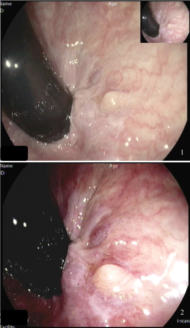
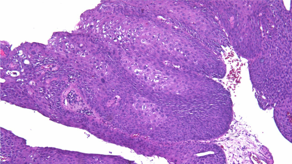
A 66-year-old male patient with hypertension and diabetes mellitus presented for a screening colonoscopy. He had no gastrointestinal complaints, namely no abdominal pain, change in bowel habits, rectal bleeding or anal itching. Colonoscopy showed diffuse diverticular disease throughout the colon. Upon retroflexion in the rectum, a 3 mm cerebriform, pale polypoid lesion was seen just proximal to the dentate line (Figure 1). After closer inspection of the lesion and use of i-scan, this lesion appeared broad based and had a rubbery surface (Figure 2). This single lesion was completely excised with a single pinch using cold biopsy forceps. What do you think this lesion is?
Answer: Rectal Condyloma Acuminatum
The polyp returned a condyloma acuminatum (CA) (Figure 3). CA is caused by herpes papillomavirus (HPV) infection. Low-risk HPV group includes HPV 6 and 11 and are the most prevalent. This group has no malignant potential. There have been cases associated with high-grade intraepithelial neoplasia and warty/basaloid squamous cell carcinoma (SCC) in cases of co-infection with high-risk HPV types (HPV 16 and 18) [1]. CA typically arises in the genitalia, anus, and surrounding areas. Rectal CA is uncommon with only few reported cases, where concomitant anal lesions are present. Our patient had isolated rectal CA. Presumed pathogenesis of rectal CA includes direct infection from anal intercourse and spread of genital or perianal warts into rectal mucosa [2]. Most rectal CA cases are found incidentally during colonoscopy. Meticulous inspection of the anorectal area by straight view and retroflexion is a key for diagnosis. Screening the patient and partners for other sexually transmitted diseases is necessary. Standard treatment for CA includes surgical or immunotherapeutic medications. Endoscopic submucosal dissection has been used in select cases [3]. There are no data for surveillance of rectal CA. It may be reasonable to apply the same surveillance recommendations for anal CA with follow-up every 6-12 months [4].
Conflicts of Interest
No conflicts of interest exist.
Sources of Funding
None.
Author's Contributions
M Shmais: Acquisition of data; analysis and interpretation of data; drafting of the manuscript. FF Francis: Drafting of the manuscript and critical review of the manuscript. JG Hashash: Study concept and design; acquisition of data; analysis and interpretation of data; drafting of the manuscript; and critical review of the manuscript.
Acknowledgement
Informed consent from the patient was obtained for publication of the case details.
References
- Chan MP (2019) Verruciform and condyloma-like squamous proliferations in the anogenital region. Arch Pathol Lab Med 143: 821-831.
- Ye Y, Sun XZ, Feng JS (2015) Woman with rectal condyloma acuminatum: A case report. Int J Clin Exp Med 8: 6365-6368.
- Suzuki K, Suzuki T, Fujita N, et al. (2013) Anorectal condyloma acuminatum treated with endoscopic submucosal dissection. Gastroenterological Endoscopy 55: 281-286.
- Long KC, Menon R, Bastawrous A, et al. (2016) Screening, surveillance, and treatment of anal intraepithelial neoplasia. Clin Colon Rectal Surg 29: 57-64.
Corresponding Author
Jana G Hashash MD, MSc, Assistant Professor of Medicine, Division of Gastroenterology, American University of Beirut, Lebanon, Tel: +961-1-374374, Fax: +961-1-365612
Copyright
© 2021 Shmais M. This is an open-access article distributed under the terms of the Creative Commons Attribution License, which permits unrestricted use, distribution, and reproduction in any medium, provided the original author and source are credited.