Intestinal Metastasis of Primary Lung Tumour as a Cause of Intestinal Wall Thickening: A Case Report
Abstract
We describe the case of a 73-year-old male patient with personal history of pulmonary malignancy, who in a follow up scan, presented multiple areas of nodular wall thickening of small bowel loops. Ultrasound revealed hypoechoic areas of mural thickening with well-defined borders and loss of the layered echostructure of the intestinal wall. The pathology study confirmed the diagnosis of metastasis. Intestinal metastases are a rare entity that should be taken into consideration to avoid under-diagnosis.
Introduction
Gastrointestinal metastases from primary lung cancer are uncommon, although there is a discrepancy between the incidence and the post-mortem studies incidence [1]. This discrepancy seems to indicate that this condition is often under under-diagnosed since most patients are asymptomatic or presenting with nonspecific symptoms [2]. Therefore, radiologic diagnosis on both first and follow up scans may be key to avoid undesired complications and to provide a proper staging.
Case Presentation
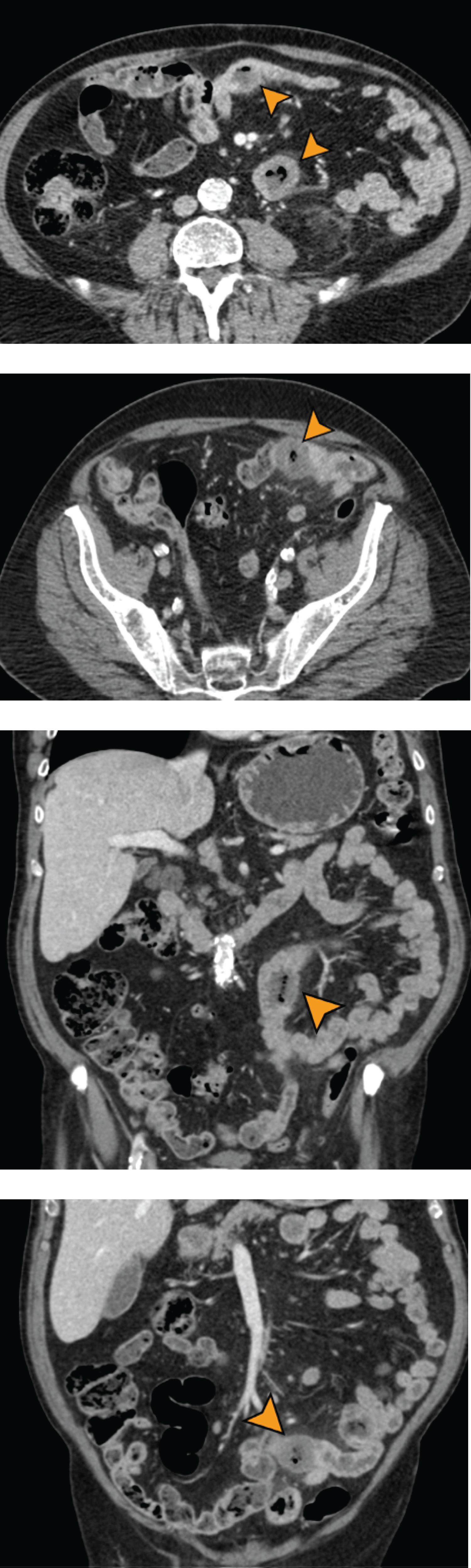
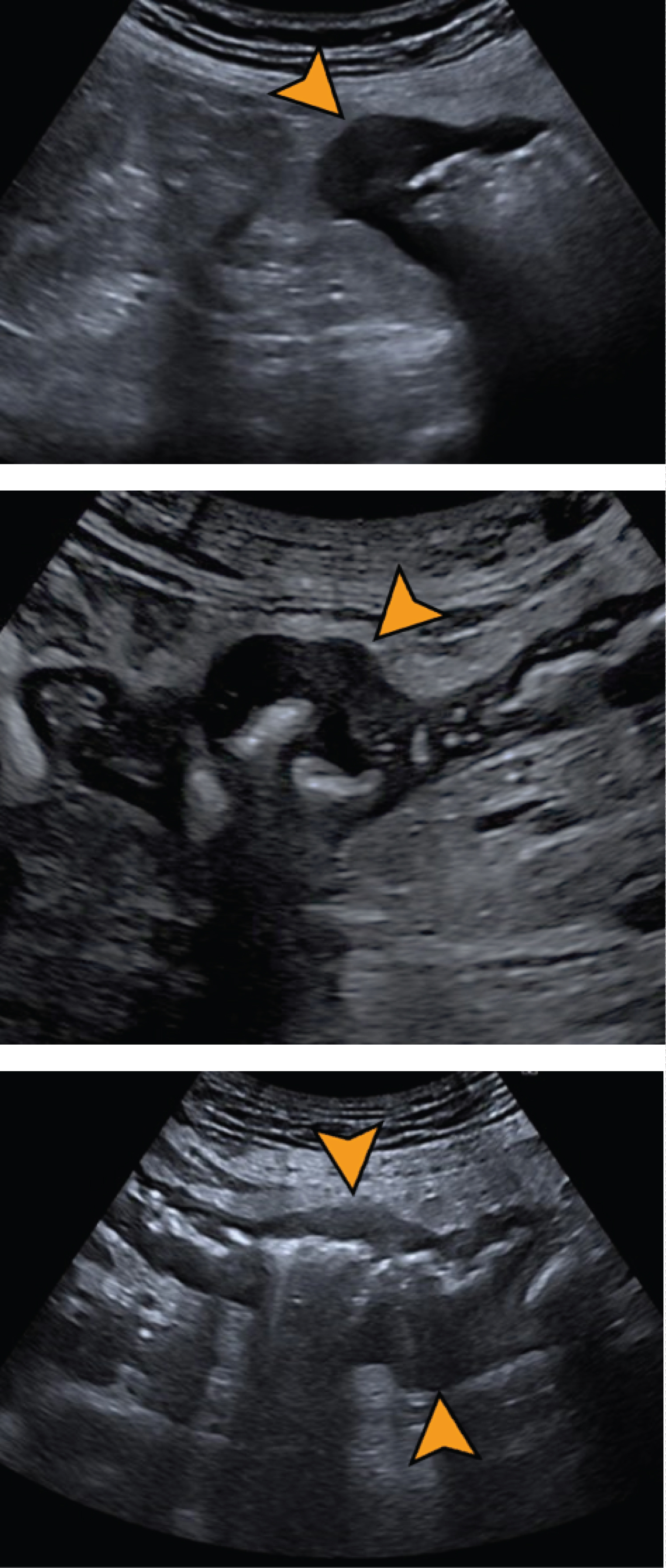
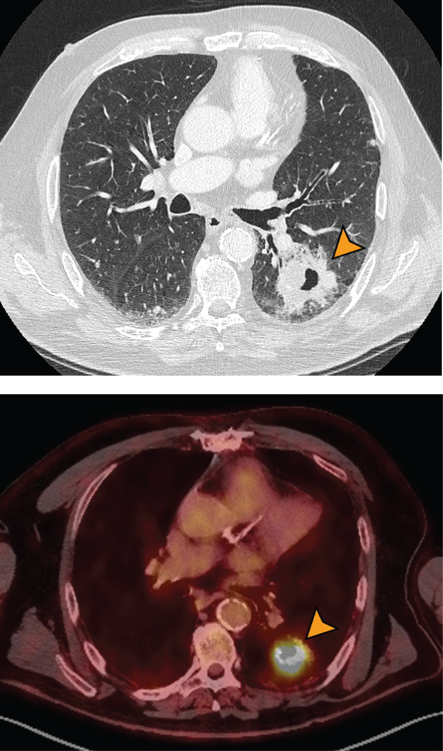
We present a case of a 73-year-old male, controlled due to pulmonary adenocarcinoma. He was treated and achieved a complete response. Whole-body CT scan was performed to follow up purposes, revealing a pulmonary mass and multifocal thickening of small bowel wall (Figure 1). Ultrasound imaging showed thickening of the wall of multiple intestinal loops with loss of layered echostructure (Figure 2). No lymphadenopathy or distant metastases were observed. No other masses were found.
The presence of small bowel metastasis may be difficult to be suspected by the clinician since patients often presents with nonspecific GI symptoms. Abdominal pain or weight loss are also present in patients without small bowel metastasis due to chemotherapy side effects. Small bowel perforation, obstruction, intussusception or acute digestive haemorrhage can be as well the initial manifestation of this condition.
In our case, the presence of the small bowel metastasis was unexpected, as the patient remained asymptomatic.
Discussion
Small bowel metastasis is an under-diagnosed condition, which may have clinical implications. It portents a poor prognosis for the patient, with a very low median survival rate [3].
The use of CT both as a diagnostic as well as a follow up tool has been proved useful in depicting bowel metastasis. Any hypodense nodular thickening of the small bowel wall should raise concern, especially with any ancillary criteria such as adjacent fat stranding or lymphadenopathy [3]. Special attention should be paid to the most frequent sites of metastasis, which would be the ileum followed by jejunum [1] (Figure 3).
We advise the assessment of the layered structure of the intestinal wall using ultrasound, as this finding is better assess using this technique, as it is the experience in our work group. PET-CT scan can also be useful.
Acknowledgement
Department of Pathology for helping with the anatomopathological findings.
Conflict of Interests
None to declare.
Role of Funding Source
Not applicable.
Ethics Committee Approval
Not applicable.
Authors Declaration
Informed consent was obtained from the patient for the case report.
References
- Balla A, Subiela J, Bollo J, et al. (2018) Gastrointestinal metastasis from primary lung cancer. Case series and systematic literature review. Cir Esp 96: 184-197.
- McNeill P, Wagman L, Neifeld J (1987) Small bowel metastases from primary carcinoma of the lung. Cancer 59: 1486-1489.
- Kim SY, Kim KW, Kim AY, et al. (2006) Bloodborne metastatic tumours to the gastrointestinal tract: CT findings with clinicopathologic correlation. AJR Am J Roentgenol 186: 1618-1626.
Corresponding Author
Edgar Lorente, Department of Radiology, Hospital Universitari Doctor Peset, Valencia, Spain
Copyright
© 2021 Lorente E, et al. This is an open-access article distributed under the terms of the Creative Commons Attribution License, which permits unrestricted use, distribution, and reproduction in any medium, provided the original author and source are credited.